Episode 7 – Heart Failure

Episode 7 (iTunes or Listen Here)

The Free Open Access Medical Education (FOAM)

We review RAGE Podcast Episode 4 that awesomely covers nearly everything under the sun. A few of our favorite pearls:

Impact apnea –people who sustain traumatic brain injury often have associated apnea.  Support their airway because this could lead to arrest. Bystander support is crucial.

Right Ventricular Myocardial Infarction (RVMI)– Think about this in any inferior MI situation. Give fluids, these patients are preload dependent and need the cath lab.

Right Ventricular Heart Failure – OH CRAP.

  • Optimize Oxygenation, Hemodynamics, Contractility, Rate/rhythm, Afterload, and Preload
  • Give fluids, but not too much. Inotropes and vasopressors are often necessary. These guys like milrinone and epinephrine.

The Bread and Butter

We summarize some key topics from the following readings,  Tintinalli (7e) Chapters 53, 57; Rosen’s (8e) Chapters 78, 81…but, the point isn’t to just take our word for it.  Go enrich your fundamental understanding yourself!

RVMI – often associated with inferior MIs and can carry increased morbidity/mortality.

ECG pearls –

  • look for ST elevation in lead III greater than lead II or lead V1 (especially with ST depression in V2.
  • Right sided leads – elevation in V4R most specific but elevation in V3R-6R are indicative of RVMI.  Keep V1 and V2
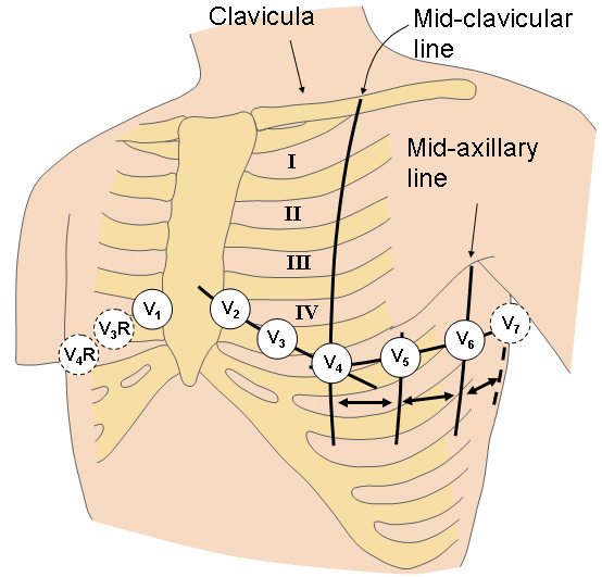
025_ECG_12leads
http://www.elin.ttu.ee/mesel/Study/Courses/3240BME/Content/1_Bioelectricity/BME_2_bioelectric_signals.htm

Treatment – RVMI is preload dependent so they need fluid.  Too much fluid may cause the RV to impinge on the LV.

Acute Heart Failure (WikEM)

  • Give nitrates, which decrease preload, before diuretics.  Diuretics are only indicated in volume overloaded patients, and many patient simply have fluid shifts and are overall euvolemic or have decreased plasma volume.  Thus, in some patients, diuretics may be harmful.
  • Use non-invasive ventilation
  • BNP.  The boards and ACEP recommend it but randomized clinical trials have not consistently demonstrated a benefit in the Emergency Department (Carpenter et al)

Right vs Left Sided Heart Failure – this distinction is someone artificial as chambers are interdependent in series.

  • Left-sided failure – pulmonary symptoms (dyspnea and orthopnea)
  • Right-sided failure have systemic venous congestion(pedal edema and hepatomegaly)

High Output Failure – conditions with excess cardiac output

  • Causes – increased preload (excess mineralocorticoids, fluid/salt retention), decreased systemic vascular resistance (pregnancy, cirrhosis, severe anemia, beriberi, thyrotoxicosis, Paget’s disease, or vasodilator medications), or tachycardia and persistent beta-adrenergic stimulation.
  • Treatment – correct underlying cause

Generously donated Rosh Review questions (scroll for answers)

Question 1.  [polldaddy poll=8168396]

Question 2. A 73-year-old man presents to the ED with progressive shortness of breath for two days without chest pain. The patient has a history of hypertension controlled with hydrochlorothiazide, but has been noncompliant with his medications. In the ED, his vital signs are BP 186/102, HR 108, RR 34, and oxygen saturation 90% on room air. On exam, the patient has pulmonary crackles midway up both lung fields, jugular venous distension, and pitting edema of his lower extremities. A chest X-ray depicts increased interstitial markings and an enlarged cardiac silhouette. An ECG shows sinus tachycardia. [polldaddy poll=8168406]

Question 3.  [polldaddy poll=8169897]

References:

O’Brien JF.  Heart Failure.  Rosen’s Emergency Medicine (8e).  Chapter 81, 1075-1090.e7

Hollander JE, Diercks DB.  Chapter 53:  Acute Coronary Syndromes: Acute Myocardial Infarction and Unstable Angina.”Tintinalli’s Emergency Medicine: A Comprehensive Study Guide, 7e. New York, NY: McGraw-Hill; 2011. p 367-385

Peacock WF.  Chapter 57: Congestive Heart Failure and Acute Pulmonary Edema. Failure. Tintinalli’s Emergency Medicine: A Comprehensive Study Guide, 7e. New York, NY: McGraw-Hill; 2011  p 404-414


Answers:

Question 1.  D. This patient presents with symptoms of acute decompensated heart failure (ADHF) and should be started on nitroglycerin therapy immediately. Patients with ADHF present with shortness of breath, increased work of breathing, tachycardia, hypoxia, crackles on lung examination and jugular venous distension. These patients experience acute worsening of left ventricular function and output secondary to a number of mechanisms including increased systemic vascular resistance. Therapy focuses on reduction of preload to decrease the flow of blood into the lungs and afterload reduction to increased the effectiveness of the left ventricle. Both of these goals can be accomplished through the administration of nitroglycerin. At lower doses, nitroglycerin acts as a peripheral vasodilator and increases venous capacitance leading to decreased preload. At higher doses, nitroglycerin causes arterial vasodilation leading to decreased afterload. Because of its rapid onset of action, nitroglycerin is the first line medication in patients with ADHF. Furosemide (A) may be useful in patients with volume overload and ADHF but this represents less than half of patients with ADHF. Additionally, the effects are delayed. Heparin (B) can be given in cases of ischemia induced ADHF but will not yield any immediate benefits. Morphine (C) was historically used as a preload reducer but has been associated with increased morbidity in patients with ADHF and is no longer recommended.

Question 2.   A .  This patient is in acute heart failure with pulmonary edema. The clinical presentation of heart failure includes shortness of breath, jugular venous distension, crackles and rales, peripheral edema, S3 gallop, orthopnea, and paroxysmal nocturnal dyspnea. A chest X-ray may show an enlarged cardiac silhouette, Kerley B lines suggesting pulmonary edema, and pulmonary vessel cephalization. Labs may show an elevated plasma brain natiuretic peptide (BNP). Management of heart failure with acute pulmonary edema begins with addressing the ABCs.Noninvasive respiratory therapy, such as bilevel positive airways pressure (BiPAP) or continuous positive pressure airway (CPAP) is the most appropriate next step in management. Noninvasive positive pressure ventilation increases oxygenation, decreases the worth of breathing, and decreases preload and afterload. In addition to BiPAP, adjunctive medications include nitrates, diuretics, morphine sulfate, and position the patient sitting up. Nitrates act as venous and arterial vasodilators and help to reduce preload and afterload. Morphine sulfate is thought to decrease oxygen consumption by decreasing catecholamines, decrease preload from mild vasodilator effects and decreases pain and anxiety. However, there are some studies that link the use of morphine sulfate to an increased mortality. Furosemide can be used in patients with evidence of fluid retention (JVD, extremity edema). Pulmonary edema secondary to heart failure usually responds well to preload and afterload reduction with noninvasive ventilation and nitroglycerin. If the patent continues to deteriorate then intubation (C) may be necessary. A myocardial infarction is less likely given the lack of chest pain and ECG that does not reveal ST-segment elevations. Therefore, activation of the cath lab (B) is unnecessary. Hydrochlorothiazide (D) has no role in the acute management of pulmonary edema. Once stabilized, the patient can resume his daily medications.


Question 3.  Correct Answer ( C ) Atrioventricular (AV) conduction blocks occur in 25%–30% of patients with acute myocardial infarction. A narrow complex third-degree AV block in the setting of an inferior wall MI is usually transient and resolves spontaneously. Other AV blocks associated with a favorable prognosis include first-degree heart block and second-degree Mobitz type I (Wenckebach). Patients with a new left bundle branch block (A) in the setting of an acute MI are more likely to develop CHF, AV block, and ventricular fibrillation and have an overall increased mortality. Left posterior hemiblock (B) is associated with a large infarct size, increased risk of cardiogenic shock, and increased mortality. A new right bundle branch (D)in the setting of an anterior wall MI is associated with an increased risk of developing complete AV block and cardiogenic shock.

Episode 5 – Psychiatry and Increased ICP

Episode 5 – Psychiatry & Increased Intracranial Pressure (iTunes or Listen Here

The Free Open Access Medical Education (FOAM)

Academic Life in Emergency Medicine -Atypical Antipsychotic Medication Re-initiation in the Emergency Department

  • Determine how long the patient has been without their atypical antipsychotic.
  • Consider dosing reductions in patients who have been without their medications for more than a few doses or in patients at higher doses.
  • Look up the recommendations.

Broome Docs – Optic Nerve Sheath Diameter (ONSD) – helpful and interesting for gauging increased ICP, but probably not quite ready for prime time.

  • ONSD under 5 mm, ICP is probably OK
  • ONSD over 6 mm, ICP is probably not OK
  • ONSD 5-6 mm utility is uncertain

The Bread and Butter

We summarize some key topics from the following readings,  Tintinalli (7e) Chapters 284, 254 ; Rosen’s (8e) Chapters 110, 29 …but, the point isn’t to just take our word for it.  Go enrich your fundamental understanding yourself!

Psych Pearls

The 1st generation antipsychotics treat the positive psychotic features

  • Hallucinations, delusions, disorganized speech, disorganized behavior

The 2nd generation antipsychotics treat  the negative psychotic features

  • Blunted affect, emotional withdrawal

Brief psychotic disorder vs schizophrenia

  • Brief psychotic disorder is the sudden onset of psychotic symptoms in response to major stress and lasts several days to 1 month (<1 month)
  • Schizophreniform is 1-6months
    •  ⅓ of pts with schizophreniform recover, ⅔ don’t and develop schizophrenia.
  • Schizophrenia is > 6 monthst.

Consider metabolic, drug, and organic causes

  • Thiamine, B12, thyroid, uremia, hepatic encephalopathy, lupus, sarcoid, syphilis, recreational drugs, medication side effects, etc

Diagnosis of Increased ICP

  • Physical exam often inadequate as papilledema often does not show up immediately.
  • ONSD for Increased ICP – How To by the Ultrasound Podcast

  • If >5mm, suggests increased ICP and better than clinical exam [Tintinalli, 8e, p 1549]
  • Cushing’s Triad (bradycardia, agonal respirations, hypertension) – only present in <1/3 of cases of life-threatening increased ICP

Treatment of Increased ICP

  • Normalize the patient – normotensive, normocarbic (35-40 mmHg), normothermic
  • Adequately sedate the patient, raise head of bed to 30 degrees
  • Impending herniation?  These patients may need repeat CT.  Consider mannitol 0.25 – 1 gram/kg IV bolus, consider very brief hyperventilation, call neurosurgery
  • Rosenalli (Rosen’s + Tintinalli) both do not recommend hypertonic saline (3% NaCl) for the treatment of increased ICP

Generously donated Rosh Review questions (scroll for answers)

Question 1.

A young man is involved in a motor vehicle collision and sustains a severe head injury. In the ED, his GCS is 7. His blood pressure is 115/70 mm Hg and heart rate is 85 beats per minute. His pupils are 3 mm and equal and reactive to light. You intubate the patient and place him on a mechanical ventilator. The FAST ultrasound is negative and there are no other obvious injuries.

A. Avoid hypotension

B. Administer mannitol

C. Hyperventilation

D. Initiate induced hypothermia

[accordion]
[toggle title=”Answer” state=”closed”]

B.  In patients with traumatic brain injury (TBI), hypotension has been shown to have a devastating effect on outcome. An early measurement of cerebral blood flow (CBF) in TBI patients demonstrates ischemic levels. This initial inadequacy of CBF may cause irreversible damage. Therefore, maintenance of adequate CBF by avoiding hypotension and maintaining a normal or even elevated blood pressure is one of the key principles in management of TBI. Mannitol (B) is an osmotic agent sometimes utilized in TBI patients when there is evidence of significantly elevated intracranial pressure and impending cerebral herniation.  Although this patient has an abnormal GCS, he does not exhibit signs of impending herniation.

[/toggle]
[/accordion]

Question 2.A 12-year-old boy presents to the emergency department with recurrent headaches. The headaches have been present for the past four weeks and are increasing in intensity. They are worse in the morning and when lying flat, and are associated with vomiting but no nausea. For the past few days, he has complained of blurry vision. His initial exam is notable for altered mental status, extensor posturing, and papilledema. Which of the following are the most likely vital sign abnormalities?

[accordion]
[toggle title=”Answer” state=”closed”]

C. The most common clinically significant traumatic herniation syndrome is uncal herniation, a form of transtentorial herniation.  As the uncus is compressed, cranial nerve III is compressed. The first signs of compression include anisocoria, ptosis, impaired extraocular movements, and a sluggish pupillary light reflex ipsilateral to the expanding mass lesion. As the compression progresses, the ipsilateral pupil dilates and becomes nonreactive.

[/toggle]
[/accordion]

Good FOAM reviews:


Episode 4 – Transfusions and Ingested Foreign Bodies

Episode 4 – Transfusion Emergencies (iTunes or Listen Here

The Free Open Access Medical Education (FOAM) –from Dr. Ryan Radecki’s erudite blog, Emergency Medicine Literature of Note..

Infections & Transfusions” – a JAMA meta-analysis found that higher hemoglobin targets were associated with an increased incidence of infection with a number needed to harm of 20-38.  The  group with a target level of 7-9 g/dL had an infection rate of 11.8% (95% CI, 7.0%-16.7%)  compared with an infectious complication rate of 16.9% (95% CI, 8.9%-25.4%) in the “liberally” transfused group.

Grilling Injuries on Memorial Day” – Grilling isn’t risk free.  Dr. Radecki reviewed a case series of six individuals who presented in one year to a hospital after ingesting meat cooked on a grill.  Three patients had neck pain with wire grill bristles removed via laryngoscopy and three had abdominal pain necessitating removal – 2 by colonoscopy and 1 with urgent surgery secondary to intestinal perforation.

The Bread and Butter

We summarize some key topics from the following readings,  Tintinalli (7e) Chapters 233; Rosen’s (8e) Chapter 7…but, the point isn’t to just take our word for it.  Go enrich your fundamental understanding yourself!

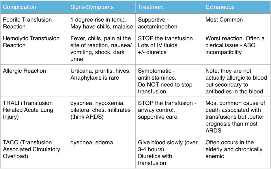
Transfusion Reactions

Immediate Reactions – great FOAM summary from Life in the Fast Lane: Transfusion Risks, Transfusion Reactions 

Screen Shot 2014-06-08 at 9.39.12 PM


The Ones Our Patients Care About (Infectious – statistics are US based)

  • Bacterial contamination is rare: 1/500,000 – 1/1,000,000
    • Most common pathogen: Yersinia Entercolitica
  • More common in platelets: 1/1000-1/2000 per Rosen, CDC, and the AABB  (Tintinalli cites 1 in 6 million)
  • Most Common virus: Parvovirus B19 (1 in 10,000).  The others are very very rare: HIV and Hepatitis C > 1 in 1 million, Hepatitis B 1 in 100,000-200,000

Ingested Foreign Bodies

  • Beware the button battery. These can cause necrosis within hours in the esophagus and must be removed ASAP.
  • Objects that are irregular, very sharp, or have dimensions greater than 2.5cm in width or 6 cm in length that are still in the stomach or duodenum – call GI to have these removed via endoscopy.

Generously donated Rosh Review questions (scroll for answers)

Question 1 A 55-year-old woman is receiving a blood transfusion due to persistent vaginal bleeding and a hemoglobin of 5 mg/dL. While receiving the transfusion, she develops fever, chills, back pain, pain at the site of transfusion, and tachycardia. [polldaddy poll=8109900]

Question 2 A 28-year-old man presents with a 1-day history of rectal bleeding. In the ED, he is hypotensive, thrombocytopenic, and is found to be passing melena. He receives a transfusion of platelets and packed red blood cells as part of his resuscitation. Twenty minutes after the start of his platelet transfusion, his BP is 90 mm Hg systolic, he becomes dyspneic, and his oxygen saturation drops from 99% on room air to 91% on 2L of oxygen supplementation. On exam, you note rales at the lung apices and that he is using accessory muscles to breathe. His chest radiograph shows diffuse interstitial infiltrates. [polldaddy poll=8109907]

Question 3 [polldaddy poll=8109908]


References:

Emery M.  Blood and Blood Products.  Rosen’s Emergency Medicine. 2014: 8th ed. p 75-80.e2

Coil CJ, Santen SA.  Transfusion Therapy  Tintinalli’s Emergency Medicine: A Comprehensive Review. 7th ed.

Hillyer CD, Josephson CD, Blajchman MA, et al. Bacterial contamination of blood components: risks, strategies, and regulation: joint ASH and AABB educational session in transfusion medicine. Hematology Am Soc Hematol Educ Program. 2003:575-89.

1. C – Up to 20% of all transfusions may lead to some type of adverse reaction. Although most of these reactions are minor, some are life-threatening. The patient is having an acute intravascular hemolytic reaction. This occurs when the recipient’s antibodies recognize and induce hemolysis of the donor’s red blood cells and may result in activation of the coagulation system and disseminated intravascular coagulation. This type of reaction typically presents with back pain, pain at the site of transfusion,headache, fever, hypotension, dyspnea, tachycardia, chills, bronchospasm, pulmonary edema, bleeding, and development of renal failure. First, stop the transfusion. Then initiate intravenous hydration to maintain diuresis.

2.D- This patient is most likely suffering from transfusion-related acute lung injury (TRALI), one of the leading causes of transfusion-related mortality. It is most closely associated with platelet and fresh frozen plasma transfusions, though cases have been reported with packed red blood cells since there is some residual plasma in the packed cells. Symptoms begin abruptly during transfusion or within 6 hours and resemble adult respiratory distress syndrome with noncardiogenic pulmonary edema, dyspnea, hypoxemia, and bilateral infiltrates on chest radiograph.

3. A- The patient is experiencing an allergic reaction without serious signs or symptoms. The transfusion does not need to be stopped for such a reaction; an antihistamine will help to relieve symptoms.

Episode 3 – Ear Emergencies

Episode 3 – Ear Emergencies (iTunes

The Free Open Access Medical Education (FOAM) – SMART EM Pseudoaxioms 2 and Literature Update

The podcast reviews:

  • Bullous Myringitis, a painful infection of the tympanic membrane, is typically caused by viruses, strep. pneumoniae and moraxella and, less commonly, mycoplasma.  The boards have caught up with this. Check out this review by Mellick.
  • PHANTOM-S trial: Use of the STEMO (Stroke Emergency Mobile – an ambulance with neurologist and a CT scanner for suspected strokes) reduced mean alarm-to-treatment time by 25 minutes (95% CI, 20-29; P < .001) without an appreciable neurologic benefit for this intensive intervention.
  • An article by Gregg et al in the NEJM discussed incredible improvements in diabetes outcomes as a result of tight glycemic control. Dr. Newman asserts that much of this is secondary to dilution, as the definition of diabetes changed in 1997.  This resulted in more people with less severe disease being diagnosed with diabetes.

The Bread and Butter

We summarize some key topics from the following readings,  Tintinalli (7e) Chapters 237; Rosen’s (8e) Chapter 92.  A good read on common ear emergencies from EBMedicine…but, the point isn’t to just take our word for it.  Go enrich your fundamental understanding yourself!

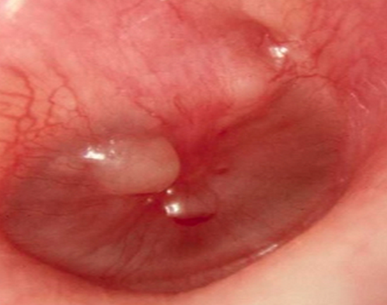
Bullous Myringitis

This is a painful infection characterized by bullae on the tympanic membrane (TM), which has a richly innervated outer epithelium (hence the severe otalgia). Patients may collect fluid behind their TM or have a concomitant otitis media.

Etiology: viruses, typical otitis media pathogens.  Mycoplasma and chlamydia have been associated with bullous myringitis but the association is unclear and these are not the most common causes.

Treatment: pain control, pain control, pain control.  Antibiotics are optional in most cases. But then again, antibiotics are not necessary in most cases of otitis media.  Both Rosen and Tintinalli are on board with this.

Perforated Tympanic Membrane

Photo:Didier Descouens (Wikimedia Commons)

Etiology: infection, trauma (q-tips, instrumentation), changes in pressure (diving, flying)

Treatment:  Keep the ear canal dry, follow up with ENT.  Most of these patients can go home.  If the injury is in the posteriorsuperior aspect of the TM or secondary to penetrating trauma, they should see ENT within 24 hours because they may have damage to the bones of the middle ear.

Auricular Hematoma

Etiology: Blunt trauma (often associated with boxing, fights, or termed “rugby ear“)

Treatment:  Incision, drainage, and compression dressing/splint.  Photo guide to repair.

Ototoxic Agents (great table in Tintialli 8 e, Ch 237, p1551)

Risk of hearing loss typically increases with exposure to medication (dose and length of use) and issues with clearance such as renal insufficiency may cause medications to hang around longer than anticipated or at higher levels.

Loop Diuretics: furosemide, bumetanide, ethacrynic acid

Salicylates (aspirin and quinine), NSAIDs

Antibiotics that end in -mycin or -micin: aminoglycosides (gentamicin), vancomycin, erythromycin

Chemotherapeutic agents: vincristine, vinblastine, cisplatin, carboplatin

Topical agents: ethanol, polymixin B, neomycin

Sudden Hearing Loss Differential Diagnosis 

Occurs over the span of three days.

Differential Diagnosis categories for any ailment can be remembered by the mnemonic VINDICATE

  • Vascular – sickle cell, polycythemia
  • Infectious/Inflammatory – viruses (zoster oticus or herpes, EBV, CMV, mumps), syphilis, labyrinthitis, temporal arteritis
  • Neoplasms – leukemia, masses,  acoustic neuroma
  • Drugs –  loop diuretics, antibiotics that end in -mycin or -micin (aminoglycosides like gentamicin, vancomycin, erythromycin), salicylates and NSAIDs, chemotherapeutic agents (cisplatin, carboplatin, vinblastine, vincristine, and topical agents (ethanol, polymyxin B, neomycin)
  • Iatrogenic/idiopathic – perforated TM, idiopathic endolymphatic hydrops (Meniere’s disease) – vertigo, hearing loss, tinnitus
  • Autoimmune -granulomatosis with polyangitis (Wegener’s)
  • Trauma – ruptured TM
  • Endocrine – diabetes, high cholesterol

Necrotizing Otitis Externa (Malignant Otitis Externa) – an infection that can turn into osteomyelitis of the skull

Presentation: otalgia, headache, and swelling and tenderness around the ear particularly in the setting of a prolonged course of otitis externa.  Diagnosis often requires CT scan to gauge involvement.

Epidemiology: Diabetics, immunocompromised

Etiology: Pseudomonas (90%)
Treatment:   Pediatrics- imipenem or an aminoglycoside and an antispeudomonal penicillin.  Adults – cephalosporin or quinolone

Dispo: Mild cases with good follow up can get oral quinolones as outpatients. More severe cases – admission, IV antibiotics, and perhaps surgical debridement.

Generously donated Rosh Review questions (scroll for answers)

Question 1.[polldaddy poll=8093105]Screen Shot 2014-06-01 at 9.28.25 PM

Question 2.

A 36-year-old woman presents to the ED complaining of decreased hearing and increased fullness to the right ear. Over the last week, she has used cotton-tipped applicators to attempt to remove cerumen from her right ear. On exam, you notice a cerumen-impacted external canal on the right. You irrigate the right ear with warm saline using an 18-gauge IV catheter and a plastic curette to remove the cerumen. During the procedure, the patient has sudden and complete hearing loss to the right ear. [polldaddy poll=8093098]


Question 3. A 16-year-old girl presents complaining of pain behind her left ear. She thought the pain was due to an ear infection and took three of her boyfriend’s leftover antibiotic tablets without seeing her primary care physician. Her ear pain improved for a couple of days, but now she is complaining of fever and discharge from the external auditory canal. Her vitals are T 38.4°C, BP 120/80, HR 108, and RR 18. On physical examination, she has postauricular tenderness, swelling, and erythema. You note purulent otorrhea through a perforated tympanic membrane. [polldaddy poll=8096505]

Answers:

1. D-Bullous myringitis was previously linked to Mycoplasma pneumoniae but it appears, based on middle ear aspirate culture results, that typical acute otitis media pathogens are the true cause. Among these, Streptococcus pneumoniae is most common.
2. C – The patient does not require admission (A) to the hospital. ENT care can be arranged for as an outpatient. Traumatic tympanic membrane perforations do not require otic antibiotics (B) unless the ear was contaminated such as from diving in seawater or the rupture is secondary to infection. The patient should receive more than ac otton ball (D) in her ear. Her management should include analgesia and ENT follow-up because complications of tympanic membrane rupture include facial nerve palsy, vertigo, and hearing loss.

3. C – This patient has necrotizing otitis externa.

References:

Silverberg M, Lucchesi M.  Common Disorders of the External, Middle, and Inner Ear.  Tintinalli’s Emergency Medicine, A Comprehensive Study Guide, ed 7. New York, McGraw-Hill, 2011, (Ch) 237:p 1556-1557.

Episode 2: Urologic Emergencies

Episode 2 – Urologic Emergencies (iTunes

The Free Open Access Medical Education (FOAM) – The Skeptics Guide to Emergency Medicine (SGEM) Episode 71

The podcast reviews: Tamsulosin for ureteral stones: a systematic review and meta-analysis of a randomized controlled trial.

  • Paper’s Conclusion:  ‘Tamsulosin is a safe and effective medical expulsive therapy choice for ureteral stones. It should be recommended for most patients with distal ureteral stones before stones are 10 mm in size. In the future, high-quality multicenter, randomized and placebo- controlled trials are needed to evaluate the outcome.”

The SGEM’s analysis:  Tamsulosin is useless in most ED patients with ureteral colic unless their stone size exceeds at least 4mm.

The Bread and Butter

We summarize some key topics from the following readings,  Tintinalli (7e) Chapters 95, 97; Rosen’s (8e) Chapter 99, but the point isn’t to just take our word for it.  Go enrich your fundamental understanding yourself!

Renal Colic

Diagnostics

  • Urinalysis demonstrating microscopic hematuria.  Note: 10-15% of patients with renal colic have no hematuria
  • Imaging isn’t needed in patient’s with a history of renal colic and symptoms consistent with their previous episodes and without signs of symptoms of significant obstruction or infection.  Non-contrast CT scan is currently the standard diagnostic imaging of choice and bedside ultrasound may be used to look for hydronephrosis, but isn’t great for picking up stones.  The Ultrasound Podcast guys have a great episode on the topic.  Plain films (KUBs) are only useful in following the location of the stone after CT scan.
  • Make sure you’re not dealing with a tricky abdominal aortic aneurysm.

Management

  • Analgesics – nonsteroidals and, if needed, narcotics
  • Anti-emetics
  • Use of alpha-antagonist such as tamsulosin is controversial, as indicated in The SGEM podcast.  Tintinalli supports the use of these agents, whereas Rosen’s cautions that use of this medication is controversial.
  • Disposition – home with a strainer to catch the stone and outpatient urology follow up if patient has adequate pain control and oral intake and lacks significant infection or obstruction.  Remember, stones <5mm are going to pass on their own most of the time (~95%), whereas patients with stones >8mm will undergo intervention 95% of the time.

Infected Kidney Stones – Suspect in patients with SIRS criteria or those that appear sick (don’t forget that temperature <36 C, 10% bands, <4K white blood cells, and elevated respiratory rate are all part of SIRS) and in those with signs of infection on their urinalysis.

Management

  • Urgent or emergent urologic consultation to evaluate the need for drainage and for relief of the obstruction.
  • Treat sepsis and shock with good sepsis care including antibiotics and fluid resuscitation but these patients may need immediate operative intervention by urology for adequate source control.  This may include a stent or percutaneous nephrostomy tube.  Call urology.

Acute Urinary Retention

Causes

  • Spontaneous – Benign Prostatic Hypertrophy (BPH)
  • Precipitated – a host of medications (pseudoephedrine, NSAIDs, anticholinergics), anesthesia, strictures, masses, spinal cord compression (most sensitive finding in cauda equina!), infection

Diagnosis

  • Palpate the patient’s abdomen, feel for a distended bladder
  • Ultrasound, checking for a post-void residual >150 cc (LxWxH x 0.52 – although, notably there are a variety of coefficients to multiply by based on the shape)
  • Check a urinalysis, BUN, creatinine
  • History and physical should guide further testing with regard to etiology

Management

  • Treat the underlying cause (stop the offending medication, treat the infection, etc)
  • Place a foley catheter to relieve the obstruction.  There is some literature on spontaneous voiding trials in the ED but this isn’t standard (see this Academic Life in Emergency Medicine article).
  • Urology follow up within 3-7 days
  • Admit patients with signs of sepsis, co-morbidities, or renal insufficiency (or those that won’t follow up otherwise).

Generously donated Rosh Review questions (scroll for answers)

Question 1 [polldaddy poll=8057572]

Question 2 [polldaddy poll=8077276]

Listen 


Answers:

1) C.  The 3 primary predictors of stone passage without the need for surgical intervention are stone size, stone location, and the degree of patient pain at discharge. The most important factor, however, is calculus size. Approximately 90% of calculi smaller than 5 mm pass spontaneously within 4 weeks.

2) A.  Indications for hospitalization: intractable nausea/vomiting, severe dehydration, pain, associated UTI, solitary or transplanted kidney, high grade obstruction.

References:

Tintinalli (8e) Chapter 97.  Tintinalli’s Emergency Medicine: A Comprehensive Study Guide, 7e. 2011.

Ban KM, Easter JS.  Selected Urologic Problems.  Chapter 99.  Rosen’s Emergency Medicine.  (8e) p 1326-1354

Yen D, Lee C. Chapter 95. Acute Urinary Retention. Tintinalli’s Emergency Medicine: A Comprehensive Study Guide, 7e. 2011.

Episode #1: EMCrit Episode #122 – Cyanide and Carbon Monoxide Toxicity

Welcome

Welcome to FOAMcast, a podcast created by residents who love Free Open Access Medical education (FOAM).  We are looking at cutting edge FOAM and distilling it down to the basics.  We’re not doing this to replace reading and hard work but increase interest and direct listeners to linking sexy FOAM with core content.  So listen, and go read it yourself.

What Did Weingart and Dr. Nelson say?

  • Patient in extremis or arrest after a fire and burn size doesn’t correlate to severity of illness?  Treat for cyanide toxicity.
  • Treatment: hydroxocobalamin 5g IV in ~250mL normal saline in adults or 70 mg/kg in pediatric patients.
  • Get labs, including lactate, carboxyhemoglobin level, and transaminases prior to giving hydroxocobalamin if possible because the drug turns everything red and interferes with these tests.

And the Basics of Chemical Asphyxiants?

If a patient presents after smoke exposure, consider cyanide and carbon monoxide toxicity (Case Quiz).  These toxicities have many similarities such as: impaired oxygen delivery and utilization, metabolic acidosis with elevated lactate, and presence in patients with smoke inhalation.

Cyanide Toxicity – Tintinalli (7e) Ch 198; Rosen’s (8e) Ch 179

Mechanism – Binds to the iron of the cytochrome a3 of complex IV in the mitochondria, the last step of oxidative phosphorylation, effectively shutting down the mitochondria and ATP production leading to tissue hypoperfusion.

Diagnostics-

  • Cyanide level is worthless in the acute setting.  If suspicious, treat without waiting for labs.
  • Labs often demonstrate a metabolic acidosis and, in a fire victim, a lactate >10 mmol/L is suspicious for cyanide toxicity.

Treatment

  • ABCs – 100% oxygen, crystalloids and vasopressors for hypotension
  • Hydroxocobalamin 5 g IV for adults or 70 mg/kg IV for pediatrics
    • Cyanide binds to hydroxocobalamin, forming cyanocobalamin (vitamin B12) which is renally excreted.  It also turns everything red, which can interfere with labs and dialysis.
    • Note: Tintinalli cautions that there’s no good evidence on hydroxocobalamin over the traditional sodium nitrite kits.
  • There’s also the traditional cyanide antidotes which include: inhaled amyl nitrite, Sodium nitrite 3% – 300 mg IV (10 mL), and sodium thiosulfate.
    •  Sodium nitrite forms methemoglobin from hemoglobin, for which cyanide has enormous affinity.  Cyanide leaves the cytochrome, setting the mitochondria free, forming cyanmethemoglobin. This is transformed to thiocyanate by an enzyme (rhodanese) and renally excreted. 
    • If using this approach in a patient with carbon monoxide poisoning, use only sodium thiosulfate given these patient already have impaired tissue oxygenation and methemoglobinemia only further exacerbates this.

Carbon Monoxide (CO) – Tintinalli (7e) Ch 217; Rosen’s (8e) Ch 159

Carbon monoxide poisoning is non-specific and may manifest as headache, flu like illness, or coma and death and occurs throughout the year, not just during heat/generator seasons.

Mechanism –

  • Most well recognized – CO has a far greater affinity for hemoglobin than oxygen, leading to impaired delivery of oxygen to tissues.
  • Causes a left shift of the hemoglobin-oxygen dissociation curve (Right shift = Removal of oxygen from hemoglobin.  Left shift = loaded hemoglobin).
  • Inhibits the cytochrome system in aerobic metabolism, akin to cyanide toxicity, leading to a shift toward anaerobic metabolism.

Diagnostics –

  • Clinical suspicion is key.
  • Labs may show a metabolic acidosis with elevated lactate.
  • Carboxyhemoglobin level is often available and for boards, remember that levels >15% in pregnant patients or >25% in other patients may be a trigger to think about hyperbaric oxygen therapy.  These levels do not correlate with symptoms.

Treatment –

  • ABCs, including high inspired oxygen which reduces the half-life of CO from ~4 hrs to 90 minutes.
  • Hyperbaric oxygen (HBO) or “diving” patients in controversial but if a patient is near-dead, pregnant with significant toxicity (level >15), consider HBO, at least on the boards.

Questions generously donated by the Rosh Review

Question 1.  [polldaddy poll=8074537]


Question 2. [polldaddy poll=8074535]


Episode 1: Cyanide and Carbon Monoxide Toxicity

(iTunes)

References:

Gresham C, LoVecchio F.  Chapter 198.  Inhaled Toxins. Tintinalli’s Emergency Medicine: A Comprehensive Study Guide, 7e. New York, NY: McGraw-Hill; 2011. p 1317-1320.

Nelson RS, Hoffman RS.  Chapter 159.  Inhaled Toxins.  Rosen’s Emergency Medicine, 8e.  2014.  p 2036-2043.

Maloney G. Chapter 217. Carbon Monoxide.  Tintinalli’s Emergency Medicine: A Comprehensive Study Guide, 7e. New York, NY: McGraw-Hill; 2011. p 1410-1413.


Answers.  1) A  2)A