Thromboelastography (TEG) Guided Resuscitation

iTunes or Listen Here

Thromboelastography (TEG) or its related counterpart rotational thromboelastometry (ROTEM) have gained in popularity over the past several years. These tests assess viscoelastic clot strength in whole blood. These tests may offer more granular and potentially reliable information on the patient’s clot formation and fibrinolytic state than traditional measures of coagulation such as International Normalized Ratio (INR), partial thromobplastin time (PTT), and prothrombin time (PT).

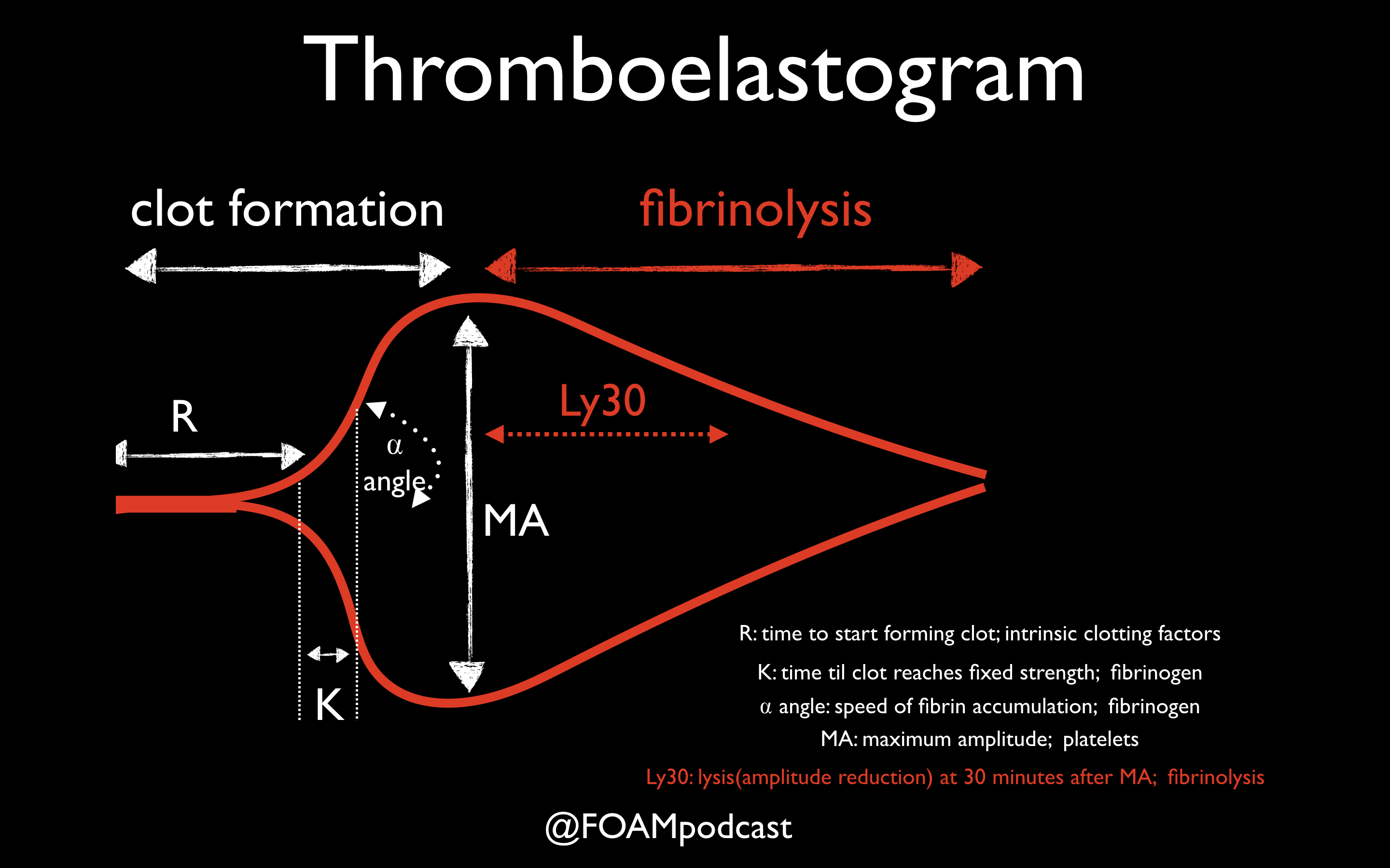
One of the primary advantages to TEG/ROTEM is the ability to target transfusion related therapies to the patient’s overall coagulation profile. Below are some common patterns that emerge and the recommended therapies.

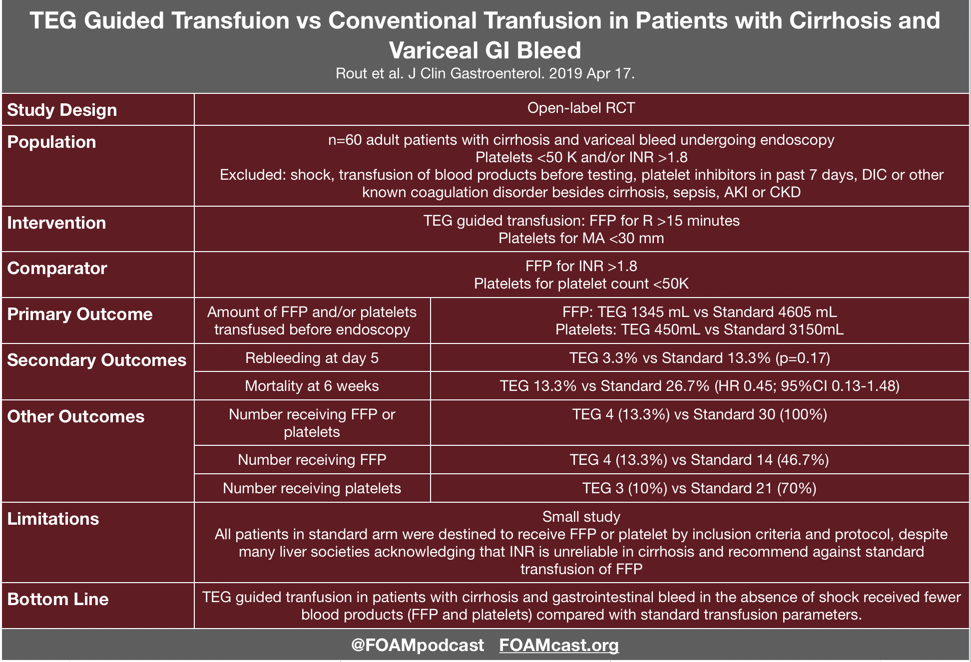
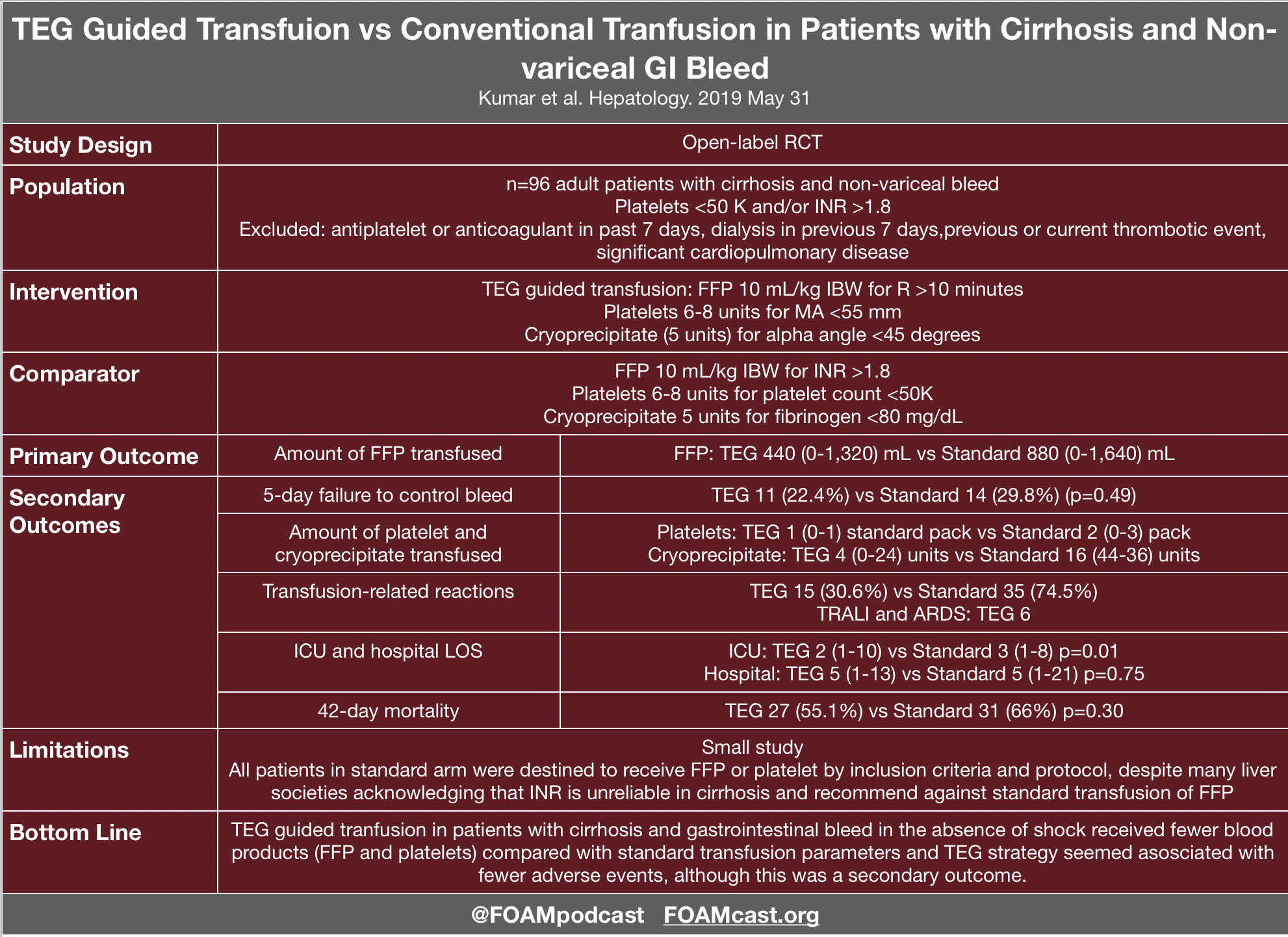
Evidence for TEG in Cirrhosis

Additional FOAM resources: PulmCrit

References:

  1. Wikkelsø A, Wetterslev J, Møller AM, Afshari A. Thromboelastography (TEG) or thromboelastometry (ROTEM) to monitor haemostatic treatment versus usual care in adults or children with bleeding. Cochrane Database Syst Rev. 2016;(8):CD007871.
  2. Kumar M, Ahmad J, Maiwall R, et al. Thromboelastography-Guided Blood Component Use in Patients With Cirrhosis With Nonvariceal Bleeding: A Randomized Controlled Trial. Hepatology. 2019; In Press
  3. Rout G, Shalimar, Gunjan D, et al. Thromboelastography-guided Blood Product Transfusion in Cirrhosis Patients With Variceal Bleeding: A Randomized Controlled Trial. J Clin Gastroenterol. 2019; In Press.
  4. Goodman MD, Makley AT, Hanseman DJ, Pritts TA, Robinson BR. All the bang without the bucks: Defining essential point-of-care testing for traumatic coagulopathy. J Trauma Acute Care Surg. 2015;79(1):117-24.

You are called to the bedside for a postoperative patient who is hypotensive, febrile, and has acute onset of respiratory distress following the initiation of a blood transfusion. Which of the following is the most appropriate initial action in the management of this patient?

A. Apply oxygen

B. Call the blood bank

C. Order steroids

D. Stop the transfusion

[accordion]
[toggle title=”Answer” state=”closed”]

Transfusion-related acute lung injury is a blood transfusion complication characterized by a rapid onset of non-cardiogenic pulmonary edema. The pathogenesis is thought to be a two-part mechanism involving neutrophil sequestration with priming in the lung microvasculature followed by neutrophil activation by a factor in the blood product. Pre-transfusion risk factors include current smoking, chronic alcohol use, liver transplantation surgery, positive fluid balance, shock, and higher ventilated peak airway pressures. Though there is an association of transfusion-related acute lung injury with all blood products, high-plasma-volume products (plasma, apheresis platelet concentrations, and whole blood) have the greatest risk. Clinical presentation may occur immediately after the initiation of the blood transfusion although it can be delayed up to six hours. The patient may rapidly develop acute respiratory distress syndrome, with symptoms that may include hypoxemia, fever, hypotension, cyanosis, pulmonary infiltrates on chest imaging, and if intubated, pink frothy secretions when suctioned. Treatment involves immediate discontinuation of the transfusion followed by supportive care of the acute respiratory distress syndrome. This includes oxygen supplementation and hemodynamic support. Apply oxygen (A), call the blood bank (B), and order steroids (C) are treatments for transfusion-related acute lung injury but are not the most appropriate in the initial management. Discontinuing the transfusion, which is the cause of the acute lung injury, is the immediate need upon identifying the condition.

[/toggle]
[/accordion]

Top Literature of 2019 – Mid Year Review

iTunes or Listen Here

Risk Stratification and D-dimer in Pregnant Patients With Suspected Pulmonary Embolism (PE)

Van der pol LM, Tromeur C, Bistervels IM, et al. Pregnancy-Adapted YEARS Algorithm for Diagnosis of Suspected Pulmonary Embolism. N Engl J Med. 2019;380(12):1139-1149.

Infectious Disease Society of America (IDSA) Guidelines for Asymptomatic

Nicolle LE, Gupta K, Bradley SF, et al. Clinical Practice Guideline for the Management of Asymptomatic Bacteriuria: 2019 Update by the Infectious Diseases Society of America. Clin Infect Dis. 2019.

Benzodiazepine dosing for seizures

Sathe AG, Tillman C, Coles LD, et al. Underdosing of benzodiazepines in patients with status epilepticus enrolled in Established Status Epilepticus Treatment Trial. Acad Emerg Med. 2019 Jun 4.

  • Outcomes after Resuscitative Endovascular Balloon Occlusion of the Aorta (REBOA) in Trauma Patients

Joseph B et al. Nationwide analysis of resuscitative endovascular balloon occlusion of the aorta in civilian trauma. JAMA Surg 2019. Mar 20.

Additional References:

  1. Brophy GM, Bell R, Claasen J, et al. Guidelines for the evaluation and management of status epilepticus. Neurocrit Care. 2012;17(1):3-13.
  2. Glauser T, Shinnar S, Gloss D, et al. American Epilepsy Society Guideline Evidence-Based Guideline: Treatment of convulsive status epilepticus in children and adults: report of the guideline committee of the American Epilepsy Society. Epilepsy Curr. 2016;16(1):48-61.

Psychogenic Non-epileptic Attacks (PNEA)

iTunes or Listen Here

Psychogenic non-epileptic attacks (PNEA) have been formally referred to as pseudoseizures or psychogenic non-epileptic seizures. Recently, this entity has been rebranded to PNEA to reflect that there is no actual seizure activity and potentially prevent confusion and mislabeling among patients.

A 10-year-old girl presents with episodes of brief staring spells that she is unaware of. The patient’s mother states that the onset and termination of the spells are abrupt. The patient is otherwise healthy. She has lots of friends at school and has excellent academic performance. Physical examination and laboratory studies are normal at this time. Which of the following is the most likely diagnosis?

  1. Absence seizure
  2. Attention deficit hyperactivity disorder
  3. Autism spectrum disorder
  4. Complex partial seizure

[accordion]
[toggle title=”Answer” state=”closed”]

  1. Absence seizure is a type of generalized seizure characterized by loss of consciousness sometimes accompanied by myotonic, atonicclonic, or tonic components, autonomic components (e.g., enuresis), or automatisms. The onset of absence seizure commonly occurs in early childhood with a predilection to the female sex. It usually terminates by the age of 20 years or may transform into another form of generalized seizure. Absence seizure may be typical or atypical. The onset and termination of a typical absence seizure are abrupt while they may be gradual in an atypical absence seizure. Additionally, atypical seizure commonly occurs in patients with multiple seizure types and demonstrates more marked changes in tone. Patients with an atypical absence seizure may also have a developmental delay or mental retardation. Patients with absence seizure may miss a few words or break off mid-sentence during an attack. The attack period is so brief that the patient is unaware of it. Physical examination may be normal. Having the child hyperventilate for three to five minutes may precipitate a staring attack. Diagnosis of typical absence seizure is with electroencephalography that shows bursts of bilateral synchronous and symmetric 3-Hertz spike-wave activity. A slower spike-wave discharge is noted in an atypical absence seizure. Treatment is with antiepileptic medications including ethosuximide and valproic acid. All patients with absence seizure should be referred to a neurologist for management. Drug levels of valproic acid should be monitored.While attention deficit hyperactivity disorder (B) has a childhood onset, it is marked by symptoms of inattentiveness, hyperactivity, and impulsivity that are not present in the vignette above. Autism spectrum disorder (C) may also have an early onset. However, patients suffer from pervasive difficulties with social communication and repetitive interests and behavior that are absent in the vignette above. In complex partial seizure (D), the duration of seizure usually lasts > 30 seconds, automatisms are present, and the termination of the seizure is gradual.

[/toggle]
[/accordion]

References:

  1. Lafrance WC, Baker GA, Duncan R, Goldstein LH, Reuber M. Minimum requirements for the diagnosis of psychogenic nonepileptic seizures: a staged approach: a report from the International League Against Epilepsy Nonepileptic Seizures Task Force. Epilepsia. 2013;54(11):2005-18.
  2. Wardrope A, Newberry E, Reuber M. Diagnostic criteria to aid the differential diagnosis of patients presenting with transient loss of consciousness: A systematic review. Seizure. 2018;61:139-148.
  3. Chen et al. Value of witness observations in the differential diagnosis of transient loss of consciousness.  Neurology. 2019 Feb 26;92(9):e895-e904. doi: 10.1212/WNL.0000000000007017. Epub 2019 Jan 25.
  4. Shmuely S, Bauer PR, Van zwet EW, Van dijk JG, Thijs RD. Differentiating motor phenomena in tilt-induced syncope and convulsive seizures. Neurology. 2018;90(15):e1339-e1346.
  5. Doğan EA, Ünal A, Ünal A, Erdoğan Ç. Clinical utility of serum lactate levels for differential diagnosis of generalized tonic-clonic seizures from psychogenic nonepileptic seizures and syncope. Epilepsy Behav. 2017;75:13-17.

Emergent Issues in Sickle Cell Disease

iTunes or Listen Here

Sickle cell disease (SCD) is a terminal disease resulting from sickled blood cells caused by an abnormally folded beta-globin chain. The sickled cells cause occlusion and hyperviscosity leading to a myriad of complications. Unfortunately, some stigma associated with SCD persists in the healthcare field leading to undertreatment of pain or marginalization of patients. The American Society of Hematology (ASH) currently has draft recommendations that are open for public comment until May 13, 2019.

Which of the following is the most common cause of an aplastic crisis in a child with sickle cell disease?

A. Epstein-Barr virus

B. Parvovirus B19

C. Salmonella

D. Streptococcus pneumoniae

[accordion]
[toggle title=”Answer” state=”closed”] 
B. Predominantly affecting individuals of African ancestry, sickle cell anemia is a genetic disease that results in the formation of sickled red blood cells. Affected patients are homozygous for sickle hemoglobin (HbSS) which results in deoxygenated red blood cells developing a sickle or crescent shape. This leads to inflexible red blood cells, increased blood viscosity, and decreased blood flow within organs or extremities. A complication of sickle cell anemia is a transient aplastic crisis. This is caused by a short-lived stoppage of erythropoiesis, resulting in acute reductions in red cell precursors in the bone marrow, severely reduced reticulocytes in the peripheral blood, and an abrupt drop in hemoglobin level. Return to normal erythropoiesis usually occurs within two to 14 days. This transient aplastic crisis is typically caused by infection, with Parvovirus B19 being the most common etiologic agent in children. Individuals with a transient aplastic crisis are managed with transfusion. [/toggle]
[/accordion]

References:

  1. Glassberg J, Tanabe P, Richardson L, Debaun M. Among emergency physicians, use of the term “Sickler” is associated with negative attitudes toward people with sickle cell disease. Am J Hematol. 2013;88(6):532-3.
  2. Aisiku IP, Smith WR, Mcclish DK, et al. Comparisons of high versus low emergency department utilizers in sickle cell disease. Ann Emerg Med. 2009;53(5):587-93.
  3. Lovett PB, Sule HP, Lopez BL. Sickle Cell Disease in the Emergency Department. Hematol Oncol Clin North Am. 2017;31(6):1061-1079.
  4. Glassberg JA, Tanabe P, Chow A, et al. Emergency provider analgesic practices and attitudes toward patients with sickle cell disease. Ann Emerg Med. 2013;62(4):293-302.e10.

Mechanical CPR, Balloon Tamponade, and Advocacy

iTunes or Listen Here

We are at #SMACC in Sydney, Australia, thanks to the Rosh Review, delivering updates from the conference to your earbuds.

Mechanical CPR vs Manual CPR – Ken Milne vs Salim Rezaie

  1. Perkins GD, Lall R, Quinn T, et al. Mechanical versus manual chest compression for out-of-hospital cardiac arrest (PARAMEDIC): a pragmatic, cluster randomised controlled trial. Lancet (London, England). 2015; 385(9972):947-55. [pubmed]
  2. Rubertsson S, Lindgren E, Smekal D, et al. Mechanical chest compressions and simultaneous defibrillation vs conventional cardiopulmonary resuscitation in out-of-hospital cardiac arrest: the LINC randomized trial. JAMA. 2014; 311(1):53-61. [pubmed]
  3. Wik L, Olsen JA, Persse D. Manual vs. integrated automatic load-distributing band CPR with equal survival after out of hospital cardiac arrest. The randomized CIRC trial. Resuscitation. 2014; 85(6):741-8. [pubmed]
  4. Gates S, Quinn T, Deakin CD, Blair L, Couper K, Perkins GD. Mechanical chest compression for out of hospital cardiac arrest: Systematic review and meta-analysis. Resuscitation. 2015;94:91-7.  [pubmed]

Massive GI Bleed and Balloon Tamponade – Dr. Sara Gray

Blakemore, Linton, and Minnesota Tube Review Video

Blakemore Placement

Advocacy in Emergency Medicine – Esther Choo and Hugh Montgomery

Dr. Choo spoke about how she became an advocate and helped start TIMES UP HEALTHCARE.  Check out her NEJM perspective article.

Hugh Montgomery spoke on ways to motivate individuals to change behavior by appealing to emotional aspects.

Roc vs Sux, The Crashing Asthmatic, and Updates from #SMACC

iTunes or Listen Here

We are at SMACC in Sydney, Australia, thanks to the Rosh Review, delivering updates from the conference to your earbuds. Today we cover resuscitation pearls.

Bougie vs Standard Stylet in emergency department (ED) rapid sequence intubation (RSI) – Brian Driver vs Rich Levitan

Driver BE, Prekker ME, Cole JB. Use of a Bougie for Intubation in an Emergency Department-Reply. JAMA. 2018;320(15):1603-1604.

Rocuronium vs Succinylcholine Debate – Billy Mallon and Reuben Strayer

  1. April MD, Arana A, Pallin DJ, et al. Emergency Department Intubation Success With Succinylcholine Versus Rocuronium: A National Emergency Airway Registry Study. Ann Emerg Med. 2018;72(6):645-653.
  2. Hiestand B, Cudnik MT, Thomson D, Werman HA. Rocuronium versus succinylcholine in air medical rapid-sequence intubation. Prehosp Emerg Care. 2011;15(4):457-63.
  3. Patanwala AE, Erstad BL, Roe DJ, Sakles JC. Succinylcholine Is Associated with Increased Mortality When Used for Rapid Sequence Intubation of Severely Brain Injured Patients in the Emergency Department. Pharmacotherapy. 2016;36(1):57-63.
  4. Levitan R. Safety of succinylcholine in myasthenia gravis. Ann Emerg Med. 2005;45(2):225-6.

The Crashing Asthmatic – Haney Mallemat

Special thanks to the Rosh Review for sponsoring us to come to SMACC to bring updates to y’all!

anti-NMDA receptor encephalitis and better communication #smacc

iTunes or Listen Here

We are at #smacc in Australia bringing y’all pearls thanks to the Rosh Review. The opening session this morning was amazing and we can’t do it justice on the podcast. Gill Hicks (@MadForPeace), a victim in the July 7, 2005 bombing attack on a London train spoke on how impactful everyone in the healthcare delivery system can be (from medics to detectives to nurses to physical therapists to physicians). Dr. Dara Kass delivered a powerful talk on vulnerability, responsibility, and lack of knowledge even in a super health care literate individual…..what it means to be a provider and a patient (and a mom and a medical home for the family and a wife) in her story of donating part of her liver to her son.

David Carr on anti-NMDA receptor encephalitis


  1. Lasoff D, Vilke G, Nordstrom K, Wilson M. Psychiatric Emergencies for Clinicians: Detection and Management of Anti-N-Methyl-D-Asparate Receptor Encephalitis. The Journal of emergency medicine. 51(5):561-563. 2016.
  2. Gurrera RJ. Frequency and temporal sequence of clinical features in adults with anti-NMDA receptor encephalitis presenting with psychiatric symptoms. Psychol Med. 2018;:1-8.
  3. Gurrera RJ. Recognizing psychiatric presentations of anti-NMDA receptor encephalitis in children and adolescents: A synthesis of published reports. Psychiatry Clin Neurosci. 2019;
  4. Wang GL, Yin F, Wang Y, et al. [Clinical analysis of 71 cases of anti-N-methyl-D-aspartate receptor encephalitis in children]. Zhonghua Er Ke Za Zhi. 2019;57(2):125-130.
  5. Young PJ, Baker S, Cavazzoni E, et al. A case series of critically ill patients with anti- N-methyl-D-aspartate receptor encephalitis. Crit Care Resusc. 2013;15(1):8-14.

Dr. Laura Rock on Communicating Better

Special thanks to the Rosh Review for supporting our trip to SMACC!

Emergency Medicine Updates – Hot Literature

iTunes or Listen Here

We co-hosted (with John Vassiliadis) the #smacc EM Updates half-day conference. We had amazing speakers. Salim Rezaie spoke on TXA for Everything, Ken Milne spoke on hot papers from 2018, and we learned about when ultrasound may be helpful in pediatric lumbar punctures. In addition, Jeremy spoke on what is usual care in sepsis and Lauren spoke on pulmonary embolism: the next generation. In this short podcast we highlight some of our other talks.

Aidan Baron (@Aidan_Baron) on Prehospital Updates in Cardiac Arrest

This talk focused on focusing on things that are most likely to make a difference in OHCA (bystander CPR and defibrillation) rather than on fun interventions like intubation and adrenaline (epinephrine). Aidan suggests that the future debates and questions in OHCA will be largely philosophical – what outcomes do we care about: neuro intact survival or ROSC or survival?

  1. Jabre P, Penaloza A, Pinero D, et al. Effect of bag-mask ventilation vs endotracheal intubation during cardiopulmonary resuscitation on neurological outcome after out-of-hospital cardiorespiratory arrest a randomized clinical trial. JAMA -2018;319(8):779–87.
  2. Benger JR, Kirby K, Black S, et al. Effect of a Strategy of a Supraglottic Airway Device vs Tracheal Intubation During Out-of-Hospital Cardiac Arrest on Functional Outcome: The AIRWAYS-2 Randomized Clinical Trial. JAMA. 2018;320(8):779-791.
  3. Wang HE, Schmicker RH, Daya MR, et al. Effect of a Strategy of Initial Laryngeal Tube Insertion vs Endotracheal Intubation on 72-Hour Survival in Adults With Out-of-Hospital Cardiac Arrest: A Randomized Clinical Trial. JAMA. 2018;320(8):769-778.

Low-Risk Chest Pain and the HEART Score by Barbra Backus

Modified Heart Score (redefining the T or troponin based on newer assays) results in a NPV of 99.8% and classifies 48% of patients as low-risk.

Clinically Relevant Adverse Cardiac Events (CRACE) is way less common than major adverse cardiac events (MACE). HEART score of ≤3 ? CRACE is 0.05%

Hot Literature in 2019

Lemkes JS, Janssens GN, van der Hoeven NW, et al. Coronary Angiography after Cardiac Arrest without ST-Segment Elevation. N Engl J Med. 2019;NEJMoa1816897

Pluymaekers NAHA, Dudink EAMP, Luermans JGLM, et al. Early or Delayed Cardioversion in Recent-Onset Atrial Fibrillation. N Engl J Med .2019;NEJMoa1900353.

Special thanks to the Rosh Review for supporting our trip to SMACC!

Supraventricular Tachycardia Treatment

iTunes or Listen Here

We have previously podcasted on tachyarrythmias (Episode 34 Tachyarrhythmias), but in this episode, we focus specifically on the treatment of supraventricular tachycardia  (SVT),  specifically paroxysmal SVT.

Adenosine and calcium channel blockers are both commonly used in the treatment of SVT; however, practice often varies by region. Advantages to adenosine are the short half-life but this comes with a trade-off of patients experiencing terrifying feelings as they have a sinus pause. Calcium channel blockers have the advantage of not causing those side effects and may prevent recurrence, but patients may infrequently experience hypotension [3,4]. Adenosine and calcium channel blockers are both contra-indicated in pre-excitation syndromes as they may precipitate ventricular fibrillation. A case series is often cited as a reason to not give calcium channel blockers in SVT; however, in these cases 4 of the 5 patients were treated for atrial fibrillation with rapid ventricular response NOT SVT and all patients had signs of preexcitation on their ECG in which use of nodal blockers are discouraged [5].  Both adenosine and calcium channel blockers are recommended by the AHA [1]. In fact, the AHA recommendation for calcium channel blockers in SVT is higher than that for epinephrine in out of hospital cardiac arrest (which is a Class IIb recommendation).

Personally, we like calcium channel blockers for the treatment of stable SVT in patients who do not have signs of pre-excitation.


A 2-month-old male infant presents with rapid heart rate from the pediatrician’s office. The baby’s blood pressure is normal for age and he appears interactive with mom. ECG confirms your suspicion of supraventricular tachycardia. What is the mechanism of action of the medication of choice?

A. Blocks accessory pathway re-entry

B. Decreases conduction through the accessory pathway

C. Decreases conduction through the atrioventricular node and blocks atrioventricular nodal re-entry

D. Increases conduction through the atrioventricular node and blocks atrioventricular nodal re-entry

[accordion]
[toggle title=”Answer” state=”closed”]

C. Adenosine is the pharmacologic treatment of choice for supraventricular tachycardia (SVT). It is a purine nucleoside that decreases the rate of conduction through the atrioventricular (AV) node and blocks AV nodal re-entry. It works directly on adenosine receptors at the AV node. By decreasing the rate of conduction, adenosine effectively slows the anterograde entry of SVT. By blocking AV nodal re-entry, it effectively stops retrograde conduction through the AV node. Thus, adenosine works well for SVT with and without accessory pathways. Adenosine is a very short acting medication and is metabolized quickly, so it should be given as close to the heart as possible. It should be administered through a large gauge peripheral IV, with a flush of 5 to 10 mL of normal saline, traditionally using a 3-way stopcock. SVT is the most common dysrhythmia in childhood. Often, children with SVT are relatively stable. Heart rates for infants are generally over 220 beats per minute and over 180 beats per minute in young children under 2 years old. In children who have normal mentation and stable blood pressures, IV access should be obtained and vagal maneuvers may be attempted. If a child is unstable, synchronized cardioversion at 0.5-1 J/kg should be performed as soon as possible. Cardiology should be consulted for further management, including accessory pathway ablation in many cases.  

Adenosine does not block accessory pathway re-entry (A). Adenosine also does not slow forward conduction of the accessory pathway (B). It only works at the AV node and blocks AV re-entry. Adenosine is effective only at the adenosine receptors on the AV node. Adenosine does not increase conduction through the atrioventricular node (D) but rather decreases conduction at the AV node.

/toggle]
[/accordion]

References:

  1. Page RL, Joglar JA, Caldwell MA, et al. 2015 ACC/AHA/HRS Guideline for the Management of Adult Patients With Supraventricular Tachycardia: A Report of the American College of Cardiology/American Heart Association Task Force on Clinical Practice Guidelines and the Heart Rhythm Society. J Am Coll Cardiol. 2016;67(13):e27-e115.
  2. Appelboam A, Reuben A, Mann C, Gagg J, Ewings P, Barton A, et al. Postural modification to the standard Valsalva manoeuvre for emergency treatment of supraventricular tachycardias ( REVERT ): a randomised controlled trial. Lancet. 2015;6736(15):1–7.
  3. Alabed, S. orcid.org/0000-0002-9960-7587, Sabouni, A., Providencia, R. et al. (3 more authors) (2017) Adenosine versus intravenous calcium channel antagonists for supraventricular tachycardia. Cochrane Database of Systematic Reviews (10). CD005154. https://doi.org/10.1002/14651858.CD005154.pub4
  4. Lim SH, Anantharaman V, Teo WS, Chan YH. Slow infusion of calcium channel blockers compared with intravenous adenosine in the emergency treatment of supraventricular tachycardia. Resuscitation. 2009;80(5):523–8.
  5. Mcgovern B, Garan H, Ruskin JN. Precipitation of cardiac arrest by verapamil in patients with Wolff-Parkinson-White syndrome. Ann Intern Med. 1986;104(6):791-4.

Fluoroquinolones and Aortic Dissection

iTunes or Listen Here

The US Food and Drug Administration (FDA) recently released another warning for fluoroquinolones, this time regarding aortic dissection and aneurysm.

Much of the evidence behind this warning comes from these studies, released in 2015 and 2018 [1-3].

We also review the ACEP Clinical Policy on thoracic aortic dissections [4].

Check out Figure 1 cases on aortic dissection (free educational app for healthcare providers)

A 75-year-old man presents to the emergency department with acute onset of chest pain that radiates to his upper back. Which of the following features would help support a diagnosis of an aortic dissection?

A. A difference of 24 mm Hg in the systolic blood pressures between the upper extremities

B. An increase in heart rate of 120 beats per minute with standing

C. Auscultation of a carotid bruit

D. Chest pain that worsens with inspiration and improves with expiration

[accordion]
[toggle title=”Answer” state=”closed”]

A. An aortic dissection is relatively rare, but it can be life-threatening, so early diagnosis and intervention are critical. They occur more commonly in men aged 60 to 80 years old. Dissections occur secondary to tears in the aortic intima. They can develop due to trauma or degeneration of the aortic media. This deficit in the aorta causes blood to pass into the aortic media, which separates the intima from the surrounding media or adventia. A false lumen then results. Death can occur if the the dissection continues into the aortic valve causing severe aortic regurgitation or if the dissection ruptures into the pericardium causing cardiac tamponade. Other factors that increase mortality include obstruction of the abdominal aortic branch vessels causing end-organ failure or obstruction of the coronary artery causing myocardial infarction. Aortic dissections may be classified as acute or chronic. The acute phase is defined as the first two weeks, and during this time period, the risk of complications or aortic rupture is much higher compared to the chronic phase. Patients will commonly complain of symptoms in the early morning hours as this may be the time of day when the blood pressure is elevated. Chest or back pain is the most common presenting symptom. Patients with a prior history of diabetes or an aortic aneurysm may present with heart failure, syncope, or stroke symptoms. The pain associated with an aortic dissection usually begins abruptly and is severe and sharp in nature with radiation to the back. Weak pulses may occur secondary to impaired blood flow to the peripheral vessels. Often, patients may maintain a variation of over 20 mm Hg in systolic blood pressure between the arms. If the dissection involves the aortic valve, a diastolic murmur associated with aortic regurgitation may be appreciated at the right sternal border. Hypertension is more common in patients with descending aortic dissection compared to hypotension, which is a more classic presentation of ascending aortic dissectionsAn increase in heart rate of 120 beats per minute with standing (B) would likely be associated with postural orthostatic tachycardia syndrome (POTS). Tachycardia may occur as a compensatory mechanism to the hypotension associated with a dissection but it would not be impacted by position changes. Postural orthostatic tachycardia syndrome tends to be more common in teenage girls and will present with anxiety, palpitations, or dizziness. The auscultation of a carotid bruit (C) would classically correlate with carotid artery disease and occurs secondary to turbulent blood flow. It is not commonly associated with an aortic dissection, however, it can correlate with an increased risk of stroke. Chest pain that worsens with inspiration and improves with expiration (D) often correlates with a pulmonary embolism. This type of pain is referred to as pleuritic. While this symptom is not sensitive or specific for an aortic dissection, any patient with chest pain should be thoroughly evaluated for potential cardiac pathology. [/toggle]
[/accordion]

  1. Lee CC, Lee MT, Chen YS, et al. Risk of Aortic Dissection and Aortic Aneurysm in Patients Taking Oral Fluoroquinolone. JAMA Intern Med. 2015;175(11):1839-47.
  2. Lee CC, Lee MG, Hsieh R, et al. Oral Fluoroquinolone and the Risk of Aortic Dissection. J Am Coll Cardiol. 2018;72(12):1369-1378.
  3. Pasternak B, Inghammar M, Svanström H. Fluoroquinolone use and risk of aortic aneurysm and dissection: nationwide cohort study. BMJ. 2018;360:k678.
  4. Clinical Policy: Critical Issues in the Evaluation and Management of Adult Patients With Suspected Acute Nontraumatic Thoracic Aortic Dissection. Ann Emerg Med. 2015; 65(1)