ACEP Non-ST Elevation Acute Coronary Syndrome Guidelines

iTunes or Listen Here

We cover the clinical policy from the American College of Emergency Physicians – Clinical Policy: Critical Issues in the Evaluation and Management of Emergency Department Patients With Suspected Non–ST-Elevation Acute Coronary Syndromes

Check out our sponsor for this show, Figure 1, a free open access application for providers using images and cases.


Rosh Review Emergency Board Review Questions

An elderly man presents with 4 episodes of angina in the past 24 hours. His medical history includes diabetes and asthma. Based on initial testing, you diagnose non-ST-elevation myocardial infarction. You are waiting for the cardiac team to admit him to the critical care unit. In the interim, which of the following is the most appropriate medication to begin?

A. Atelplase

B. Clopidogrel

C. Digoxin

D. Metoprolol

[accordion]
[toggle title=”Answer” state=”closed”]

B. Non-ST-elevation myocardial infarction (NSTEMI) treatment begins with a basic anti-ischemic regimen consisting of oxygen, morphine, nitrates, and possibly beta-blockers and ACE-inhibitors. Antiplatelet medications are then considered. Choices include aspirin, clopidogrel, and prasugrel. NSTEMI treatment is rounded out with anticoagulants such as enoxaparin, bivalirudin, and fondaparinux. Based on risk stratification, definitive treatment may include medications-alone, angiography, percutaneous cardiac intervention or coronary artery bypass surgery. Clopidogrel acts by irreversibly inhibiting a platelet receptor that is needed for activation, thereby inhibiting platelet function. Thrombolytics (fibrinolytics), such as alteplase (A), reteplase and tenecteplase, are contraindicated in the treatment of NSTEMI, as they have shown worse outcomes with their use. Digoxin (C), a cardiac glycoside, is used in treating certain dysrhythmias and heart failure, not myocardial infarction. Beta-blockers such as metoprolol (D) carry a relative contraindication in patients with severe COPD, asthma, atrioventricular block, hypotension or bradycardia.

[/toggle]
[/accordion]

Medicated Assisted Therapy

iTunes or Listen Here

We discussed Medicated Assisted Treatment for opioid use disorder (OUD), focusing on buprenorphine initiated MAT from the ED. In the United States, providers must obtain a DEA X waiver in order to prescribe buprenorphine for OUD For $199, physicians can take a fully online training course, advanced practitioners require additional training. 

Protocol specifics vary by institution. Below is an example of a protocol. The clinical opiate withdrawal scale (COWS) can be found on MDCalcYale’s protocol, along with extensive resources for MAT from the ED can be found here.  Washington ACEP also has online resources .

Check out our sponsor for this show, Figure 1, a free open access application for providers using images and cases.

References:

  1. Berg ML, Idrees U, Ding R, Nesbit SA, Liang HK, and McCarthy ML. Evaluation of the use of buprenorphine for opioid withdrawal in an emergency department. Drug Alcohol Depend. 2007;86(2-3):239-44. doi:10.1016/j.drugalcdep.2006.06.014.
  2. Mattick RP, Breen C, Kimber J, Davoli M. Buprenorphine maintenance versus placebo or methadone maintenance for opioid dependence. Cochrane Database Syst Rev. 2014;(2):CD00220
  3. Pierce M, Bird SM, Hickman M, Marsden J, Dunn G, Jones A, and Millar T. Impact of treatment for opioid dependence on fatal drug-related poisoning: a national cohort study in England. Addiction. 2016;111(2):298-308. doi:10.1111/add.13193.
  4. Clark RE, Samnaliev M, Baxter JD, and Leung GY. The evidence doesn’t justify steps by state Medicaid programs to restrict opioid addiction treatment with buprenorphine. Health Aff (Millwood). 2011;30(8):1425-33. doi:10.1377/hlthaff.2010.0532
  5. Martin SA, Chiodo LM, Bosse JD, Wilson A. The Next Stage of buprenorphine Care for Opioid Use disorder. Annal of Internal Medicine

Magnesium

iTunes or Listen Here

The Free Open Access Medical Education (FOAM) world is often obsessed with magnesium. There are two situations that get unequivocal, high recommendations to give intravenous (IV) magnesium – eclampsia and polymorphic ventricular tachycardia. In this show we address the use of magnesium for various applications.

Magnesium for Acute Atrial Fibrillation with Rapid Ventricular Response (RVR)

  1. Bouida et al. LOw dose MAGnesium sulfate versus HIgh dose in the early management of rapid atrial fibrillation: randomised controlled double blind study. Acad Emerg Med. 2018 Jul 19.
  2. Ho KM, Sheridan DJ, Paterson T. Use of intravenous magnesium to treat acute onset atrial fibrillation: a meta-analysis. Heart. 2007;93(11):1433-40.
  3. Davey MJ, Teubner D. A randomized controlled trial of magnesium sulfate, in addition to usual care, for rate control in atrial fibrillation. Ann Emerg Med. 2005;45(4):347-53.

Magnesium for Migraine

  1. Corbo J, Esses D, Bijur PE, Iannaccone R, Gallagher EJ. Randomized clinical trial of intravenous magnesium sulfate as an adjunctive medication for emergency department treatment of migraine headache. Ann Emerg Med. 2001;38(6):621–7.
  2. Bigal ME, Bordini CA, Tepper SJ, Speciali JG. Intravenous magnesium sulphate in the acute treatment of migraine without aura and migraine with aura. A randomized, double-blind, placebo-controlled study. Cephalalgia. 2002;22(5):345-53.
  3. 1Cete Y, Dora B, Ertan C, Ozdemir C, Oktay C. A randomized prospective placebo-controlled study of intravenous magnesium sulphate vs. metoclopramide in the management of acute migraine attacks in the Emergency Department. Cephalalgia. 2005;25(3):199–204.
  4. Shahrami A, Assarzadegan F, Hatamabadi HR, Asgarzadeh M, Sarehbandi B, Asgarzadeh S. Comparison of therapeutic effects of magnesium sulfate vs. dexamethasone/metoclopramide on alleviating acute migraine headache. J Emerg Med. 2015;48(1):69–76.
  5. Orr SL, Friedman BW, Christie S, Minen MT, Bamford C, Kelley NE, et al. Management of Adults with Acute Migraine in the Emergency Department: The American Headache Society Evidence Assessment of Parenteral Pharmacotherapies. Headache. 2016;56(6):911–40.

Magnesium for Acute Asthma Exacerbation

Griffiths B, Kew KM. Intravenous magnesium sulfate for treating children with acute asthma in the emergency department. Cochrane Database Syst Rev. 2016;4:CD011050.
Kew KM, Kirtchuk L, Michell CI. Intravenous magnesium sulfate for treating adults with acute asthma in the emergency department. Cochrane Database Syst Rev. 2014;(5):CD010909.

2018 Literature Review

iTunes or Listen Here

In this episode we highlight key articles in Emergency Medicine that came out, thus far, in 2018.

D’souza et al. Effects of prophylactic anticholinergic medications to decrease extrapyramidal side effects in patients taking acute antiemetic drugs: a systematic review and meta-analysis Emerg Med J. 2018;35(5):325-331.

Driver et al. Effect of Use of a Bougie vs Endotracheal Tube and Stylet on First-Attempt Intubation Success Among Patients With Difficult Airways Undergoing Emergency Intubation: A Randomized Clinical Trial. JAMA. 2018;319(21):2179-2189.Atkinson PR, Milne J, Diegelmann L, et al. Does Point-of-Care Ultrasonography Improve Clinical Outcomes in Emergency Department Patients With Undifferentiated Hypotension? An International Randomized Controlled Trial From the SHoC-ED Investigators. Ann Emerg Med. 2018; In press.

Perkins GD et al. A Randomized Trial of Epinephrine in Out-of-Hospital Cardiac Arrest. N Engl J Med 2018; 379:711-721

Meltzer et al. Effect of Tamsulosin on Passage of Symptomatic Ureteral Stones: A Randomized Clinical Trial. JAMA Intern Med. 2018 Aug 1;178(8):1051-1057

Kabrhel et al Multicenter Evaluation of the YEARS Criteria in Emergency Department Patients Evaluated for Pulmonary Embolism. Acad Emerg Med. 2018;25(9):987-994

Myths in imaging and pediatric emergency medicine

iTunes or Listen Here

We are in San Diego for the American College of Emergency Physicians (ACEP) Scientific Assembly and are bringing you daily pearls from the lectures.

References

  1. Chen F, Shen YH, Zhu XQ, Zheng J, Wu FJ. Comparison between CT and MRI in the assessment of pulmonary embolism: A meta-analysis. Medicine. 2017;96(52):e8935.
  2. Akhter M et al. Ruling out Pulmonary Embolism in Patients with High Pretest Probability. Western Journal of Emergency Medicine. 2018; (18)3.
  3. Hogg K, Brown G, Dunning J, et al. Diagnosis of pulmonary embolism with CT pulmonary angiography: a systematic review. Emerg Med J. 2006;23(3):172-8.
  4. Mccrindle BW, Rowley AH, Newburger JW, et al. Diagnosis, Treatment, and Long-Term Management of Kawasaki Disease: A Scientific Statement for Health Professionals From the American Heart Association. Circulation. 2017;135(17):e927-e999.
  5. Schwartz RH, Kim D, Martin M, Pichichero ME. A Reappraisal of the Minimum Duration of Antibiotic Treatment Before Approval of Return to School for Children With Streptococcal Pharyngitis. Pediatr Infect Dis J. 2015;34(12):1302-4.

Problems with CYA medicine and biomarkers

iTunes or Listen Here

We are in San Diego for the American College of Emergency Physicians (ACEP) Scientific Assembly and are bringing you daily pearls from the lectures.

We cover a lecture by Nathan Schlicher on problems with CYA medicine including admissions (which can be harmful), imaging (also can be harmful), and polypharmacy.

We also cover a lecture by Haney Mallemat on the utility of common biomarkers.

Severe pediatric head trauma, cutting edge aortic dissection diagnostics, and chest tube pearls

iTunes or Listen Here

We are in San Diego for the American College of Emergency Physicians (ACEP) Scientific Assembly and are bringing you daily pearls from the lectures.

Nazerian P, Mueller C, Dematosoeiro A, et al. Diagnostic Accuracy of the Aortic Dissection Detection Risk Score Plus D-Dimer for Acute Aortic Syndromes: The ADvISED Prospective Multicenter Study. Circulation. 2018 Jan 16;137(3):250-258

Check out this post from Highland on regional anesthesia for rib fractures

Lyme Disease (Borreliosis)

iTunes or Listen Here

We cover a JAMA Clinical Reviews podcast on lyme disease, including some myth-busters.

  • Doxycycline can be used, safely, in kids < 8 years old [1].
  • Testing for lyme is a mess because :
    • (1)  we test patients with an ultra low probability of disease
    • (2) we test patients who shouldn’t be tested (i.e. have erythema migrans and thus very high probability)
    • (3) the tests are a pain to interpret and many clinicians (42.4% in lyme endemic Vermont) misinterpret tests, most commonly as false positive [2].
  • Lyme disease is spreading further south and west in the US, into Canada, and it’s also increasingly found in Europe [3,4].


Rosh Review Emergency Board Review Questions

A 32-year-old man with a history of hypertension and sickle cell disease presents to the ED for intermittent fevers. He has been feeling ill for the past few weeks with intermittent headaches, night sweats, and abdominal pain. He recently returned from Maine after a trip to see the fall colors. His vital signs are only remarkable for a temperature of 100.7oF. Physical exam reveals mild scleral icterus and hepatomegaly. The patient’s Wright stain shows intraerythrocytic rings. What co-infection is common in this disease?

A. Babesia microti

B. Borrelia burgdorferi

C. Francisella tularensis

D. Rickettsia rickettsii

[accordion]
[toggle title=”Answer” state=”closed”]

A.This patient’s presentation is consistent with acute babesiosis infection. Patients are commonly co-infected with Borrelia burgdorferi (Lyme disease) or ehrlichiosis. Risk factors include functional or surgical asplenia, immunocompromised state, and advanced age. This patient likely has functional asplenia given his age and history of sickle cell disease. Diagnosis is confirmed with a Wright or Giemsa stain showing intraerythrocytic rings, similar to malaria. Babesiosis is due to Babesia parasite and transmitted via the Ixodes deer tick. Patients present with a wide array of symptoms from a vague viral syndrome-type symptoms and spiking fevers to hepatomegaly and hemolytic anemia. Patients with risk factors tend to have more severe features including severe hemolysis, jaundice, renal failure, and acute respiratory distress syndrome. Treatment is with atovaquone and clindamycin or azithromycin.

[/toggle]
[/accordion

Which of the following would be the best antibiotic choice for first-line treatment of a 5-year-old who presents to your office with multiple erythema migrans lesions but no cardiac or neurological symptoms?

A. Amoxicillin PO for 14 to 21 days

B. Azithromycin PO for 14 to 21 days

C. Doxycycline PO for 10 to 21 days

D. Ceftriaxone IM for 14 days

[accordion]
[toggle title=”Answer” state=”closed”]

***This is an example of the difference between practice and board exams, which tend to lag behind current knowledge.

Correct-Answer: Amoxicillin PO for 14 to 21 days. Lyme disease is a tick-borne illness that is caused by Borrelia burgdorferi, making it a spirochetal infection. When deciding the medical therapy for Lyme disease, staging is important. Early localized disease usually presents with a “bullseye rash,” otherwise known as erythema migrans. Patients in this stage of disease may have a few constitutional symptoms, such as fever, fatigue, headache, and myalgias. Early disseminated disease is present if patients have multiple erythema migrans, cardiac or neurologic findings. Late disease usually involves persistent arthritis of a large joint or more severe neurological findings such as encephalopathy or polyneuropathy. In the case above, amoxicillin is the best choice of treatment because the patient is under 8 years of age, and, while multiple lesions are present, the patient does not have neurological or cardiac involvement and should be treated with the same therapy as if he had a single lesion. Recommended duration of therapy is between 14 and 21 days. The goal of therapy is to reduce the risk of developing late Lyme disease and to shorten the duration of symptoms. Azithromycin PO for 14 to 21 days (B) is not recommended as a first-line treatment for Lyme disease because it is less effective than amoxicillin. There has been documented resistance to macrolides by some strains of Borrelia burgdorferi. Doxycycline PO for 10 to 21 days (C) is not recommended in this case because of the patient’s age. Tetracyclines are not recommended for children under 8 years of age because they can lead to permanent staining of their teeth. Ceftriaxone IM for 14 days (D) is not recommended because oral antibiotics, such as doxycycline, are just as effective for treatment of erythema migrans and make for easier administration.

[/toggle]
[/accordion

References:

  1. Todd S et al. No Visible Dental Staining in Children Treated with Doxycycline for Suspected Rocky Mountain Spotted Fever. The Journal of Pediatrics. May 2015. Volume 166, Issue 5, Pages 1246–1251
  2. Conant JL, Powers J, Sharp G, Mead PS, Nelson CA. Lyme Disease Testing in a High-Incidence State: Clinician Knowledge and Patterns. Am J Clin Pathol. 2018;149(3):234-240.
  3. European Centre for Disease Prevention and Control Accessed 9.22.2018
  4. Infectious Disease Society of America’s  2006 Lyme Guidelines
  5.  Min Han J et al. Comparative effectiveness and toxicity of oral antibiotics for early Lyme disease associated with erythema migrans: a systematic review and network meta-analysis. ECCMID abstract . 2017.
  6. Tintinalli’s Emergency Medicine: A Comprehensive Review. Chapter 160 “Zoonotic infections.”
  7. Rosen’s Emergency Medicine. (8 ed) . Chapter 126 “Tickborne Illnesses”

Penile fracture and priapism

iTunes or Listen Here

We review the American Urological Association guidelines on penile fracture and priapism. We supplement this with core content from Tintinalli and Rosen’s.

Rosh Review Emergency Board Review Questions

A 16-year-old boy with sickle cell disease presents to the emergency department having intermittent but frequent short and painful erections. He denies current pain but reports having had four unwanted painful erections beginning this morning, each becoming progressively longer with the most recent lasting one hour. On exam he is well appearing and in no distress with a flaccid non-tender penis on genitourinary exam. Which of the following is the most appropriate next step?

A. Corporal aspiration

B. Oxygen

C. Pseudoephedrine

D. Warm penile compress

[accordion]
[toggle title=”Answer” state=”closed”]

C. Priapism is a painful erection unrelated to sexual stimulation. It is most common in sickle cell patients and is usually an ischemic, lowflow process resulting in venous stasis and sickling of cells. Irreversible tissue damage begins at six hours, so patients with a painful erection are taught to seek medical attention with erections lasting more than four hours. Optimal management is not well defined and local expert opinion may vary. Brief episodes can often be managed at home with oral analgesics, brief exercise, warm/cold compresses, or showering. Recurrent episodes of priapism such as seen in our patient are referred to as stuttering priapism which may herald a longer event. The first-line treatment for stuttering priapism is with an oral alpha-adrenergic agent such as pseudoephedrine. Patients with priapism lasting more than four hours are treated with a combination of intravenous fluids, intravenous analgesia, and emergent urology consult for corporal aspiration. Conscious sedation may be indicated depending on patient age and anxiety. Once low-flow priapism is confirmed, intracavernosal injection of an alpha-adrenergic receptor agent such as phenylephrine can help to achieve detumescence. If multiple attempts at corporal aspiration fail, a surgical shunt may be indicated.

Corporal aspiration (A) is not recommended in patients with current detumescence as in our patient. Oxygen (B) is recommended if hypoxic but has not been shown to assist in achieving detumescence. Warm penile compresses (D) are a recommended home therapy to achieve detumescence early in a painful erection but will not help between recurrent stuttering priapism.

[/toggle]
[/accordion]

A 21-year-old man presents to the Emergency Department with sudden onset penile pain that occurred while having intercourse with his wife. Physical examination reveals an angulated circumcised penis with fusiform swelling and ecchymosis. Which of the following is the next best step in management?

A. Analgesic therapy and outpatient urology follow up

B. Foley catheter placement

C. Intracavernosal phenylephrine injection

D. Urologic consultation for surgical repair

[accordion]
[toggle title=”Answer” state=”closed”]

D. A urologic consultation for surgical repair is required to optimize functional outcome in the case of penile fracture. Penile fractures occur during direct penile trauma and involves rupture of Buck’s fascia, tunica albuginea, or corpora cavernosa. Symptoms include acute pain and swelling, angulation of the penis, and loss of erection. The penis may take on a characteristic “eggplant deformity” due to swelling and ecchymosis from extravasated blood that collects within Buck’s fascia. Complications include deep dorsal vein injury and partial or complete urethral rupture. Sexual intercourse is the most common cause, but other causes include animal bites, ballistic injuries, stab wounds, and vigorous masterbation. Management involves early surgical repair of the tunica albuginea (within 24-36 hours after the injury) to improve functionality.

[/toggle]
[/accordion]

Corneal Emergencies

iTunes or Listen Here

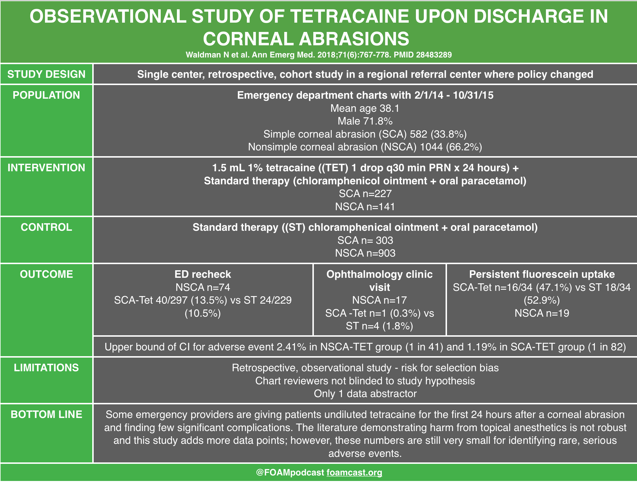
We review one of the editors top picks from Annals of Emergency Medicine, which are free for 6 months from issue release.  The article covered in this podcast is Waldman et al, is a study of tetracaine in corneal abrasions.

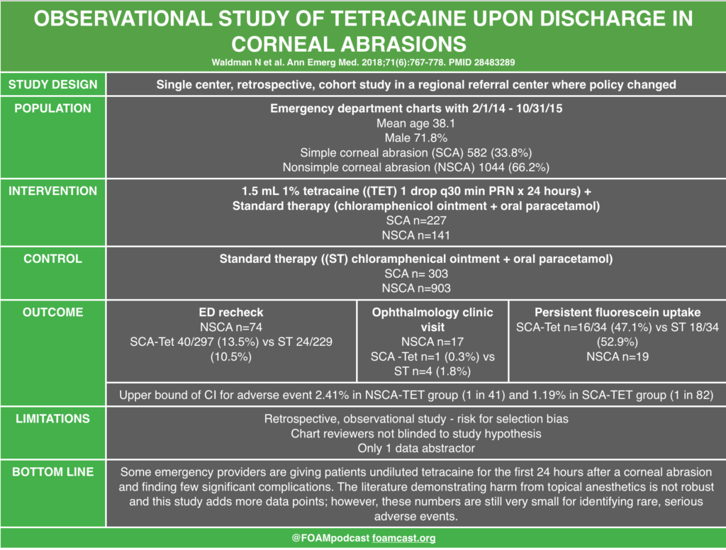
Review from First10EM on topical anesthetics and corneal abrasions.

Rosh Review Emergency Board Review Questions

A 21-year-old man presents to the emergency department complaining of left eye pain. Fluorescein staining reveals a dendritic lesion over the cornea. What is the most likely diagnosis based on this finding?

A. Bacterial conjunctivitis

B. Corneal abrasion

C. Herpes keratitis

D. Retinal detachment

[accordion]
[toggle title=”Answer” state=”closed”]

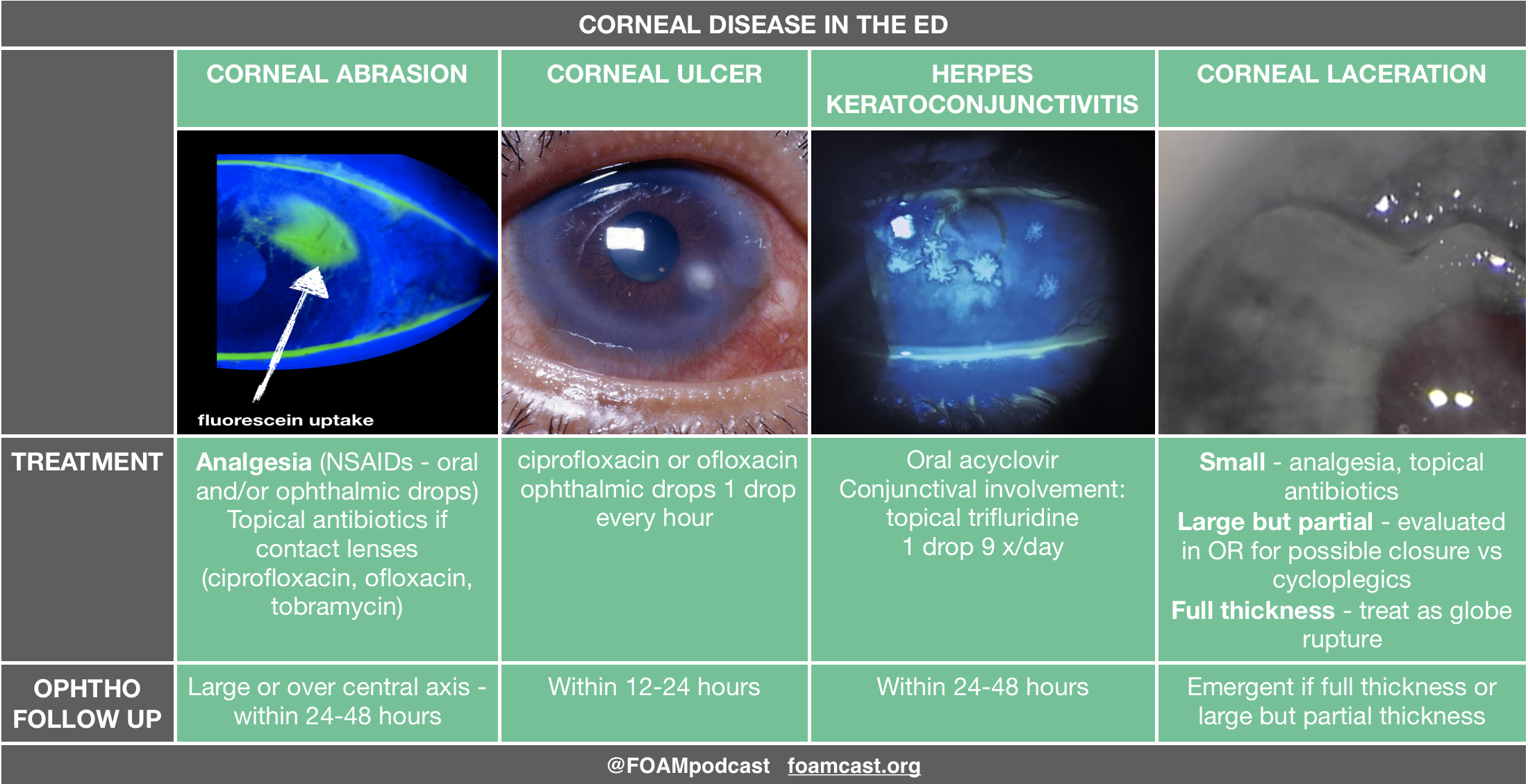
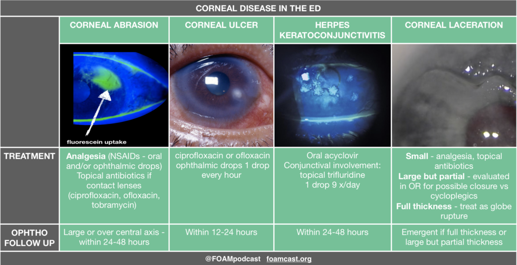
A dendritic lesion of the cornea detected by fluorescein staining indicates the presence of herpetic keratitis. Herpetic keratitis is caused by herpes simplex virus and can lead to blindness because of scarring and opacification of the cornea. A typical presentation includes a painful eye, blurred vision or excessive tearing. Diagnosis can be made with fluorescein staining which gives the classic dendritic appearance. The presence of a dendritic lesion indicates active replication of the virus. Superficial epithelial herpetic keratitis can be successfully treated in two to three weeks with topical antivirals such as ganciclovir. Topical steroids must be avoided in patients with herpetic keratitis as the herpetic lesions can become much deeper and threaten the patient’s vision. Oral antivirals have not been studied comparatively with topical antivirals. Risk of transmission of ocular herpes simplex virus to another person is thought to be extremely low.

Bacterial conjunctivitis (A) can cause eye discomfort but typically presents with copious purulent discharge from one or both eyes. Fluorescein staining of the eye will be negative for any defect. Corneal abrasions (B) present with a superficial epithelial lesion that is usually linear in shape. The cornea is expected to be clear. Retinal detachment (D) is painless and does not cause a change in the appearance of the surface of the eye. Fundoscopic exam will often show rugae indicating the edge of the retina floating in the vitreous humor.

[accordion]
[toggle title=”Answer” state=”closed”]

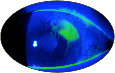
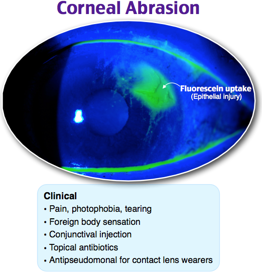
A 20-year-old woman presents to the emergency department for left eye pain. Yesterday she scratched her eye while putting in her contact lens. She has had constant left eye pain and tearing since. She has no foreign body sensation. On exam, her visual acuity is at baseline and equal in both eyes. Her fluorescein exam findings are shown below. Which of the following is the most appropriate treatment for this patient’s condition?

A. Erythromycin ophthalmic ointment

B. Gentamicin-prednisolone ophthalmic suspension

C. Tetracaine ophthalmic solution

D. Tobramycin ophthalmic ointment

[accordion]
[toggle title=”Answer” state=”closed”]

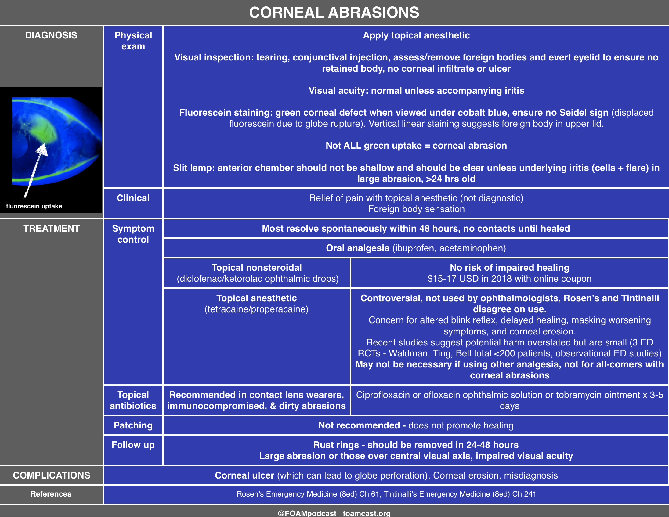
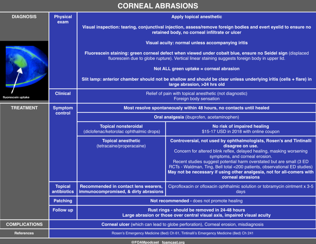
D. This patient’s fluorescein exam is consistent with a corneal abrasion. It is caused by direct mechanical damage, leading to a partial-thickness corneal injury. Athletes, contact lens wearers, welders, and glass workers may present more often with these injuries. Diagnosis is confirmed when fluorescein dye highlights the usually linear or punctate abrasions. Multiple vertical corneal abrasions may indicate a retained foreign body beneath the eyelid. Corneal abrasion fluorescein exam findings are not to be confused with open globe injuries (streaking of the dye from the site of injury), corneal ulcers (circular patches of dye uptake with ragged, “heaped up” edges), and herpes simplex keratitis (dendritic pattern uptake). Patients commonly present with eye pain and tearing with a usually known mechanism of injury. The mainstay of treatment includes pain control, infection prophylaxis, and preventative care to avoid abrasions in the future. Pain control includes cycloplegics like homatropine and cyclopentolate and nonsteroidal anti-inflammatory medications. Commonly prescribed antibiotic ointments include erythromycin, ciprofloxacin, tobramycin, and gentamicin. Notably, erythromycin does not cover pseudomonal infections, which are more likely to occur in contact lens wearers.  Erythromycin (A) does not have appropriate pseudomonal coverage for this patient who wears contact lenses and has a corneal abrasion. Ophthalmic medications containing steroids, like gentamicin-prednisolone (B), should not be prescribed in corneal abrasions or ulcers as they may decrease healing rates. While in the emergency department, tetracaine (C) may be used to more easily evaluate the patient’s eye. However, ocular anesthetics should not be prescribed as they decrease healing rates and block the basic corneal reflex to avoid further injury.

[/toggle]
[/accordion]

References:

  1. Waldman N, Winrow B, Densie I, et al. An Observational Study to Determine Whether Routinely Sending Patients Home With a 24-Hour Supply of Topical Tetracaine From the Emergency Department for Simple Corneal Abrasion Pain Is Potentially Safe. Ann Emerg Med. 2018;71(6):767-778.
  2. Ball IM, Seabrook J, Desai N, Allen L, Anderson S. Dilute proparacaine for the management of acute corneal injuries in the emergency department. CJEM. 2010;12:(5)389-96.
  3. Ting JY, Barns KJ, Holmes JL. Management of Ocular Trauma in Emergency (MOTE) Trial: A pilot randomized double-blinded trial comparing topical amethocaine with saline in the outpatient management of corneal trauma. J Emerg Trauma Shock. 2009;2:(1)10-4.
  4. Swaminathan A, Otterness K, Milne K, Rezaie S. The safety of topical anesthetics in the treatment of corneal abrasions: A review. J Emerg Med 2015
  5. Calder LA, Balasubramanian S, Fergusson D. Topical nonsteroidal anti-inflammatory drugs for corneal abrasions: meta-analysis of randomized trials. Acad Emerg Med. 2005;12(5):467-73.
  6. Galuma K, Lee J. “Ophthalmology.” Rosen’s Emergency Medicine: Concepts and Clinical Practice, Chapter 61, 790-819.e3