Episode 21 – Acute Kidney Injury

(ITUNES OR LISTEN HERE)

The Free Open Access Medical Education (FOAM)

Dr. Josh Farkas of the PulmCrit blog has produced a couple of blog posts on the importance of renal protection in sepsis, Renoresuscitation: Sepsis resuscitation designed to avoid long-term complications and Renal microvascular hemodynamics in sepsis: a new paradigm.  Much of this is theoretical and certainly not something that is standard practice, rathery a theory extrapolated from subgroups of several trials.

Suggested renoresuscitation measures:

(1) Avoid renal failure – avoid nephrotoxins (many antibiotics, NSAIDs, ace-inhibitors), avoid hyperchloremic metabolic acidosis.

(2) Avoid volume overload – treating decreased urine output by flooding a patient with fluids is not necessarily the best move.

(3) Protect the glycocalyx of the endothelium – this suggestion proffers more questions than answers. Steroids? Albumin? Certain vasopressors?  Stay tuned, as we’re not really certain what this entails.

The Bread and Butter

We summarize some key topics from Rosenalli, that’s Tintinalli (7e) Chapter 91; Rosen’s (8e) Chapter 97.  But, don’t just take our word for it.  Go enrich your fundamental understanding yourself.

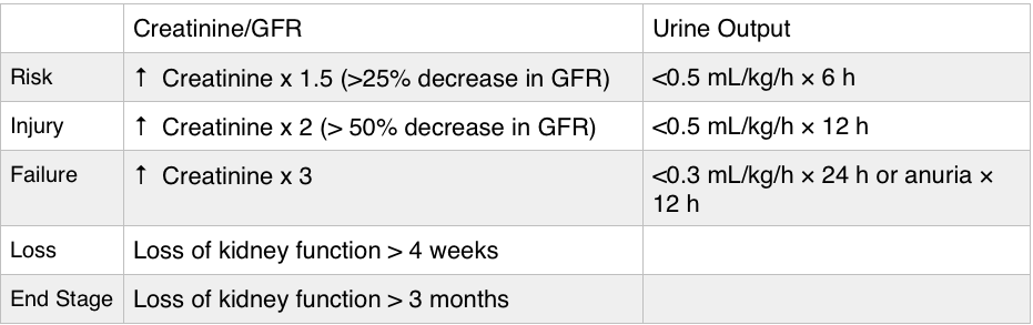
Acute Kidney Injury – typically a creatinine 1.5-2x the patient’s baseline is classified as acute kidney injury.  Urine output can be increased initially but determine whether a patient is making urine and how much, as urine output <0.5 mL/kg/h qualifies as AKI.

RIFLE criteria
RIFLE criteria

Importance – AKI is associated with worse outcomes, although it’s unclear as to whether this is merely a marker of

  • Found in 35-65% of admissions to the intensive care unit, in 5-20% of hospital admissions.  Furthermore, AKI is associated with higher mortality.
  • Renal failure can also cause significant problems for the patient such as electrolyte abnormalities (hyperkalemia the most worriesome, but also hyperphosphatemia) and pulmonary edema.

Etiology – many causes of AKI are reversible or amenable to treatment.

Prerenal – this is one of the most common causes of acute kidney injury and basically is caused by decreased blood flow to the kidney.  Associated with a high BUN/creatinine ratio, increased urine osmolality, a urine sodium concentration less than 20 mEq/L, and FENa less than 1% (this is why getting urine sodium and a concurrent chemistry panel is key).

  • Hypovolemia – volume depleted, hemorrhage, intravascular volume depletion from congestive heart failure or cirrhosis.
  • Hypotension – poor cardiac output (heart failure, valvular problems), shock
  • Decreased flow through the renal artery disease – Nonsteroidal anti-inflammatories: inhibit prostaglandins in the afferent arteriole.  ACE inhibitors prevent the conversion of angiotensin I to angiotensin II, leading to decreased levels of angiotensin II, which when absent decreases the GFR because of dilatation of the efferent arteriole.

Post Renal (Obstructive) – Check out Episode 2 on urologic emergencies.

  • Benign prostatic hypertrophy (BPH) is the most common cause but medications such as anticholinergics and pseudoephedrine. Trauma, stones, strictures, and malignancy can also cause obstruction.

Intrinsic acute renal failure divided into: tubular disease (most common), glomerular disease, vascular disease and interstitial disease.

  • Least common form of AKI in the ED, more common in inpatients.
  • Acute Tubular Necrosis (ATN) most common cause – via nephrotoxins such as aminoglycosides and contrast.
  • Granular “muddy brown” casts – think of necrosis from the “N” in ATN and necrosis tends to be dark.

Indications for emergent dialysis – AEIOU

A- Acidosis

E- Electrolyte emergencies (hyperkalemia!)

I-  Intoxication with dialyzable toxins (ethylene glycol)

O- Overloaded with volume

U- Uremia

 Generously Donated Rosh Review Questions 

Question 1. A 72-year-old man is brought to the ED from a nursing home for evaluation of oliguria. He is found to have an acutely elevated BUN and plasma creatinine from baseline. A Foley catheter is placed; his urine sodium (UNa) is measured below 20 mEq/L and fractional excretion of sodium (FENa) below 1%. [polldaddy poll=8545511]

Question 2.  A 54-year-old man presents to the ED in acute renal failure (ARF). [polldaddy poll=8545512]

Answer 1.  D. This patient’s oliguria with acutely elevated BUN and plasma creatinine suggest that he is in acute renal failure (ARF). His UNa <20 mEq/L and FENa <1% indicate that he has intact reabsorptive function and is able to conserve sodium. This is consistent with prerenal azotemia as the cause for his ARF.

Acute tubular necrosis (ATN) (A), loop diuretics (e.g., furosemide) (B), and osmotic diuresis (e.g., mannitol) (C)all lead to UNa >20 mEq/L and FENa >1% because there is impairment in the ability to concentrate the urine. In such cases, a high-sodium load is excreted.

Answer 2. A.   Acute tubular necrosis (ATN) is a severe form of impairment of tubular epithelial cells caused by ischemia or toxic injury. It is a leading cause of ARF. One of its hallmarks is the presence of brown granular casts on urinalysis. These contain cellular debris rich in cytochrome pigments. In contrast, hyaline casts (B) are usually nonspecific but present after exercise; red cell casts (C) are indicative of glomerular hematuria (e.g., glomerulonephritis); and white cell casts (D) imply renal parenchymal inflammation (e.g., acute interstitial nephritis, pyelonephritis).

Episode 20 – Anticoagulation

(ITUNES OR LISTEN HERE)

The Free Open Access Medical Education (FOAM)

We review Dr. Rory Spiegel’s, A Case of Identity Part Two, post on EMNerd which is essentially a take down of dual antiplatelet therapy (DAT) in acute coronary syndrome (ACS).  The bottom line?  There’s no demonstrable and clinically significant benefit from DAT as demonstrated in the trials below.

CURE trial – composite endpoints of questionable clinical significance and an enormous sample size.

  • 2.1% absolute decrease in cardiovascular death and myocardial infarction (MI), completely powered by the 1.5% absolute difference in MIs. Almost all of these MIs were Type IV and peri-procedural. Mortality between groups was identical at 30 days and end of follow up (1.0% vs 1.1% and 2.3% vs 2.4%, respectively).

ACCOAST – RCT of prasugrel or placebo prior to angiography

  • No difference in cardiovascular death, myocardial infarct, stroke, urgent revascularization or glycoprotein IIb/IIIa rescue therapy (10.8% vs 10.8%)
  • Approximately 1% increase in major bleeding

CREDO – RCT with placebo or clopidogrel 3-24 hours prior to urgent cardiac catheterization

  • No statistical difference  in the rates of death, stroke or MI at 28 days
  • Statistical significance of a secondary endpoint of the 1-year outcomes with a 2% absolute reduction in the rate of death, MI, and stroke, largely the result of a 1.9% reduction of MIs.
  • 1% increase in major bleeding events

Thienopyridine Meta-Analysis

  • In patients with non-ST elevation ACS, pretreatment with thienopyridines is not associated with reduced mortality but comes at a cost of a significant excess of major bleeding.

Composite endpoints are problematic (see this post, “Would You Rather“) and statistical significance claimed in these trials is largely a product of composite outcomes rather than patient oriented measures.

The Bread and Butter

We summarize some key topics from the following readings, Goldfrank (10th ed) Chapter 60, EMPractice October 2013 (there’s almost nothing in Rosenalli on this topic) but, the point isn’t to just take our word for it.  Go enrich your fundamental understanding yourself!

Aspirin

  • Irreversibly inhibits platelets (for the duration of platelet’s life)

Ibuprofen

  • Reversibly inhibits platelets

Novel Oral Anticoagulants (NOACs)

NOACs have gained increased popularity and are slowly supplanting warfarin for common anticoagulation indications such as non-valvular atrial fibrillation (NVAF) as well as treatment of venous thromboembolisms such as pulmonary embolism (PE) and deep venous thrombosis (DVT).

Direct thrombin inhibitor – dabigatran (Pradaxa).  This drug was the first to supplant warfarin for NVAF in the United States, billed as more patient friendly given the lack of purported need for routine monitoring.  Recent investigations by Cohen et al, however, demonstrate that monitoring may, in fact, be safer.  Further, in a real world, retrospective cohort of Medicare beneficiaries given either dabigatran or warfarin for atrial fibrillation, major bleeding of the dabigatran cohort was higher than in the warfarin cohort 9.0% (95% CI 7.8 – 10.2) versus 5.9% (95% CI 5.1 – 6.6) after propensity matching [Hernandez].  For more on the problems with dabigatran, check out Emergency Medicine Literature of Note.

  • Predominantly renal excretion
    • Caution with impaired renal function (can cause dabigatran to stick around longer)
    • Hemodialysis an option in acute overdose; however, most people would probably not want to put a dialysis catheter in a coagulopathic patient.
  • Half-life ~ 15 hours
  • Can elevate the PTT. If the PTT is normal, likely not coagulopathic secondary to dabigatran [Dager et al].
  • No reversal agent

Factor XA inhibitors – these have XA in the name….rivaroXAban, apiXAban, edoXAban.

Rivaroxaban – approved for NVAF and treatment of DVT/PE.  Half life approximately 6-9 hours.

Apixaban – approved for NVAF and treatment of DVT/PE. Half life about 12 hours.

Edoxaban – approved for NVAF. Half life about 10-14 hours.

Bleeding Duration from ACCP
  • Cleared by liver and kidneys.
  • Can elevate the prothrombin time (PT), but not reliably. Specific assays exist but are not widely available and are expensive.
  • No specific reversal agent although andexanet alfa is in the pipeline.  It’s a Factor Xa decoy (Andexanet Alfa) that binds up the F10A inhibitors like a sponge. Read more here.
  • In the setting of major bleeding, guidelines recommend 4 factor PCCs.  A recent study demonstrates reduction in bleeding using 4 factor PCCs on healthy patients given edoxaban [Zahir et al, EMLitofNote].  The benefit of 4 factor PCCs is predominantly based on improvement in numbers, not patient oriented benefit and is discussed in these posts by Dr. Spiegel The Sign of Four, The Sign of Four Part 2.

More FOAM on Anticoagulation Reversal

Generously Donated Rosh Review Questions 

Question 1. A 65-year-old man with a metal aortic valve presents with hematemesis. His vitals are BP 95/50 and HR 118. The patient is on warfarin and has an INR of 7.3. [polldaddy poll=8501352]

Question 2. A 66-year-old woman with atrial fibrillation on warfarin presents with dark stools for 2 days. Her vitals are T 37.7°C, HR 136, BP 81/43, RR 24, and oxygen saturation 94%. Her labs reveal a hematocrit of 19.4% (baseline 33.1%) and an INR of 6.1. [polldaddy poll=8505511]


Answers.

1. D.  The patient presents with life-threatening bleeding and an elevated INR from warfarin use requiring immediate anticoagulant reversal regardless of the indication for anticoagulation. Warfarin acts by inhibiting vitamin K recycling thus limiting the effectiveness of vitamin K dependant clotting factors (factors II, VII, IX and X). The effect of warfarin can be measured using the prothrombin time or the INR. Warfarin is indicated for anticoagulation for a number of disorders including the presence of a metal valve. Patients with metal valves are at a higher 1-year risk of clot formation around the valve and subsequent embolic stroke. The therapeutic goal of warfarin in a patient with a metallic valve is usually between 2.5 –  3.5 or 3.0 – 4.0. Despite the increased stroke risk, patients with life-threatening bleeding should always have their warfarin reversed by administration of vitamin K and fresh frozen plasma (FFP). Alternatively, prothrombin complex concentrates can be given instead of FFP.

Warfarin is not amenable to hemodialysis (A) for removal or reversal. Although patients with a mechanical valve are at an increased stroke risk (increased 1 year risk) reversal should not be delayed (B), as the patient is more likely to die in the immediate situation from their gastrointestinal bleed. Platelet transfusion (C) will not help, as warfarin does not inhibit platelet function.

2. C.  The patient has a life-threatening gastrointestinal bleed in the setting of anticoagulation with warfarin, a vitamin K antagonist. Warfarin acts by inhibiting the synthesis of vitamin K-dependant factors in the coagulation cascade (II, VII, IX, X, protein C, and protein S). The anticoagulant effect of warfarin should be reversed as part of the patient’s emergent treatment. Fresh frozen plasma (FFP) contains all factors in the coagulation cascade and should be given in patients with major bleeding and elevated INR. Vitamin K should be given IV in critically ill patients with elevated INR because it shortens the time to effect.

Vitamin K should not be given intramuscularly (B) because absorption via this route is highly variable. Vitamin K should also not be given orally (D) in critically ill patients because the onset of action will be delayed. Additionally, absorption in patients with gastrointestinal bleeding may be variable. Vitamin K should be given along with FFP (A) because the factors inhibited rely on vitamin K for function.