Episode 52 – The Esophagus

ITUNES OR LISTEN HERE

The Free Open Access Medical Education (FOAM)

We cover a Scancrit post on the Back Up Head Elevated (BUHE) intubation position.  This post details a multicenter retrospective observational study by Khandelwal et al in Anesthesia & Analgesia

FullSizeRender (4)

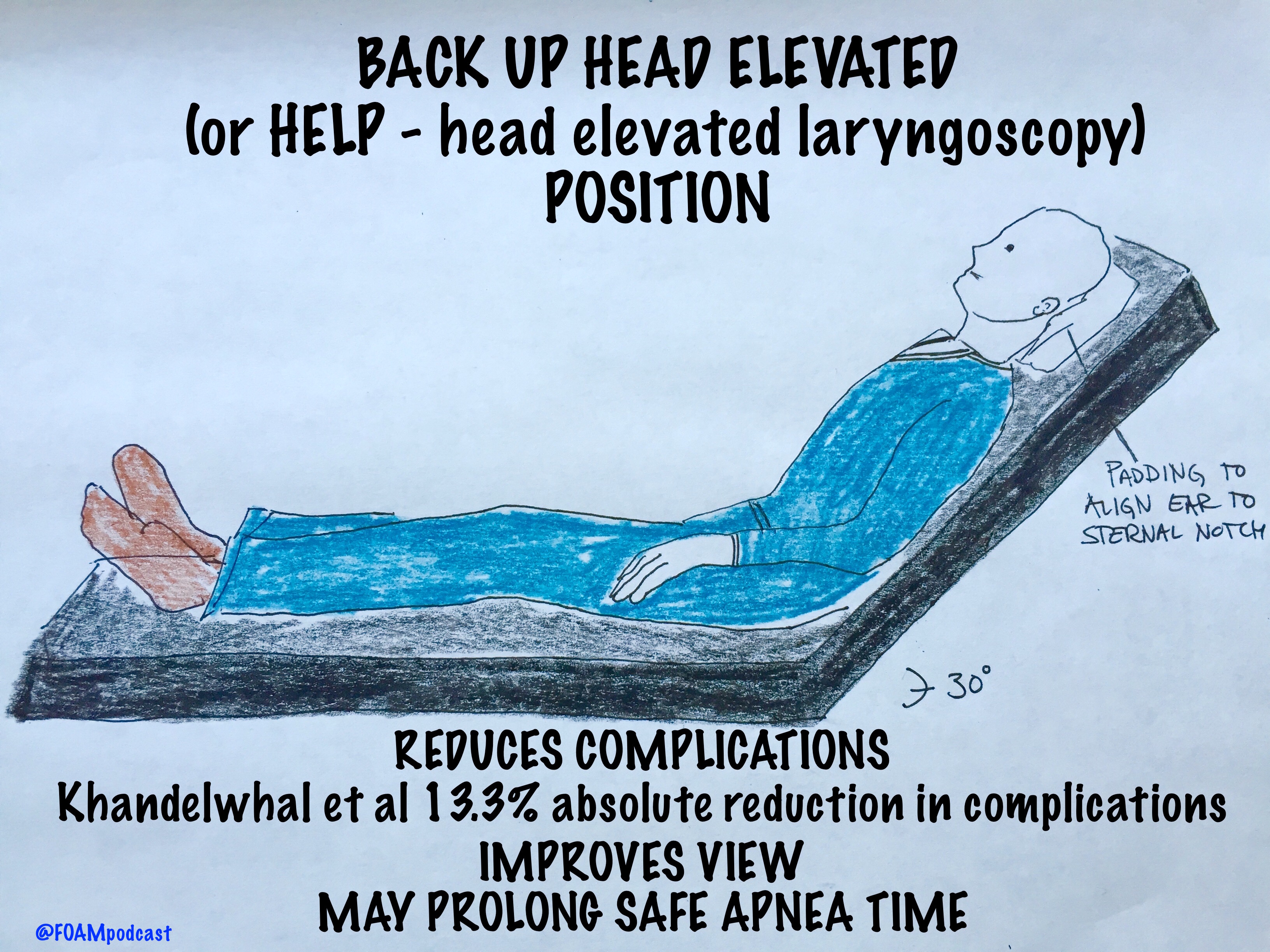
Population: 528 adults undergoing emergent intubation

Intervention: Head up at least 30 degrees during intubation

Control: Supine intubation

Primary Outcome: Occurrence of intubation related complication (difficult intubation: >3 attempts or prolonged intubation, hypoxemia, esophageal intubation, or pulmonary aspiration) – 22.6% in supine group vs 9.3% in the head elevated position.  Absolute difference of 13.3%

Limitations: Did not look at emergency department intubation.  More experienced intubators used the BUHE positioning, which could confound the reduction in intubation related complications [1].

BUHE or Head Elevated Laryngoscopy Position (HELP) has also been found to

  • Improve laryngeal view [2,3]
  • Prolong safe apnea time [4]

Note: in patients with possible spinal injuries, one may use reverse trendelenberg (or forego the back up head elevated position)

Core Content

We delve into core content on the esophagus using Rosen’s (8th ed) Chapter 71 and Chapter 77 in Tintinalli (8th ed)
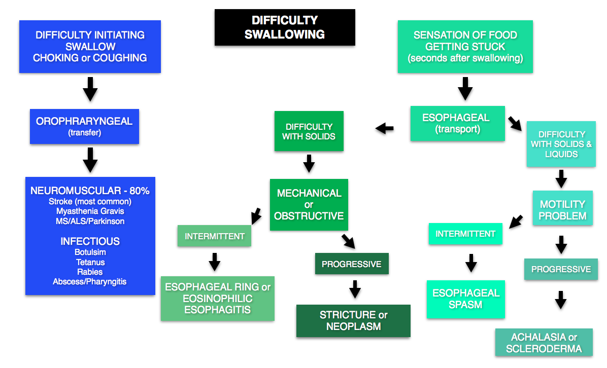
DYSPHAGIA

Emergent conditions may include stroke (most common cause), myasthenia/botulism/or other neuromuscular problems (may also have concomitant respiratory failure). Many causes do not need emergent workup.

@FOAMpodcast
@FOAMpodcast

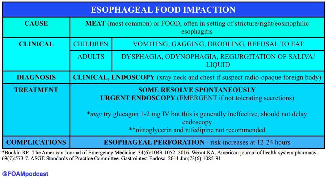
ESOPHAGEAL FOOD BOLUS

Screen Shot 2016-06-27 at 5.29.03 AM

Generously Donated Rosh Review Questions

An 87-year-old woman presents to the ED after her caregiver witnessed the patient having difficulty swallowing over the past 2 days. The patient is having difficulty with both solids and liquids. She requires multiple swallowing attempts and occasionally has a mild choking episode. She has no other complaints. Your exam is unremarkable. Which of the following is the most likely cause of her condition?

A. Achalasia

B. Cerebrovascular accident

C. Esophageal neoplasm

D. Foreign body

[accordion]
[toggle title=”Answer” state=”closed”]

B. Cerebrovascular accident.  Dysphagia can be divided into two categories: transfer and transport. Transfer dysphagia occurs early in swallowing and is often described by the patient as difficulty with initiation of swallowing. Transport dysphagia occurs due to impaired movement of the bolus down the esophagus and through the lower sphincter. This patient is experiencing a transfer dysphagia. This condition is most commonly due to neuromuscular disorders that result in misdirection of the food bolus and requires repeated swallowing attempts. A cerebrovascular accident (stroke)that causes muscle weakness of the oropharyngeal muscles is frequently the underlying cause. Achalasia (A) is the most common motility disorder producing dysphagia. It is typically seen in patients between 20 and 40 years of age and is associated with esophageal spasm, chest pain, and odynophagia. Esophageal neoplasm (C) usually leads to dysphagia over a period of months and progresses from symptoms with solids to liquids. It is also associated with weight loss and bleeding. Foreign bodies (D) such as a food bolus can lead to dysphagia, but patients are typically unable to tolerate secretions and are often observed drooling. These patients do not have difficulty in initiating swallowing.

[/toggle]
[/accordion]

A 33-year-old man presents with dysphagia to both solids and liquids, with solids being worse than liquids. He describes a sensation of the food getting stuck in his chest. Occasionally, he needs to raise his arms above his head to help food pass into his stomach. His primary care doctor has been treating him for GERD over the previous six months, but his symptoms are getting worse. Which of the following is the most likely diagnosis?

A. Achalasia

B. Diffuse esophageal spasm

C. Schatzki ring

D. Zenker’s diverticulum

[accordion]

[toggle title=”Answer” state=”closed”]

Achalasia This patient most likely has achalasia, which is an esophageal dysmotility disorder due to failure of the lower esophageal sphincter to relax. Dysphagia is the most common symptom. While all patients have dysphagia to solids, only two-thirds have liquid dysphagia. By standing after eating, straightening one’s back, or raising the arms above the head, the esophageal pressure increases, which can help emptying into the stomach. Symptoms usually begin with mild dysphagia in patients who are 20 to 40 years old; symptoms are usually progressive. Diffuse esophageal spasm (B) is a hypermotility disorder causing strong, uncoordinated peristaltic contractions that do not propel food effectively to the stomach. Dysphagia, regurgitation of food, and chest pain are commonly present. The dysphagia is intermittent and does not progress over time. A Schatzki ring (C) is a fibrous band-like structure in the distal esophagus that is the most common cause of dysphagia with solids. Patients typically do not experience difficulty with liquids. Zenker’s diverticulum (D) is an acquired disease that is due to an out-pouching in the mucosa of the pharynx. It typically occurs in individuals older than 50 years and can cause regurgitation, cough, and halitosis from food that becomes stuck in the diverticulum.

[/toggle]
[/accordion]

[accordion]
[toggle title=”What are three common treatments for achalasia?” state=”Closed”]

Nitroglycerin to reduce lower esophageal sphincter tone, endoscopic injection of botulinum toxin into the muscle of the sphincter, and surgical myotomy.  Diffuse esophageal spasm (B) is a hypermotility disorder causing strong, uncoordinated peristaltic contractions that do not propel food effectively to the stomach. Dysphagia, regurgitation of food, and chest pain are commonly present. The dysphagia is intermittent and does not progress over time. A Schatzki ring (C) is a fibrous band-like structure in the distal esophagus that is the most common cause of dysphagia with solids. Patients typically do not experience difficulty with liquids. Zenker’s diverticulum (D) is an acquired disease that is due to an out-pouching in the mucosa of the pharynx. It typically occurs in individuals older than 50 years and can cause regurgitation, cough, and halitosis from food that becomes stuck in the diverticulum.

[/toggle]
[/accordion]

References

  1. Khandelwal et al. Head-elevated Patient Positioning Decreases Complications of Emergent Tracheal Intubation in the Ward and Intensive Care Unit. Anesthesia and Analgesia. Apr 2016.
  2. Lee et al. Laryngeal exposure during laryngoscopy is better in the 25 degree back-up position than in the supine position. British Journal of Anaesthesia. July 2007.
  3. Levitan RM, Mechem CC, Ochroch EA, Shofer FS, Hollander JE. Head-elevated laryngoscopy position: improving laryngeal exposure during laryngoscopy by increasing head elevation. Annals of emergency medicine. 41(3):322-30. 2003.
  4. Ramkumar V, Umesh G, Philip FA. Preoxygenation with 20º head-up tilt provides longer duration of non-hypoxic apnea than conventional preoxygenation in non-obese healthy adults. Journal of anesthesia. 25(2):189-94. 2011. [pubmed]
  5. Bodkin RP, Weant KA, Baker Justice S, Spencer MT, Acquisto NM. Effectiveness of glucagon in relieving esophageal foreign body impaction: a multicenter study. The American Journal of Emergency Medicine. 34(6):1049-1052. 2016.
  6. Weant KA, Weant MP. Safety and efficacy of glucagon for the relief of acute esophageal food impaction. American journal of health-system pharmacy : AJHP : official journal of the American Society of Health-System Pharmacists. 69(7):573-7. 2012
  7. ASGE Standards of Practice Committee, Ikenberry SO, Jue TL, Anderson MA, Appalaneni V, Banerjee S, Ben-Menachem T, Decker GA, Fanelli RD, Fisher LR, Fukami N, Harrison ME, et al. Management of ingested foreign bodies and food impactions. Gastrointest Endosc. 2011 Jun;73(6):1085-91

FOAMcastini – The Aorta and No Analgesia Will #!&?% You Up

ITUNES OR LISTEN HERE

We cover pearls from smaccDUB (Social Media and Critical Care Conference in Dublin, Ireland), Day 3. We are here thanks to the Rosh Review.

Dr. Scott Weingart – “Post-Intubation Sedation

  • Analgesia first. Try a hydromorphone 1mg push while you’re waiting for the fentanyl drip. The endotracheal tube is uncomfortable.
  • Minimize sedation. There’s this principle: eCASH: early Comfort using Analgesia, minimal Sedatives and maximal Humane care [1]. 
  • Sedation: go for dexmedetomidine if you have it (but it’s expensive) or propofol. This is supported by the Society of Critical Care Medicine (SCCM) Pain, Agitation, and Delirium guidelines [2].
  • Be careful with rocuronium.  The long duration of rocuronium means that you can’t assess for pain or discomfort so you must be responsible and get these

Dr. David Carr – “The Aorta Will #!&?% You Up”

Screen Shot 2016-06-16 at 6.57.56 PM

Dr. Kathleen Thomas – “Oh Sh**! They’re bombing the hospital!”

We should not need a website entitled STOPBOMBINGHOSPITALS.ORG but, unfortunately, over the past 4 years, 400 hospitals have been bombed. This passionate, wrenching talk is a “must see” and “must listen” when the free talks are released on the SMACC podcast over the course of the next year.

References

  1. Vincent J, Shehabi Y, Walsh TS et al. Comfort and patient-centred care without excessive sedation: the eCASH concept. Intensive Care Med. 42(6):962-971. 2016. [article]
  2. Barr J, Fraser GL, Puntillo K et al. Clinical Practice Guidelines for the Management of Pain, Agitation, and Delirium in Adult Patients in the Intensive Care Unit. Critical Care Medicine. 41(1):263-306. 2013. [article]
  3. Watt JM, Amini A, Traylor BR, Amini R, Sakles JC, Patanwala AE. Effect of paralytic type on time to post-intubation sedative use in the emergency department. Emergency medicine journal : EMJ. 30(11):893-5. 2013. [pubmed]
  4. Imamura H, Sekiguchi Y, Iwashita T et al. Painless Acute Aortic Dissection. Circ J. 75(1):59-66. 2011. [article]
  5. Diercks DB, et al. Clinical policy: critical issues in the evaluation and management of adult patients with suspected acute nontraumatic thoracic aortic dissection. Ann Emerg Med. 2015 Jan;65(1):32-42.e12. PMID: 25529153.
  6. Hagan PG, Nienaber CA, Isselbacher EM. The International Registry of Acute Aortic Dissection (IRAD): new insights into an old disease. JAMA. 283(7):897-903. 2000. [pubmed]

FOAMcastini – Undifferentiated Agitation and Cured Pork

ITUNES OR LISTEN HERE

We cover pearls from smaccDUB (Social Media and Critical Care Conference in Dublin, Ireland), Day 2. We are here thanks to the Rosh Review.

Dr. Reuben Strayer (@emupdates) – “Disruption, Danger, and Droperidol: Emergency Management of the Agitated Patient”  

Dr. Strayer presented a brilliant talk on dealing with the quintessential Emergency Medicine patient – the undifferentiated acutely agitated patient. These patients are high risk and require emergent stabilization and resuscitation.  

Dr. Reuben Strayer's (@emupdates) algorithm for agitation in the ED
Dr. Reuben Strayer’s (@emupdates) algorithm for agitation in the ED
  • Oh, and, droperidol is not dangerous [1]. See this post on the controversial black box warning.

Dr. Haney Mallemat – “The PEA Paradox”

  • The typical way we think about PEA, the “H’s and T’s,” is overly complicated. Further, we are horrendous at pulse palpation (see this for more) [2,3], and so what we think is PEA may not actually be PEA.  Dr. Mallemat proposed something along the lines of the following the following.

Screen Shot 2016-06-15 at 6.43.52 PM

  • For some FOAM commentary on the limitations of this approach, see this post by Dr. Rory Spiegel [4].

Dr. Michele Dominico – “How Usual Resuscitative Maneuvers Can Kill Paediatric Cardiac Patients”

Interventions we jump to in sick patients – oxygenation, ventilation, vasopressors – these can kill pediatric patients with cardiac pathology. She gave examples of some high yield pearls in these already terrifying patients.

Screen Shot 2016-06-15 at 6.59.08 PM

EM Literature update by Drs. Ashley Shreves and Ryan Radecki

 Ridiculous Research Pearls from Drs. Ashley Shreves and Ryan Radecki

  • Perception of dyspnea and pulmonary function tests change with stress – and rollercoaster rides.  Rietveld S, van Beest I. Rollercoaster asthma: when positive emotional stress interferes with dyspnea perception. Behaviour research and therapy. 45(5):977-87. 2007. [pubmed]
  • Cured pork for epistaxis? Possibly. Researchers will try everything, especially if it involves bacon. Humphreys I, Saraiya S, Belenky W, Dworkin J. Nasal packing with strips of cured pork as treatment for uncontrollable epistaxis in a patient with Glanzmann thrombasthenia. The Annals of otology, rhinology, and laryngology. 120(11):732-6. 2011. [pubmed]

References:

  1. Calver L, Page CB, Downes MA et al. The Safety and Effectiveness of Droperidol for Sedation of Acute Behavioral Disturbance in the Emergency Department. Annals of Emergency Medicine. 66(3):230-238.e1. 2015. [article]
  2. Tibballs J, Weeranatna C. The influence of time on the accuracy of healthcare personnel to diagnose paediatric cardiac arrest by pulse palpation. Resuscitation. 81(6):671-5. 2010. [pubmed]
  3. Eberle B, Dick WF, Schneider T, Wisser G, Doetsch S, Tzanova I. Checking the carotid pulse check: diagnostic accuracy of first responders in patients with and without a pulse. Resuscitation. 33(2):107-16. 1996. [pubmed]
  4. Bergum D, Skjeflo GW, Nordseth T. ECG patterns in early pulseless electrical activity-Associations with aetiology and survival of in-hospital cardiac arrest. Resuscitation. 104:34-9. 2016.

Foamcastini – Do We Make Saves?

ITUNES OR LISTEN HERE

We are at SMACC in Dublin – thanks to the Rosh Review, an excellent board review question bank.

Do We Make Saves?

Dr. Mervyn Singer “Is Survival Predetermined in the Critically  Ill?”

  • Many critical care studies are negative, but in some cases this may be because critically ill patients don’t have a uniform prognosis.
  • Dr. Singer argues that some people may be “destined to die” and some may be “destined to live.”  Interventions may be harmful or futile in one group but beneficial in the other.  For example, Dr. Singer references the CORTICUS trial of steroids in septic shock [1]. This was a negative trial.  Dr. Singer asserts that some evidence (of not great quality), purports that the sickest patients could benefit from steroids, while this same intervention could be deleterious in the healthier ones.
  • Problem: many of the studies that go back and re-analyze these groups looking at the sickest or least sick patients? They perform subgroup analyses, a form of data dredging that must be taken with a huge grain of salt.

Favorite Pearls

Dr. Suzanne Mason – “Acute Care of the Elderly”

  • Hospital admission may not benefit geriatric patients – interdisciplinary interventions involving nurses, consultants, pharmacists, physical therapy may be best.
  • If there is a single, free intervention that providers can do it’s assessing for polypharmacy. Polypharmacy in the elderly is a huge problem.  Check the patient’s medication list and beware adding new medications that may not be absolutely necessary.

Dr. Victoria Brazil – “So You Think You’re a Resuscitationist?”

  • The Dunning-Kruger effect is real in medicine and this is why we MUST have and provide feedback.  A review of the Dunning-Kruger effect can be found here.  Essentially, people tend to overestimate what they know (i.e. overly confident in their knowledge). The exception?  Masters tend to underestimate their knowledge.

Screen Shot 2014-03-15 at 9.36.53 AM

  • Our perception of reality is very skewed so, again, feedback is crucial. For example, Cemalovic and colleagues found that intubators underestimated the time they took to intubate: they estimated 23.5 seconds on average vs the 45.5 seconds intubation actually took. Additionally, they thought 13% of their patients desaturated during intubation but 23% actually desaturated [2].

Also, there was an excellent tribute to the late Dr. John Hinds, a reminder that by living profoundly, you can impact people across the world.

References

  1. Sprung CL, Annane D, Keh D et al. Hydrocortisone Therapy for Patients with Septic Shock. N Engl J Med. 358(2):111-124. 2008. [article]
  2. Cemalovic N, Scoccimarro A, Arslan A, Fraser R, Kanter M, Caputo N. Human factors in the emergency department: Is physician perception of time to intubation and desaturation rate accurate? Emergency medicine Australasia : EMA. 28(3):295-9. 2016. [pubmed]

Episode 51 – Ocular Trauma

(ITUNES OR LISTEN HERE)

The Free Open Access Medical Education (FOAM)

We cover this short video from EMRAP on lateral canthotomies.  This is one of those rare procedures that is vision saving; hence, it is worthy of frequent review.

Lateral Canthotomy and Cantholysis

Indication:  Suspect orbital compartment syndrome -afferent pupillary defect (APD), “tense eye” with taught lids, and high intraocular pressure (IOP) ≥ 40 mmHg. The APD is really a critical feature, as it is your indicator that there is optic nerve compromise from the intraorbital pressure

Clinical presentation:  History of orbital trauma, often in the setting of orbital fractures with decreased visual acuity, proptosis, chemosis

Procedure: Call ophthalmology.

  • Analgesia – inject lidocaine with epinephrine into the area of the lateral canthus
  • Devascularize – use straight kelly clamps to crush the lateral canthus for 1-2 minutes
  • Incise – Use scissors to cut the lateral canthus 1-2 cm. Then,  find the inferior crus of the lateral canthus ligament (looks kind of like a wishbone from a turkey but you rarely are going to be able to see the tendon so you’re “strumming” it with the closed tips of your scissors inferomedially toward the nose) – cut the inferior crus of the ligament to release pressure (this is the key step)

When calling ophthalmology, ensure you have the “Vital Signs of the Eye”

  • Vital Signs of the Eye - @FOAMpodcast
    Vital Signs of the Eye – @FOAMpodcast

Core Content

We delve into core content on eye trauma using Rosen’s (8th edition) Chapter 71 and Chapter 241 in Tintinalli (8th edition)

Orbital Fractures

Orbital Fractures

Note: entrapment is a clinical diagnosis.  A CT cannot comment on function, only structure.  Extraocular movements are a critical part of the fracture exam.

Pearl: young people are prone to “greenstick” fractures where the floor can actually pinch the muscle and trigger the oculocardiac reflex, which can be deadly.  They often have a “white eye” without too much impressive on exam but refuse to look in a certain direction due to nausea.

Hyphema

(and things confused with hyphema)

Hyphema

Burns

Ocular Burns

Pearl: An important thing to keep in mind is that a “white” eye after such an exposure is actually an ominous sign, as it implies ischemia of the limbal blood vessels, which portends a pretty bad prognosis.  People usually think that the more red an eye, the worse when, in truth, it’s often the other way around.

Generously Donated Rosh Review Questions

A 43-year-old construction worker presents with right eye pain. He states he was using a nail gun when he felt something hit his eye. Visual examination reveals a small nail penetrating the globe. What management should be pursued?

A. CT scan of the orbit and ophthalmology consultation

B. Measure intraocular pressure and consult ophthalmology

C. Perform lateral canthotomy and consult ophthalmology

D. Remove the foreign body, start topical antibiotics and send to ophthalmology for follow up

[accordion]
[toggle title=”Answer” state=”closed”]

This patient presents with a globe injury and should have a protective shield placed, intravenous antibiotics started, CT scan of the orbit performed and ophthalmology consulted emergently. Foreign body penetration of the globe is often associated with hammering, drilling, mechanical grinding or sanding. Any patient who presents with a foreign body sensation after one of these activities should increase suspicion for a penetrating injury of the globe. CT scan, MRI and ultrasound can all be used for diagnosis but MRI should be avoided if the suspected foreign body is metal containing. Many of these patients will require operative management and should be kept NPO. A protective shield should be placed to protect the eye but patching should be avoided as it may increase pressure on the eye. Emergent ophthalmologic consultation should be obtained

[/toggle]
[/accordion]

A 23-year-old man presents after a fight. His eye is seen below. Physical examination reveals intact extraocular movements, normal fluorescein staining, normal intraocular pressure, and normal visual acuity.

content_image-_medial_lower_lid_laceration

What management is indicated?

A. Delayed closure

B.  Laceration repair by the Emergency Physician

C. Ophthalmology consultation for repair

D. Tissue adhesive for repair

[accordion]
[toggle title=”Answer” state=”closed”]

C. Ophthalmology consultation for repair. This patient presents with a complex eyelid laceration possibly involving the canalicular system and should have a consultation with either ophthalmology or plastic surgery regarding repair. It is important to search for a penetrating globe injury in any patient with an eyelid laceration because of the proximity of structures. Simple horizontal and partial thickness lid lacerations can be repaired primarily by an Emergency Physician. However, more complicated lacerations should be considered for specialist repair because of the high likelihood of cosmetic or functional complications, or both. In general, lacerations through the orbital septum, lacerations with tissue loss, lacerations involving the lid margins, lacerations involving the levator or canthal tendons and those involving the canalicular system should be repaired by a skilled ophthalmologist or plastic surgeon. Injury to the canalicular system should be suspected in any laceration involving the medial lower eyelid. Tissue adhesive (D) is contraindicated this close to the eye and should not be used in an injury that requires precision alignment of tissue. Delayed closure (A) will likely lead to worse cosmetic outcomes. Primary repair by the Emergency Physician (B) is not recommended if the canalicular system may be involved.

[/toggle]
[/accordion]


Thanks to our peer reviewer, Michael Westafer, MD – Ophthalmologist and Glaucoma Fellow at Mayo Clinic

References

  1. “Eye Emergencies.”  Chapter 241.  Tintinalli’s Emergency Medicine: A Comprehensive Review. 8th edition.  
  2. Sharma R and Brunette D.  “Ophthalmology.”  Chapter 71.  Rosen’s Emergency Medicine.  8th edition, 909-930.