Episode 59 – Syncope (and the PESIT study)

ITUNES OR LISTEN HERE

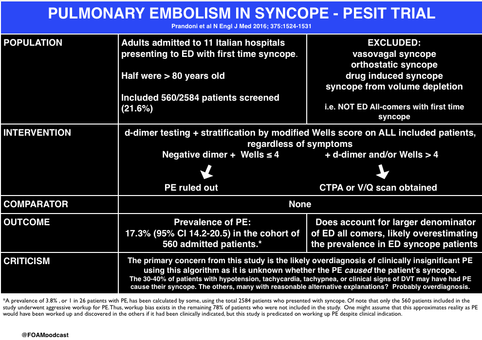
The PESIT study in the New England Journal of Medicine stirred up controversy in the FOAM world earlier in October 2016.  In this episode we cover the following posts on this article on pulmonary embolism in syncope:

screen-shot-2016-10-25-at-7-52-02-pm

Core Content

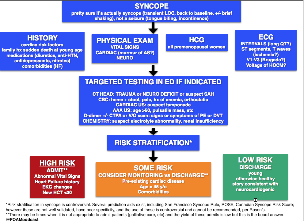
We delve into core content on syncope usingRosen’s Emergency Medicine (8th edition) and Tintinalli’s Emergency Medicine (8th edition) Chapter 52

screen-shot-2016-10-31-at-4-41-19-pm

 Rosh Review Emergency Board Review Questions

An 83-year-old is being evaluated in the emergency department after an episode of syncope. The woman was preparing dinner when she felt her heart start to race. The next thing she remembers is waking up on the floor. She experienced a similar episode about three weeks ago. She has never had anything like this before. Her past medical history is remarkable for hypertension, hyperlipidemia and hypothyroidism. Her medications include lisinopril, atorvastatin and levothyroxine. On physical exam her blood pressure is 142/83, heart rate 76/min, and respiration rate 13/min. Cardiac auscultation reveals no murmur. The remainder of her physical exam is normal. Electrocardiogram reveals normal sinus rhythm with left axis deviation. No cardiac rhythm abnormalities are detected. What is the most likely etiology of this patient’s syncope?

A. Aortic stenosis

B. Cardiac dysrhythmia

C. Orthostatic hypotension

D. Vasovagal

[accordion]
[toggle title=”Answer” state=”closed”]

B. Cardiac dysrhythmia is the most likely cause of this woman’s syncope. Cardiac dysrhythmias are a common cause of syncope in the elderly population. It is characterized by a brief or absent prodrome and palpitations immediately preceding the event. Several episodes over a short period of time in someone with no history of syncope suggest a dysrhythmia. Given this patient’s short prodrome, palpitations and history of a previous similar event makes a cardiac dysrhythmia the most likely etiology.

Aortic stenosis (A) is unlikely the cause of her syncope. Aortic stenosis is associated with a crescendo-decrescendo systolic ejection murmur. Syncope related to aortic stenosis typically occurs during exertion and is associated with very severe disease. This patient’s syncopal episode occurred while stationary. Additionally, she has no systemic symptoms of aortic stenosis.Vasovagal (D) is the most common cause of syncope in the general population. It is usually triggered by provoking factors such a blood draw or an intense emotion. Prodromal symptoms include feeling warm, sweating, nausea, and pallor. This woman does not report any of these symptoms. Orthostatic hypotension (C) causes syncope upon assuming an upright position from supine or sitting. It is often caused by hypovolemia, medications or autonomic nervous system disorders. This woman was standing while preparing dinner making orthostatic hypotension unlikely.


[/toggle]
[/accordion]

An 18-year-old woman presents after having a syncopal episode. She is complaining of a 2-day history of lower abdominal pain and vaginal spotting. Her BP is 86/42, HR is 128, RR is 18 breaths, and oxygen saturation is 99% on room air. She is drowsy, but answers questions appropriately. What is the most appropriate next step in management?


A. Establish large-bore IV access and administer an IV fluid bolus

B. Initiate rapid sequence induction and orotracheal intubation

C. Perform a bedside urine pregnancy testing

D. Perform an ultrasound of the abdomen to assess for free fluid

[accordion]
[toggle title=”Answer” state=”closed”]

A. The patient is hypotensive and tachycardic. She is suffering from hypovolemic shock secondary to a ruptured ectopic pregnancy. Therefore she requires immediate intravenous access and volume resuscitation with Lactated Ringer’s or normal saline.  Emergency Department management of unstable patients includes rapid assessment of the ABC’s (Airway, Breathing, Circulation). This patient is phonating, has a respiratory rate of 18 breaths per minute and an oxygen saturation of 99% on room air.  There is no concern that her airway or breathing is in immediate jeopardy, therefore she would not require immediate rapid sequence induction and orotracheal intubation (B). Although a bedside pregnancy test (C) and abdominal ultrasound (D) would help make a diagnosis of ruptured ectopic pregnancy, the next step would be to resuscitate the patient.

[/toggle]
[/accordion]

References

  1. De Lorenzo RA. “Syncope.” Chapter 15. Rosen’s Emergency Medicine (8 ed). pp 131-145
  2. Chapter 52. Tintinalli’s Emergency Medicine: A Comprehensive Review (8 ed).
  3. Serrano LA, Hess EP, Bellolio MF et al. Accuracy and Quality of Clinical Decision Rules for Syncope in the Emergency Department: A Systematic Review and Meta-analysis. Annals of Emergency Medicine. 56(4):362-373.e1. 2010.
Bell’s Palsy and Burns

(ITUNES OR LISTEN HERE)

We are in Las Vegas at ACEP 2016 thanks to Annals of Emergency Medicine and ACEPnow and discuss high yield and cutting edge lectures each day.

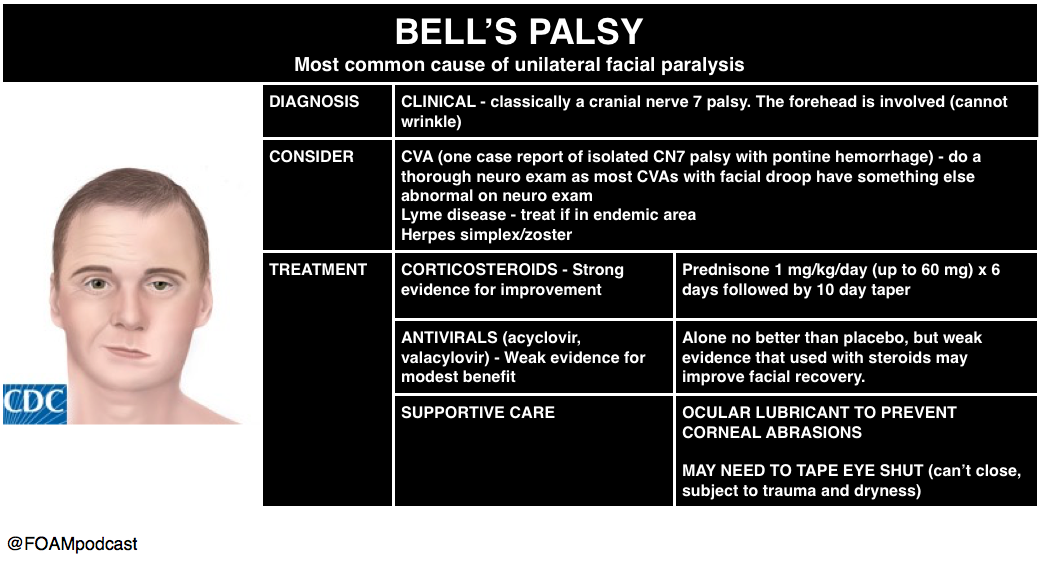
Dr. Megan Osborn – Bell’s Palsy or Stroke?

Traditional teaching: we can differentiate Bell’s palsy (lower motor neuron) from a stroke (upper motor neuron by assessing forehead involvement.  If the patient can wrinkle their forehead? Think stroke.  Dr. Megan Osborn tackled the question: does this actually work all the time in her talk in the New Speakers Forum.

screen-shot-2016-10-19-at-8-19-31-am

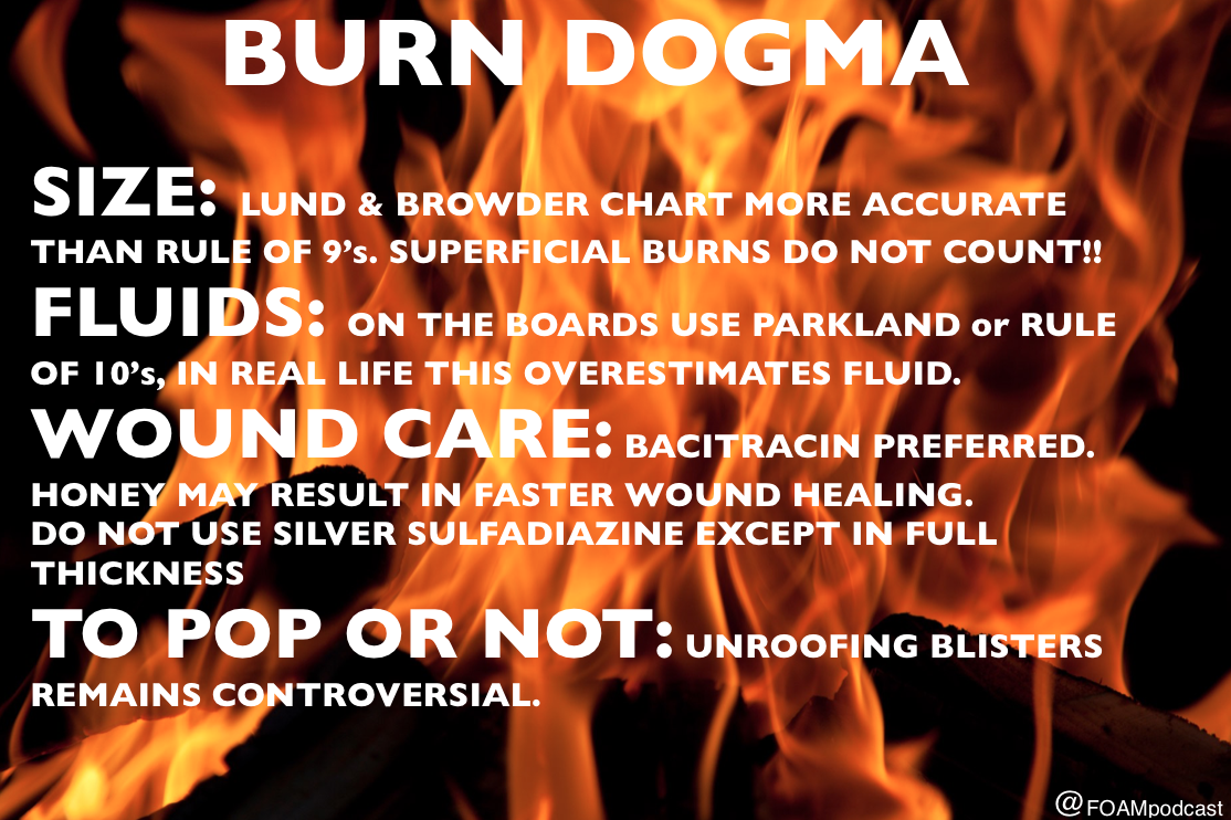
Dr. Toree McGowan – Burns

Check out this podcast for more on burns

screen-shot-2016-10-19-at-8-33-56-am

  1. Sherman SC, Thompson TM, Thompson TT. Pontine hemorrhage presenting as an isolated facial nerve palsy. Annals of emergency medicine. 46(1):64-6. 2005. [pubmed]
  2. AAN Bell’s Palsy Guideline Update
  3. Fahimi J, Navi BB, Kamel H. Potential Misdiagnoses of Bell’s Palsy in the Emergency Department. Annals of Emergency Medicine. 63(4):428-434. 2014. [article]
  4. Madhok VB, Gagyor I, Daly F. Corticosteroids for Bell’s palsy (idiopathic facial paralysis). The Cochrane database of systematic reviews. 7:CD001942. 2016. [pubmed]
  5. Gagyor I, Madhok VB, Daly F. Antiviral treatment for Bell’s palsy (idiopathic facial paralysis). The Cochrane database of systematic reviews. 2015. [pubmed]
  6. Wasiak J, Cleland H, Campbell F. Dressings for superficial and partial thickness burns. The Cochrane database of systematic reviews. 2008. [pubmed]
  7. Ringh M, Rosenqvist M, Hollenberg J et al. Mobile-Phone Dispatch of Laypersons for CPR in Out-of-Hospital Cardiac Arrest. N Engl J Med. 372(24):2316-2325. 2015. [article]

Vaccine Side Effects and Placebos

(ITUNES OR LISTEN HERE)

We are in Las Vegas at ACEP 2016 thanks to Annals of Emergency Medicine and ACEPNOW and discuss high yield or cutting edge lectures each day.

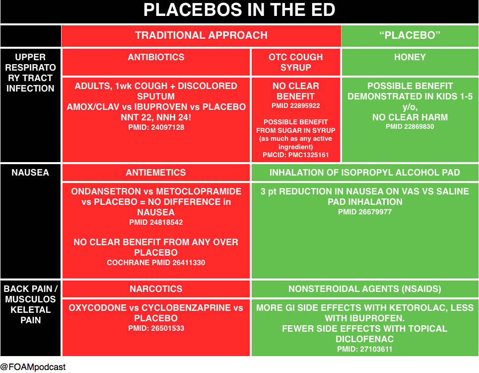
Dr. Matthew DeLaney – 21st Century Snake Oil (Placebos)

screen-shot-2016-10-17-at-5-30-30-pm

SGEM http://thesgem.com/2013/03/sgem26-honey-honey/

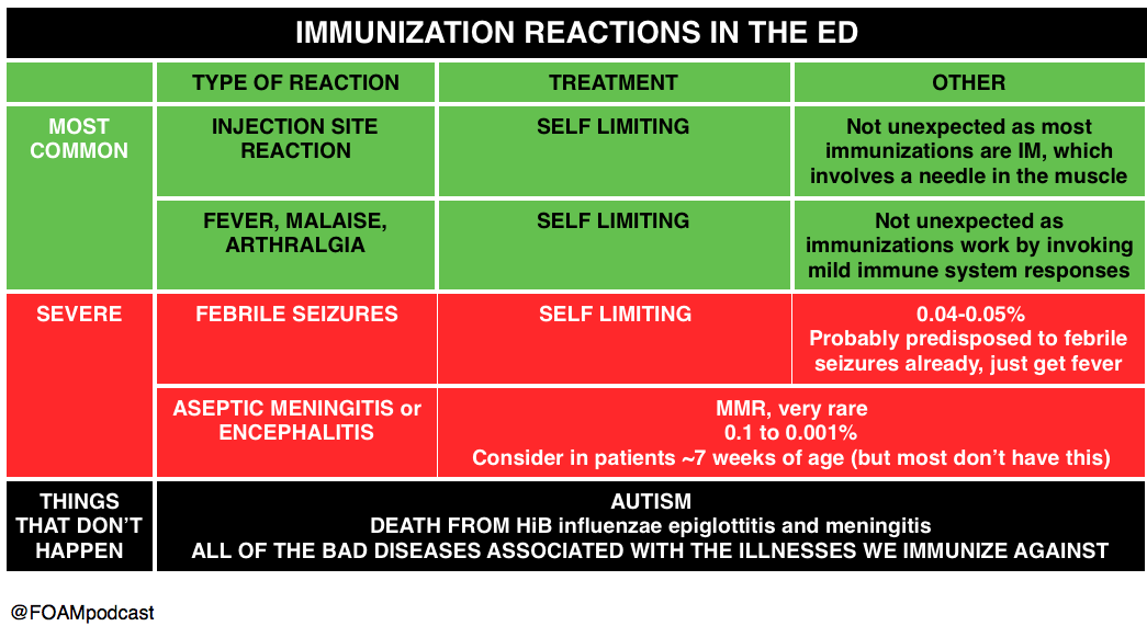
Dr. Al Sachetti – Immunization Reactions in the Emergency Department

check out the WHO fact sheets

screen-shot-2016-10-18-at-5-34-43-am

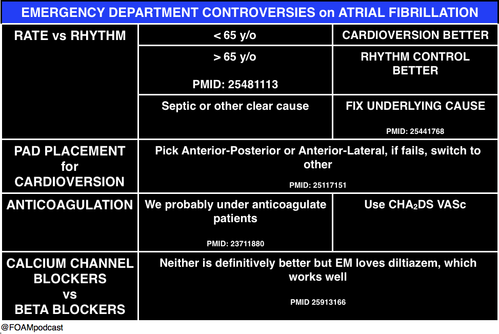
Dr. Corey Slovis – Atrial Fibrillation

screen-shot-2016-10-18-at-6-01-08-am

References:

  1. Llor C, Moragas A, Bayona C. Efficacy of anti-inflammatory or antibiotic treatment in patients with non-complicated acute bronchitis and discoloured sputum: randomised placebo controlled trial. BMJ (Clinical research ed.). 347:f5762. 2013. [pubmed]
  2.  Smith et al. Over-the-counter (OTC) medications for acute cough in children and adults in ambulatory settings. Cochrane Database of Systematic Reviews 2012, Issue 8. Art. No.: CD001831. DOI: 10.1002/14651858.CD001831.pub4.22895922
  3. Dobson R. Cough medicines’ effect is mainly placebo. BMJ. 2006 Jan 7; 332(7532): 8. PMCID: PMC1325161
  4. Cohen et al. Effect of Honey on Nocturnal Cough and Sleep Quality: A Double-blind, Randomized, Placebo-Controlled Study Pediatrics; originally published online August 6, 2012; PMID:22869830
  5. Egerton-Warburton D, Meek R, Mee MJ, Braitberg G. Antiemetic use for nausea and vomiting in adult emergency department patients: randomized controlled trial comparing ondansetron, metoclopramide, and placebo. Annals of emergency medicine. 64(5):526-532.e1. 2014. [pubmed]
  6. Furyk JS, Meek RA, Egerton-Warburton D. Drugs for the treatment of nausea and vomiting in adults in the emergency department setting. The Cochrane database of systematic reviews. 2015. [pubmed]
  7. Beadle KL, Helbling AR, Love SL, April MD, Hunter CJ. Isopropyl Alcohol Nasal Inhalation for Nausea in the Emergency Department: A Randomized Controlled Trial. Annals of emergency medicine. 68(1):1-9.e1. 2016. [pubmed]
  8.  Friedman BW, Dym AA, Davitt M. Naproxen With Cyclobenzaprine, Oxycodone/Acetaminophen, or Placebo for Treating Acute Low Back Pain: A Randomized Clinical Trial. JAMA. 314(15):1572-80. 2015. [pubmed]
  9. Derry S, Conaghan P, Da Silva JA, Wiffen PJ, Moore RA. Topical NSAIDs for chronic musculoskeletal pain in adults. The Cochrane database of systematic reviews. 4:CD007400. 2016. [pubmed]
  10. Silberban. Placebos Are Getting More Effective. Drugmakers Are Desperate to Know Why.

Care of transgender patients + more from ACEP

Listen Here or iTunes

We are in Las Vegas at ACEP 2016 and discuss high yield or cutting edge lectures each day.

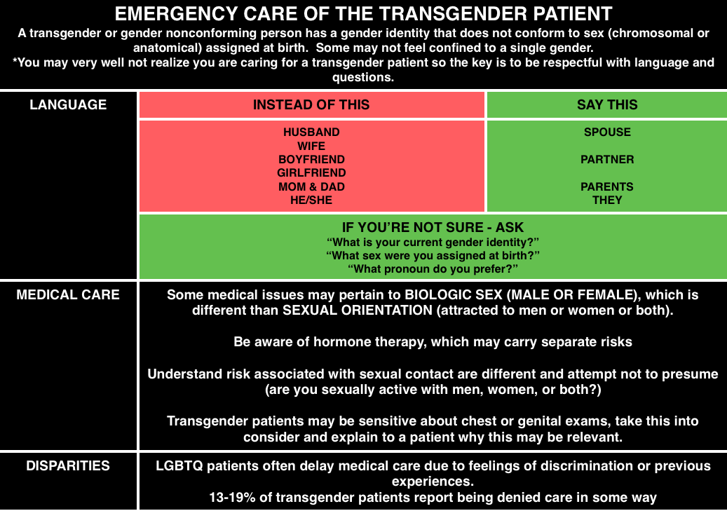
Dr. Anne Daul – Emergency Care of the Transgender Patient

Most emergency medicine physicians and trainees lack training in caring for LGBTQ patients, let alone transgender patients [1].  Many members of the LGBTQ community may have delays in medical care including 21% of transgender patients in a Canadian survey[2].  Also, according to a 2010 task force, 19% of transgender patients report being denied care in some way [3].

@FOAMpodcast
@FOAMpodcast

Here is a video from SMACC Dublin from Thom O’Neill on caring for LGBT youth

https://www.youtube.com/watch?v=7ZsYgftEv2U&feature=youtu.be

Check out these FOAM resources from EPMonthly and Mayo.

Dr. David Callaway – Active Shooter

2% of active shooter events take place in the health care setting.

Plan of Action in Active Shooter Scenario: AVOID DENY DEFEND TREAT

  • Avoid – stay away from the shooter.
  • Deny – deny them access to you or the area. Lock doors, block pathways, turn off the lights, make it more difficult for them.
  • Defend -.if necessary, defend yourself.
  • Treat – once you are safe, and the scene is safe, treat and care for your patients.

Dr. Kevin Klaur – Lawsuits

Documentation and discharge instructions repeatedly come up in lawsuits.

  • Documentation: If you document after the fact, particularly if there was a bad outcome – be straightforward that you are documenting after the fact. Do not document as though you do now know the outcome
  • Discharge – lawsuits often come up because discharge instructions or documentation were not sufficient. Klauer argues that it is not sufficient to state “patient improved, discharged home.” He urges us to document a repeat exam or show HOW they are improved.

Dr. Klauer also gave some general pearls on lawsuits – high numbers for orthopedics/missed fractures and administration of RhoGham.  An additional pearl he gave was for cauda equina.

  • These patients often have small post void residuals because it’s a neurogenic problem, not a mechanical obstruction.  Thus, if a patient has other features and has a post void residual of 100 cc, it’s not necessarily not cauda equina.


One ACEP16 lecturer talked about magnesium use in alcohol withdrawal – probably not ready for prime time, Cochrane agrees  [4].

References:

  1. Moll J, Krieger P, Moreno-Walton L. The prevalence of lesbian, gay, bisexual, and transgender health education and training in emergency medicine residency programs: what do we know? Academic emergency medicine : official journal of the Society for Academic Emergency Medicine. 21(5):608-11. 2014. [pubmed]
  2. Bauer GR, Scheim AI, Deutsch MB, Massarella C. Reported emergency department avoidance, use, and experiences of transgender persons in Ontario, Canada: results from a respondent-driven sampling survey. Annals of emergency medicine. 63(6):713-20.e1. 2014. [pubmed]
  3. Grant JM, Mottet LA, Tanis JD et al. National Transgender Discrimination Survey Report on health and health care Findings of a Study by the National Center for Transgender Equality and the National Gay and Lesbian Task Force.  October 2010
  4. Tejani SM. Magnesium for the prevention or treatment of alcohol withdrawal syndrome in adults. June 2013

Episode 58 – Ophthalmology

The Free Open Access Medical Education (FOAM) 

Listen Here or iTunes

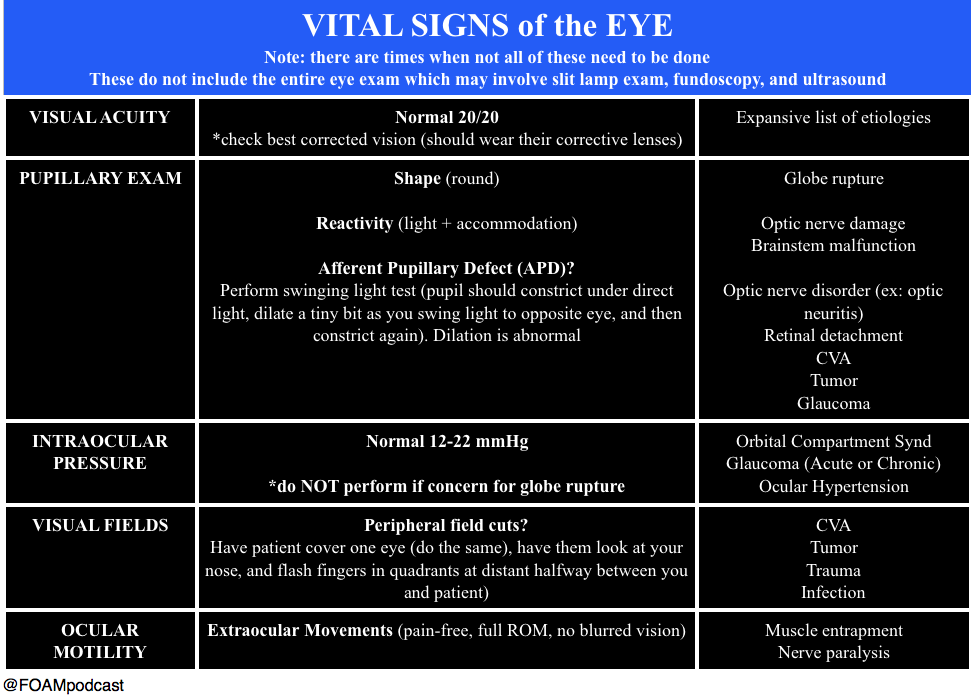
We cover an incredible ophthalmology resource, OphthoBook.com, by Dr. Tim Roots.  This resource has a free book and excellent free video lectures.  Specifically, we detail a hilarious video on eye exam tricks, especially targeting individuals who “can’t see.”

[table id=3 /]

  • Core Content
  • We previously reviewed eye trauma in this podcast. In this episode, we review ophthalmology basics using Tiintinalli’s Emergency Medicine Chapter 241.
  • screen-shot-2016-10-09-at-5-22-35-pm

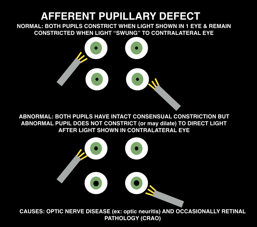
When doing the pupillary exam, it is important to assess for an afferent pupillary defect (APD). Ophthalmologists will want “there is or is NOT an APD” when you consult them for essentially any reason.  Normal pupils constrict when the light is shown in either pupil (direct and consensual constriction). To assess for an APD, perform the “swinging light test.”

screen-shot-2016-10-10-at-9-56-49-am

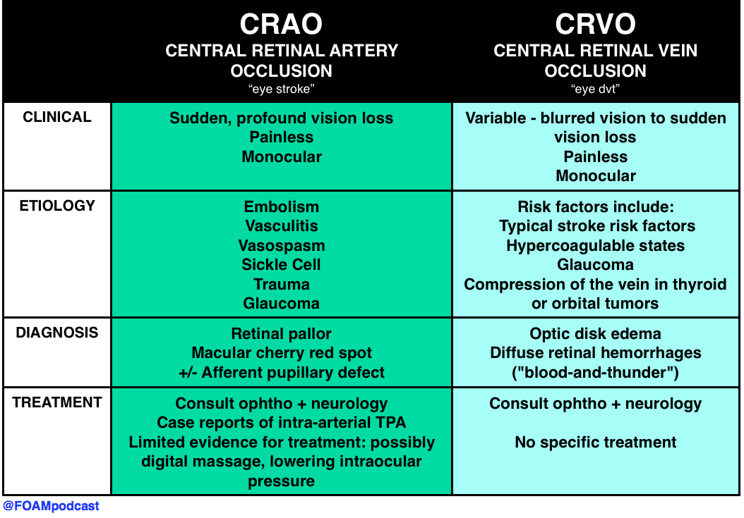
Causes: optic nerve pathology (ex: optic neuritis) or occsaionally, retinal pathology (CRAO)

screen-shot-2016-10-09-at-8-34-25-pm

Peer reviewed by Michael Westafer, MD ophthalmologist and glaucoma fellow at Cleveland Clinic.

Generously Donated Rosh Review Questions

A 72-year-old man presents with a painful red eye and visual loss worsening over the last 24 hours. He recently had cataract surgery. Examination of the eye reveals the image above. Which of the following is the most likely?

A. Endophthalmitis

B. Hyphema

C. Uveitis

D. Vitreous hemorrhage

[accordion]
[toggle title=”Answer” state=”closed”]

A. Endophthalmitis is an infection involving the anterior, posterior and vitreous chambers of the eye. It results from trauma (blunt globe rupture, penetrating injury, foreign bodies) and alsoiatrogenically after ocular surgery like cataract repair. Patients complain of severe pain in the eye and visual impairment or loss. Examination of the eye reveals decreased visual acuity, injected conjunctiva, chemosis and haziness of the infected chambers. Infections are treated with both systemic and intraocular antibiotics.

A hyphema (B) is blood in the anterior chamber usually caused by trauma. When the patient is in an upright position, blood will layer along the inferior aspect of the anterior chamber. As the hyphema increases in size, it elevates intraocular pressure. In some cases admission is warranted for patients with large hyphemas (>50%), decreased vision, sickle cell disease and elevated intraocular pressure. Treatment is aimed at decreasing pressure with topical (beta-blocker, alpha agonist or carbonic anhydrase inhibitors) and systemic therapy (carbonic anhydrase inhibitor, mannitol). Uveitis (C) occurs after blunt trauma in which the iris and ciliary body are inflamed causing ciliary spasm. Patients complain of significant photophobia with significant eye pain. Examination of the eye reveals perilimbal conjunctival injection (also called ciliary flush) and a small, poorly dilating pupil. Photophobia occurs with light shone on both the affected and unaffected eye. On slit lamp, cells (white and red) and flare (protein) are noted in the anterior chamber. Treatment is with a topical cycloplegic agent to minimize spasm. Vitreous hemorrhage (D) occurs as a result of injuries to the retina, uveal tract and their associated vascular structures. Common associated conditions include diabetic retinopathy, retinal vein occlusion and trauma. Patients complain of decreased visual acuity and floaters. The condition is not typically painful. Diagnosis is made with ocular ultrasound showing blood products in the posterior chamber.

[/toggle]
[/accordion]

What is a dependent pocket of pus seen in the anterior chamber called?

  • [accordion]
  • [toggle title=”Answer” state=”closed”]
  • Hypopyon.
  • [/toggle]
  • [/accordion]

A 51-year-old man walks into a movie theater and experiences acute onset of right eye pain associated with nausea, vomiting, and cloudy vision. Which of the following is expected during the ophthalmologic exam?

A. Cherry-red spot in the macular area

B. Deep anterior chamber

C. Intraocular pressure >21 mm Hg

D. Miotic pupil

[accordion]
[toggle title=”Answer” state=”closed”]

The patient has acute angle-closure glaucoma. This condition results in optic nerve damage from increased intraocular pressure. In patients with a narrow anterior chamber angle, reduced illumination (like entering a dark movie theater) causes mydriasis. Subsequently, folds of the peripheral iris can block the angle, which prevents aqueous humor outflow. This leads to a rapid elevation of intraocular pressure causing ocular pain, a hazy cornea, ciliary flush, a firm globe, and optic nerve damage if the pressure is not promptly relieved. An intraocular pressure >21 mm Hg is considered elevated. Pressures can elevate quickly to >60 mm Hg. The higher the pressure, the quicker damage occurs to the optic nerve and the poorer the prognosis. The diagnosis is often delayed due to the associated symptoms of nausea, vomiting, and abdominal pain. Treatment involves reducing aqueous humor production with IV acetazolamide, topical beta-blockers (timolol), and topical alpha-agonists (apraclonidine). Topical miotic agents (pilocarpine) are used to reverse the angle closure. Topical steroidshelp to reduce inflammation. Hyperosmotic agents (mannitol, glycerol) can also be administered for further reduction in intraocular pressure.

The funduscopic finding of a pale retina with a cherry-red spot in the macular area (A) is consistent with central retinal artery occlusion. This condition is associated with sudden unilateral vision loss that is painless. It is caused by a thrombotic plaque or embolus of the central retinal artery. A deep anterior chamber (B) is protective against acute angle-closure glaucoma. Individuals with a narrow chamber are at increased risk. The pupil in acute angle-closure glaucoma is most commonly fixed and mid-dilated, rather than miotic (D). Miotic pupils are associated with opiate use, cholinergic toxicity, and pontine strokes.

[/toggle]
[/accordion]

References:

“Eye Emergencies.”  Chapter 241.  Tintinalli’s Emergency Medicine: A Comprehensive Review. 8th ed.

Episode 57 – Lithium Toxicity & Rhabdomyolysis

The Free Open Access Medical Education (FOAM) 

Listen Here or iTunes

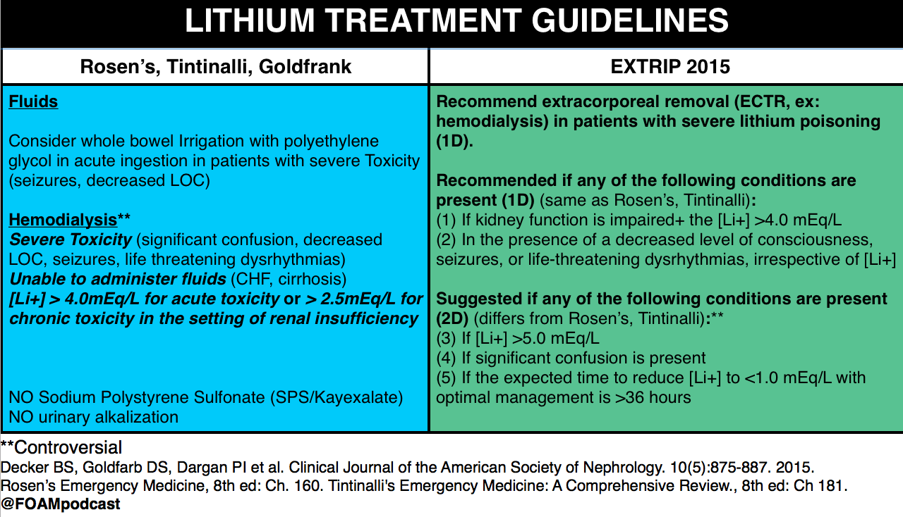
We cover a post from Dr. Josh Farkas on PULMcrit on lithium toxicity. The key message from the post is: a single serum lithium level doesn’t necessitate dialysis, despite a recommendation from the EXTRIP working group  to initiate dialysis in patients with a lithium level > 5 mEq/L [1].  Dr. Farkas advocates for aggressive management in asymptomatic patients with chronic lithium toxicity and patients without impaired renal function.

Lithium neurotoxicity does not correlate with serum lithium levels; it depends on the concentration in the CNS.

  • The argument is that the neurologic effects, among the most worrisome sequelae of lithium toxicity, results from lithium crossing the blood-brain-barrier (BBB) into the central nervous system (CNS). Thus, in an acute ingestion, patients may have a higher serum lithium level, but given the acuity, the lithium may not cross into the CNS.  Conversely, a patient with chronic toxicity may have a mildly elevated serum lithium level but the lithium has had time to cross the BBB into the CNS so more neurotoxicity can result from lower serum lithium concentrations.

Differentiating between acute, chronic, and acute on chronic toxicity is important in management.

  • Acute – expect the levels will rise rapidly as the bolus of lithium is absorbed.
  • Chronic – expect the levels will not rise rapidly given a lack of lithium bolus.
  • Acute on chronic – again, expect levels to rise rapidly and may see more neurotoxicity as some lithium may have passed through the BBB into the CNS.

screen-shot-2016-09-30-at-6-19-25-pm

screen-shot-2016-09-30-at-6-02-28-pm

Core Content

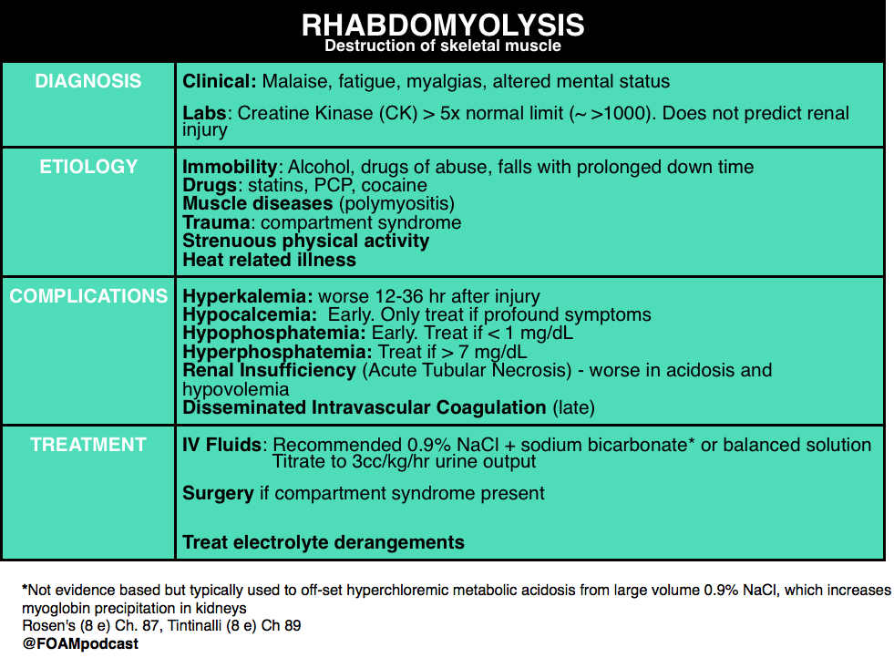
We review rhabdomyolysis using Rosen’s Emergency Medicine (8e)  Chapter 160 and Tintinalli’s Emergency Medicine (8e), Chapter 181.

screen-shot-2016-10-01-at-12-05-27-pm

Generously Donated Rosh Review Questions

A 17-year-old man presents to the ED from a correctional facility complaining of general malaise with nausea and vomiting one day after a weightlifting competition. Vital signs are T 37.2°C, BP 100/65 mm Hg, HR 125, and RR 22. Physical exam reveals an uncomfortable, fatigued male who has diffuse muscle soreness. Urinalysis shows 3+ blood without red blood cells. What is the most important next test to direct the acute management of this patient?

A. Creatine kinase assay

B. Electrocardiogram

C. Electrolyte panel with blood urea nitrogen and creatinine

D.Microscopic urinalysis

[accordion]
[toggle title=”Answer” state=”closed”]

B. Electrocardiogram. The patient presents with exercised-induced rhabdomyolysis. Although the causes of rhabdomyolysis are manifold, potential complications are independent of etiology. Of all the complications, hyperkalemia is the most concerning and, undiagnosed, can lead to sudden cardiac death. ECG changes indicative of hyperkalemia are thus critical to identify early in the course of management. Hyperkalemia results from impaired calcium transport with increased intracellular calcium accumulation, cellular necrosis, and expulsion of intracellular contents (including potassium) into the bloodstream. Rhabdomyolysis can also lead to acute tubular necrosis and kidney failure, which will exacerbate developing hyperkalemia by decreasing renal potassium clearance. Hyperkalemia is an immediate life-threatening condition that develops shortly after muscle injury. Absent point-of-care electrolyte analyzers, the most rapid way to screen for hyperkalemia is through an electrocardiogram.

Rhabdomyolysis is not defined by a specific creatine kinase (A) level. But, in general, a serum CK >5 times the upper limit of normal (a threshold that may differ by lab) is considered indicative of rhabdomyolysis. Although kidney injury can occur at any level, higher CKs correlate with an increased likelihood for the development of acute renal failure. In the absence of cerebral or myocardial infarction, a CK >5 times is diagnostic for serious muscle injury. Urinalysis (D) typically shows brownish discoloration with “large” blood on dipstick but few, if any, red blood cells on microscopic evaluation. This occurs because most dipstick tests cannot distinguish myoglobinuria from hematuria or hemoglobinuria. Protein, brown casts, and renal tubular epithelial cells may also be present. Measures of renal function and an electrolyte panel (C) should be obtained in all patients with suspected rhabdomyolysis. But, as mentioned, waiting for results may lead to a delay in the identification of life-threatening complications. In addition to hyperkalemia, hyperphosphatemia and hypocalcemia may also be seen. Additional (though less worrisome) laboratory abnormalities include elevated uric acid and low albumin.

[/toggle]
[/accordion]

One Step Further Question: What is the late complication of rhabdomyolysis associated with thrombocytopenia, hypofibrinogenemia, and an elevated D-dimer?

[accordion]
[toggle title=”Answer” state=”closed”]

Disseminated intravascular coagulopathy may occur and is a result of muscle necrosis with liberation of activating substances from injured cells.

[/toggle]
[/accordion]


  • References:
  • 1. Decker BS, Goldfarb DS, Dargan PI et al. Extracorporeal Treatment for Lithium Poisoning: Systematic Review and Recommendations from the EXTRIP Workgroup. Clinical Journal of the American Society of Nephrology. 10(5):875-887. 2015.
  • 2. “Chapter 160. Lithium.”  Rosen’s Emergency Medicine.  8th edition.
  • 3. “Chapter 181. Lithium.” Tintinalli’s Emergency Medicine: A Comprehensive Review. 8th edition.
  • 4. Grellar H. “Chapter 72. Lithium.” Goldfrank’s Toxicology. 10th edition.
  • 5.  “Chapter 127. Rhabdomyolysis. Rosen’s Emergency Medicine.  8th edition.
  • 6. “Chapter 89. Rhabdomyolysis.” Tintinalli’s Emergency Medicine: A Comprehensive Review. 8th edition.