Episode 65 – Contrast-Induced Nephropathy and Genitourinary Trauma

iTunes or Listen Here

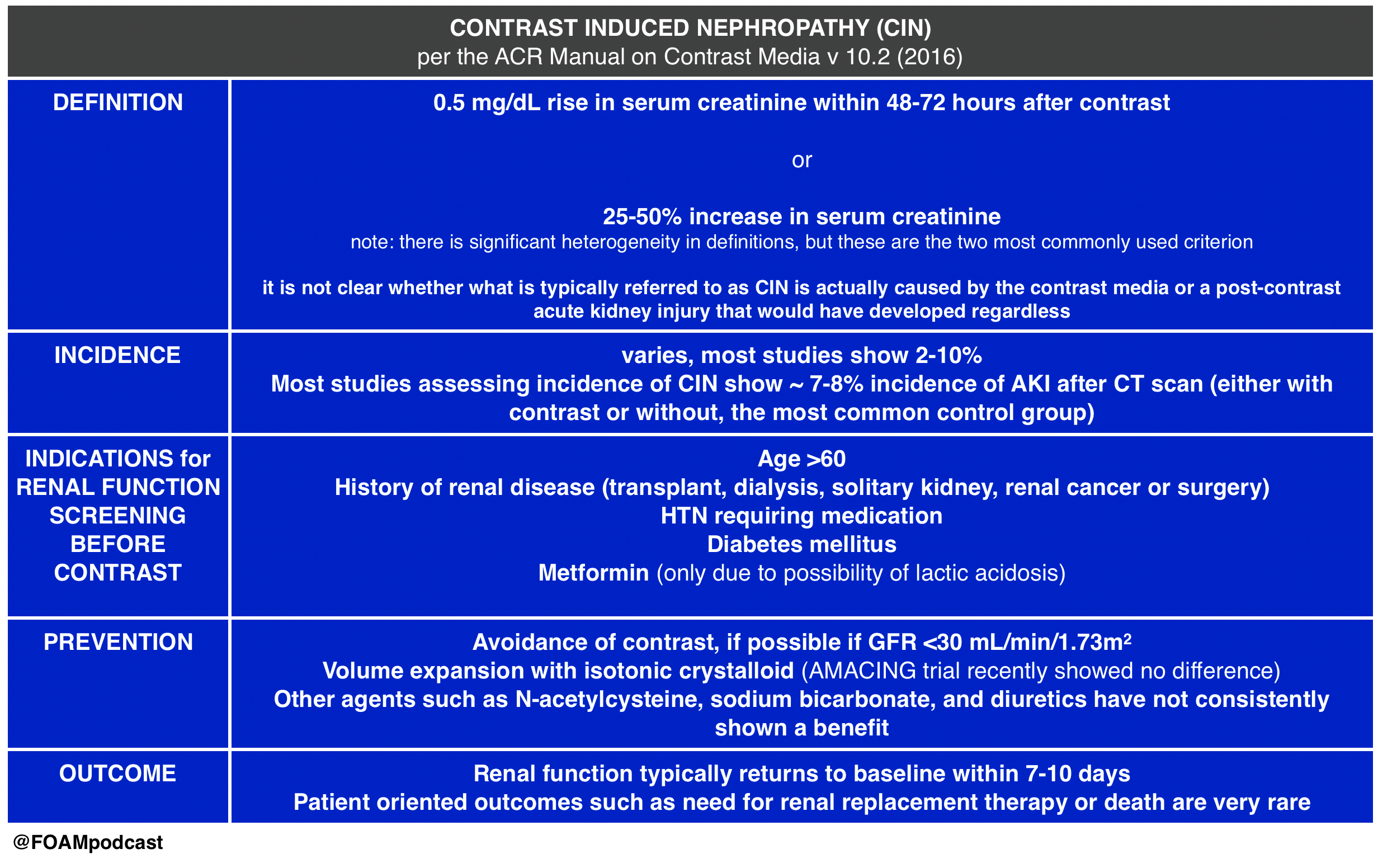
We cover Free Open Access Medical Education (FOAM) on Contrast-Induced Nephropathy (CIN).  A large retrospective study by Hinson and colleagues in Annals of Emergency Medicine, reignited enthusiasm in the FOAM world about the questionable entity (and clinical significance) of CIN. The data on CIN is somewhat difficult to parse out as the data consists entirely of retrospective cohort and case-control studies. The highest risk of CIN seems to come from large volume contrast procedures such as percutaneous coronary angiography. Studies looking at the risk of CIN after contrast-enhanced CT scan have been less conclusive.

The American College of Radiology (ACR) Manual on Contrast Induced Nephropathy

CIN

This Emergency Medicine Literature of Note post covers the AMACING  trial, which looks at intravenous fluid administration (0.9% NaCl) versus usual care in patients “at risk” for CIN undergoing contrast-enhanced CT scan.   The study found a difference of -0.1% (95% CI -2.25 to 2.06), which was below the non-inferiority margin. Prior literature shows similar results, with no clear-cut efficacy from prevention strategies for CIN.

This post by Dr. Joel Topf (nephrologist @kidneyboy) on the Precious Bodily Fluids blog discusses a nephrologist’s take on CIN.

EM topics post on fluids in CIN.

Core Content

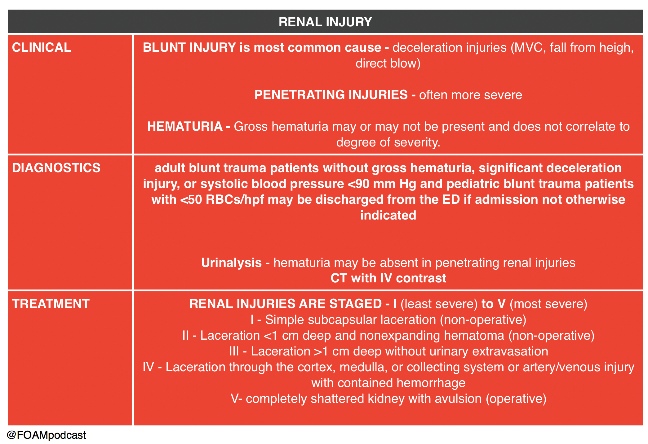
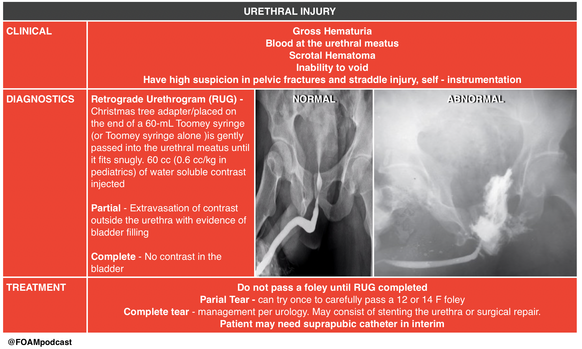
We then delve into core content on genitourinary trauma using Rosen’s Chapter 47 and Tintinalli’s Chapter265 as a guide.

Screen Shot 2017-02-28 at 1.43.40 PM Screen Shot 2017-02-28 at 1.44.33 PM

 Rosh Review Emergency Board Review Questions

An 18-year-old man involved in a motor vehicle collision is transferred from a rural facility after being diagnosed with a pelvic fracture. After your initial assessment and stabilization, a secondary survey is performed. On examination, he is noted to have blood at his urethral meatus as well as a scrotal hematoma. Which of the following is the most appropriate next step?

A. Retrograde cystogram

B. Retrograde urethrogram

C. Suprapubic catheter placement

D. Transurethral urinary catheter placement

[accordion]
[toggle title=”Answer” state=”closed”]

B. The patient should undergo a retrograde urethrogram to rule out an underlying urethral injury. In patients with a pelvic fracture and signs of urethral injury, a retrograde urethrogram should be performed in the supine position prior to urethral instrumentation. If a partial disruption is identified, one attempt to place a 12- or 14-French Foley or coude catheter can be made. If unsuccessful or a complete tear is diagnosed, then a suprapubic catheter will need to be placed. Pelvic fractures with displacement of the pubic symphysis can cause laceration or avulsion of the prostatic urethra. The three classic findings of urethral injury include blood at the urethral meatus, a high-riding prostate and a scrotal or perineal hematoma. Failure to recognize a urethral injury can lead to urethral stricture formation and urinary incontinence.

Urethral manipulation can convert a partial urethral tear to a complete tear, therefore transurethral urinary catheter placement (D) should be avoided until urethral integrity is known. Suprapubic catheter placement (C) may ultimately be necessary if complete urethral injury is diagnosed, however in this case a retrograde urethrogram should be performed prior to proceeding with suprapubic placement. The patient may also have a bladder injury and may require a retrograde cystogram (A), however urethral integrity must be evaluated by a retrograde urethrogram prior to performing a cystogram

[/accordion]

A 29-year-old man presents to the ED after a MVC. A pelvic fracture is identified on radiography. His vital signs are stable. The decision is made to place a Foley catheter, but blood is noted at the urethral meatus. Which of the following is an appropriate next step?

A. Consult a urologist

B. Obtain a CT scan to evaluate for urethral injury

C. Perform a retrograde urethrogram

D. Place a condom catheter

[accordion]
[toggle title=”Answer” state=”closed”]

C. Perform a retrograde urethrogram. In general, a Foley catheter should not be placed in the setting of suspected urethral injury. In such cases, it is recommended that further testing be performed to evaluate for urethral injury. A retrograde urethrogram should be performed. If there is no contrast extravasation, then a Foley catheter can safely be inserted.  A urologist should be consulted (A) if a urethral injury is confirmed by the retrograde urethrogram. A CT scan (B) is a poor study to identify urethral injuries. A condom catheter (D) does not allow for accurate urine output measurements and may delay identification of a urethral injury.

[/accordion]

References:

  1. Hinson JS, Ehmann MR, Fine DM, et al. Risk of Acute Kidney Injury After Intravenous Contrast Media Administration. Ann Emerg Med. 2017
  2. ACR Manual on Contrast Media.  v10.2. 2016. p33-40
  3. “Genitourinary System.”  Rosen’s Emergency Medicine, 8th ed. Chapter 47, 479-499.e1
  4. “Genitourinary Trauma.” Tintinalli’s Emergency Medicine: A Comprehensive Review.  8th ed.  Chapter 265
Episode 64 – Lumbar Puncture and Central Nervous System Infections

iTunes or Listen Here

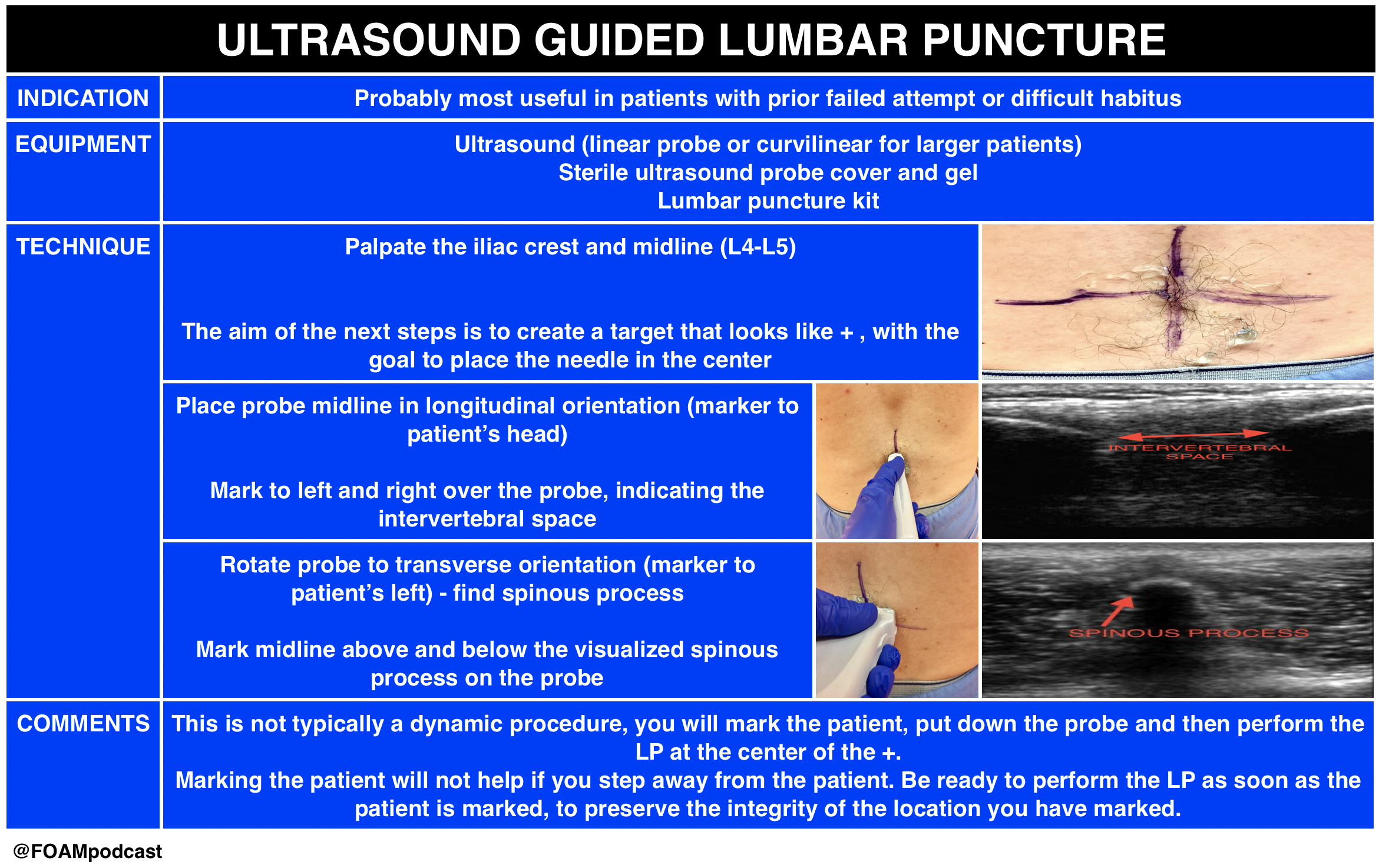
Ultrasound is gaining popularity in assisting lumbar punctures (LPs). We review this technique as well as Free Open Access Medical Education (FOAM) from the following sites:  5 Min SonoSinai EM, and PEM pearls from ALiEM.

US guided LP

  • This technique is likely most helpful in difficult patients. A recent study demonstrated 27% absolute increase in first attempt success using ultrasound in infants <6 months old undergoing LP; however, the success rate in both arms was abysmal with only 57% success rate in the ultrasound arm.
  • Core Content

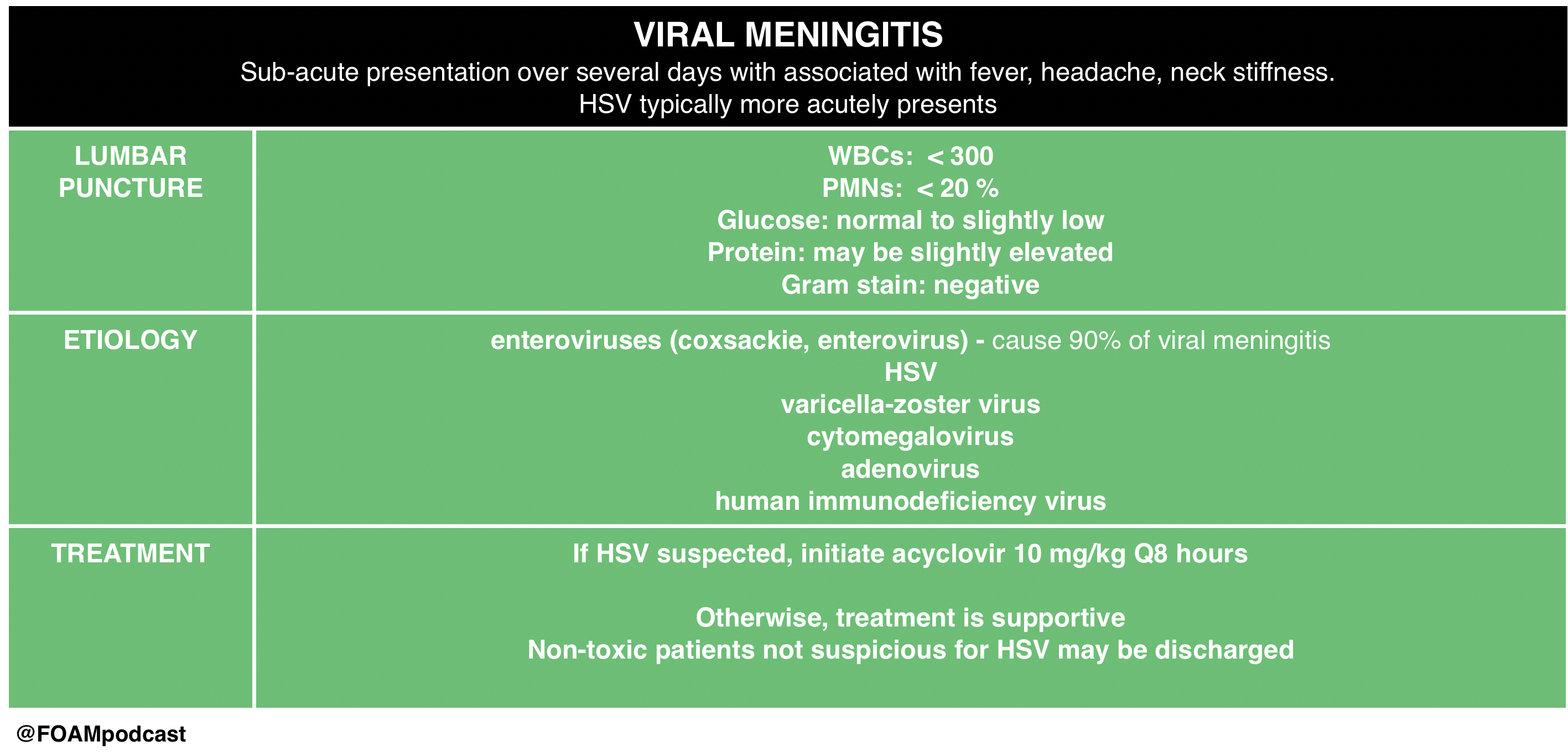
We then delve into core content on meningitis, encephalitis, and antiNMDA receptor encephalitis using Rosen’s Chapter 109 and Tintinalli’s Chapters 117 and 174 as a guide.

 Rosh Review Emergency Board Review Questions

A 40-year-old man with HIV presents with two weeks of progressive headache, malaise, and fever. On examination, he has mild nuchal rigidity, confusion, and a temperature of 38.2oC. Cerebrospinal fluid analysis shows a white blood cell count 360 cells/mL with a monocyte predominance, glucose 28 mg/dL, and protein 220 mg/dL. What is the treatment of choice?

A. Acyclovir

B. Amphotericin B

C. Ceftriaxone

D. Vancomycin

[accordion]
[toggle title=”Answer” state=”closed”]

B. Amphotericin B . Cryptococcal meningoencephalitis is an opportunistic infection that occurs primarily in patients with advanced AIDS, although it can be seen in immunocompromised transplant patients as well. The majority of HIV-related cases occur when the CD4 count is < 100 cells/mm3. Patients present with progressive headache, nausea, malaise, and fever over the course of 1 – 2 weeks. Examination findings are typical for meningitis and include altered mental status, photophobia, and fever. Meningismus is less commonly seen. Presentations can be subtle and a high index of suspicion is needed for diagnosis. A CT scan of the brain is indicated if there are signs of increased intracranial pressure or focal neurologic deficits. Lumbar puncture should be performed with careful measurement of the opening pressure. Cerebrospinal fluid will show a mildly elevated white blood cell count with a monocyte predominance, decreased glucose, and mildly elevated protein. However, in some cases the cerebrospinal fluid will only show minor, if any, abnormalities. Cryptococcal antigen testing of the CSF is nearly 100% sensitive and specific. India ink staining will show budding organisms. Treatment of choice is intravenous amphotericin B in addition to oral flucytosine for 14 days followed by an 8 week course of oral fluconazole.  Acyclovir (A) is used in the treatment of herpes meningoencephalitis. Ceftriaxone (C) and vancomycin (D) are indicated in the treatment of bacterial meningitis.

[/toggle]
[/accordion]

A previously healthy 18-year-old woman presents to the emergency department with complaints of fever, headache, and neck stiffness. She is accompanied by her sister, who expresses concern because the patient seems suddenly confused and cannot remember what she did yesterday. After you administer empiric intravenous antibiotics, which of the following is the next best step?

A. Chest X-ray

B. Complete blood count with differential

C. Lumbar puncture

D. Urinalysis

[accordion]
[toggle title=”Answer” state=”closed”] C. 
 Lumbar puncture.  Meningitis is an inflammation of the tissues surrounding the brain and spinal cord (meninges) and may be of infectious (bacterial, viral, or fungal) and various other etiologies. The classic clinical manifestations include nuchal rigidity, fever and altered mental status. Patients often present with headache as well. All patients with suspected meningitis should have lumbar puncture (LP) to evaluate the cerebrospinal fluid (CSF) unless this procedure is contraindicated. There are no absolute contraindications to LP. Relative contraindications include patients with evidence of increased intracranial pressure, thrombocytopenia, bleeding diathesis or spinal epidural abscess. Acute bacterial meningitis is a medical emergency and left untreated or treated late is almost universally fatal. Treatment involves addressing systemic complications and initiating empiric antibiotic therapy as soon as possible.

Up to half of patients with pneumococcal meningitis may have evidence of pneumonia on chest X-ray (A), but this is not part of the initial workup of patients with suspected meningitis. Complete blood count with differential (B) is often ordered in the workup for bacterial meningitis and generally shows increased white blood cell count, but is not as important to order initially as an LP. Urinalysis (D) is generally not a helpful test in the diagnosis of meningitis and therefore not recommended in the workup for patients with suspected bacterial meningitis.

[/toggle]
[/accordion]

References:

  1. Attia J, Hatala R, Cook DJ, Wong JG. The rational clinical examination. Does this adult patient have acute meningitis?. JAMA. 1999;282(2):175-81.
  2. Nakao JH, Jafri FN, Shah K, Newman DH. Jolt accentuation of headache and other clinical signs: poor predictors of meningitis in adults. Am J Emerg Med. 2014;32(1):24-8.
  3. Neal JT, Kaplan SL, Woodford AL, Desai K, Zorc JJ, Chen AE. The Effect of Bedside Ultrasonographic Skin Marking on Infant Lumbar Puncture Success: A Randomized Controlled Trial. Ann Emerg Med. 2016.