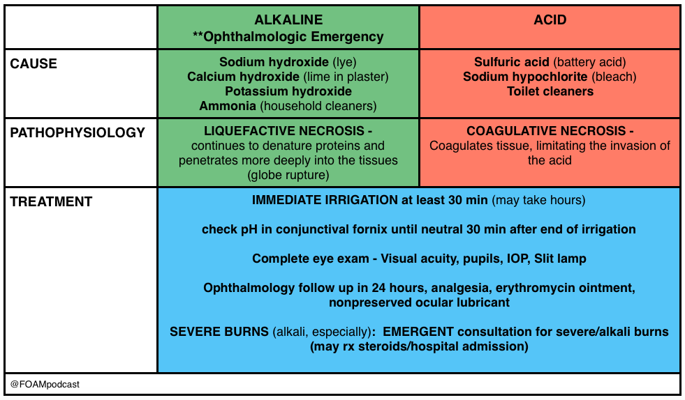
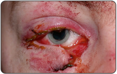
Corneal Emergencies

iTunes or Listen Here

We review one of the editors top picks from Annals of Emergency Medicine, which are free for 6 months from issue release.  The article covered in this podcast is Waldman et al, is a study of tetracaine in corneal abrasions.
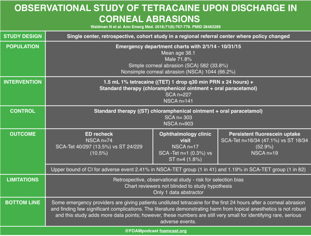
Review from First10EM on topical anesthetics and corneal abrasions.

Rosh Review Emergency Board Review Questions

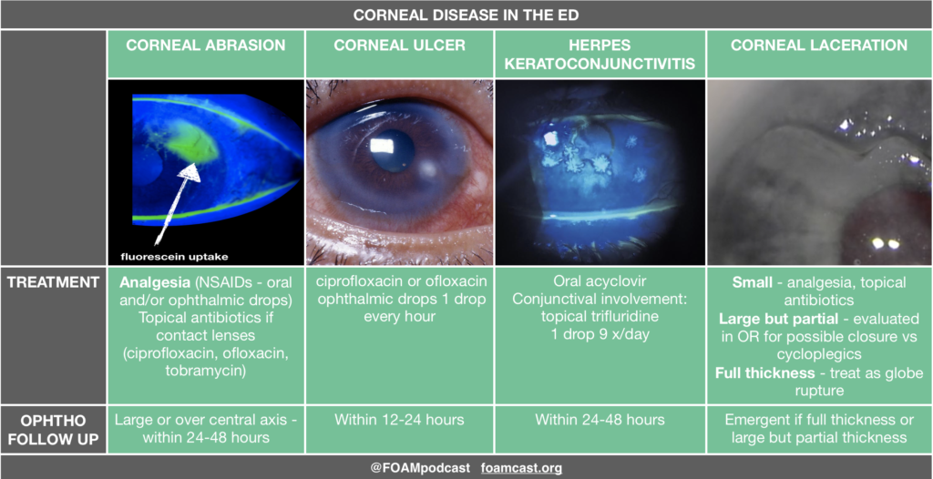
A 21-year-old man presents to the emergency department complaining of left eye pain. Fluorescein staining reveals a dendritic lesion over the cornea. What is the most likely diagnosis based on this finding?

A. Bacterial conjunctivitis

B. Corneal abrasion

C. Herpes keratitis

D. Retinal detachment

[accordion]
[toggle title=”Answer” state=”closed”]

A dendritic lesion of the cornea detected by fluorescein staining indicates the presence of herpetic keratitis. Herpetic keratitis is caused by herpes simplex virus and can lead to blindness because of scarring and opacification of the cornea. A typical presentation includes a painful eye, blurred vision or excessive tearing. Diagnosis can be made with fluorescein staining which gives the classic dendritic appearance. The presence of a dendritic lesion indicates active replication of the virus. Superficial epithelial herpetic keratitis can be successfully treated in two to three weeks with topical antivirals such as ganciclovir. Topical steroids must be avoided in patients with herpetic keratitis as the herpetic lesions can become much deeper and threaten the patient’s vision. Oral antivirals have not been studied comparatively with topical antivirals. Risk of transmission of ocular herpes simplex virus to another person is thought to be extremely low.

Bacterial conjunctivitis (A) can cause eye discomfort but typically presents with copious purulent discharge from one or both eyes. Fluorescein staining of the eye will be negative for any defect. Corneal abrasions (B) present with a superficial epithelial lesion that is usually linear in shape. The cornea is expected to be clear. Retinal detachment (D) is painless and does not cause a change in the appearance of the surface of the eye. Fundoscopic exam will often show rugae indicating the edge of the retina floating in the vitreous humor.

[accordion]
[toggle title=”Answer” state=”closed”]

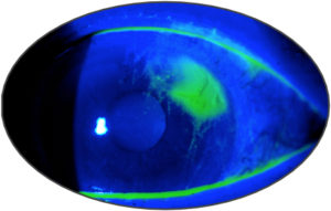
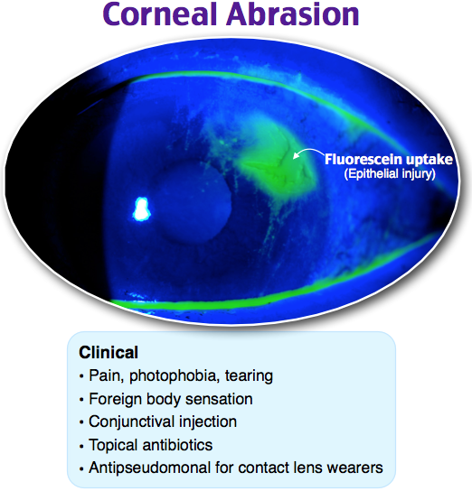
A 20-year-old woman presents to the emergency department for left eye pain. Yesterday she scratched her eye while putting in her contact lens. She has had constant left eye pain and tearing since. She has no foreign body sensation. On exam, her visual acuity is at baseline and equal in both eyes. Her fluorescein exam findings are shown below. Which of the following is the most appropriate treatment for this patient’s condition?

A. Erythromycin ophthalmic ointment

B. Gentamicin-prednisolone ophthalmic suspension

C. Tetracaine ophthalmic solution

D. Tobramycin ophthalmic ointment

[accordion]
[toggle title=”Answer” state=”closed”]

D. This patient’s fluorescein exam is consistent with a corneal abrasion. It is caused by direct mechanical damage, leading to a partial-thickness corneal injury. Athletes, contact lens wearers, welders, and glass workers may present more often with these injuries. Diagnosis is confirmed when fluorescein dye highlights the usually linear or punctate abrasions. Multiple vertical corneal abrasions may indicate a retained foreign body beneath the eyelid. Corneal abrasion fluorescein exam findings are not to be confused with open globe injuries (streaking of the dye from the site of injury), corneal ulcers (circular patches of dye uptake with ragged, “heaped up” edges), and herpes simplex keratitis (dendritic pattern uptake). Patients commonly present with eye pain and tearing with a usually known mechanism of injury. The mainstay of treatment includes pain control, infection prophylaxis, and preventative care to avoid abrasions in the future. Pain control includes cycloplegics like homatropine and cyclopentolate and nonsteroidal anti-inflammatory medications. Commonly prescribed antibiotic ointments include erythromycin, ciprofloxacin, tobramycin, and gentamicin. Notably, erythromycin does not cover pseudomonal infections, which are more likely to occur in contact lens wearers.  Erythromycin (A) does not have appropriate pseudomonal coverage for this patient who wears contact lenses and has a corneal abrasion. Ophthalmic medications containing steroids, like gentamicin-prednisolone (B), should not be prescribed in corneal abrasions or ulcers as they may decrease healing rates. While in the emergency department, tetracaine (C) may be used to more easily evaluate the patient’s eye. However, ocular anesthetics should not be prescribed as they decrease healing rates and block the basic corneal reflex to avoid further injury.

[/toggle]
[/accordion]

References:

  1. Waldman N, Winrow B, Densie I, et al. An Observational Study to Determine Whether Routinely Sending Patients Home With a 24-Hour Supply of Topical Tetracaine From the Emergency Department for Simple Corneal Abrasion Pain Is Potentially Safe. Ann Emerg Med. 2018;71(6):767-778.
  2. Ball IM, Seabrook J, Desai N, Allen L, Anderson S. Dilute proparacaine for the management of acute corneal injuries in the emergency department. CJEM. 2010;12:(5)389-96.
  3. Ting JY, Barns KJ, Holmes JL. Management of Ocular Trauma in Emergency (MOTE) Trial: A pilot randomized double-blinded trial comparing topical amethocaine with saline in the outpatient management of corneal trauma. J Emerg Trauma Shock. 2009;2:(1)10-4.
  4. Swaminathan A, Otterness K, Milne K, Rezaie S. The safety of topical anesthetics in the treatment of corneal abrasions: A review. J Emerg Med 2015
  5. Calder LA, Balasubramanian S, Fergusson D. Topical nonsteroidal anti-inflammatory drugs for corneal abrasions: meta-analysis of randomized trials. Acad Emerg Med. 2005;12(5):467-73.
  6. Galuma K, Lee J. “Ophthalmology.” Rosen’s Emergency Medicine: Concepts and Clinical Practice, Chapter 61, 790-819.e3
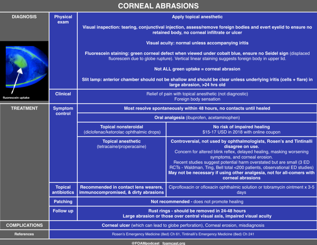
Episode 58 – Ophthalmology

The Free Open Access Medical Education (FOAM) 

Listen Here or iTunes

We cover an incredible ophthalmology resource, OphthoBook.com, by Dr. Tim Roots.  This resource has a free book and excellent free video lectures.  Specifically, we detail a hilarious video on eye exam tricks, especially targeting individuals who “can’t see.”

[table id=3 /]

  • Core Content
  • We previously reviewed eye trauma in this podcast. In this episode, we review ophthalmology basics using Tiintinalli’s Emergency Medicine Chapter 241.
  • screen-shot-2016-10-09-at-5-22-35-pm

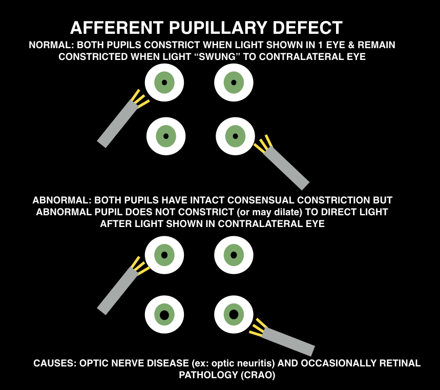
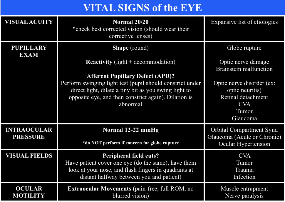
When doing the pupillary exam, it is important to assess for an afferent pupillary defect (APD). Ophthalmologists will want “there is or is NOT an APD” when you consult them for essentially any reason.  Normal pupils constrict when the light is shown in either pupil (direct and consensual constriction). To assess for an APD, perform the “swinging light test.”

screen-shot-2016-10-10-at-9-56-49-am

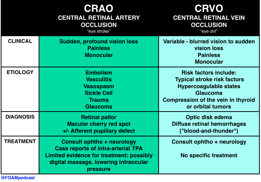
Causes: optic nerve pathology (ex: optic neuritis) or occsaionally, retinal pathology (CRAO)

screen-shot-2016-10-09-at-8-34-25-pm

Peer reviewed by Michael Westafer, MD ophthalmologist and glaucoma fellow at Cleveland Clinic.

Generously Donated Rosh Review Questions

A 72-year-old man presents with a painful red eye and visual loss worsening over the last 24 hours. He recently had cataract surgery. Examination of the eye reveals the image above. Which of the following is the most likely?

A. Endophthalmitis

B. Hyphema

C. Uveitis

D. Vitreous hemorrhage

[accordion]
[toggle title=”Answer” state=”closed”]

A. Endophthalmitis is an infection involving the anterior, posterior and vitreous chambers of the eye. It results from trauma (blunt globe rupture, penetrating injury, foreign bodies) and alsoiatrogenically after ocular surgery like cataract repair. Patients complain of severe pain in the eye and visual impairment or loss. Examination of the eye reveals decreased visual acuity, injected conjunctiva, chemosis and haziness of the infected chambers. Infections are treated with both systemic and intraocular antibiotics.

A hyphema (B) is blood in the anterior chamber usually caused by trauma. When the patient is in an upright position, blood will layer along the inferior aspect of the anterior chamber. As the hyphema increases in size, it elevates intraocular pressure. In some cases admission is warranted for patients with large hyphemas (>50%), decreased vision, sickle cell disease and elevated intraocular pressure. Treatment is aimed at decreasing pressure with topical (beta-blocker, alpha agonist or carbonic anhydrase inhibitors) and systemic therapy (carbonic anhydrase inhibitor, mannitol). Uveitis (C) occurs after blunt trauma in which the iris and ciliary body are inflamed causing ciliary spasm. Patients complain of significant photophobia with significant eye pain. Examination of the eye reveals perilimbal conjunctival injection (also called ciliary flush) and a small, poorly dilating pupil. Photophobia occurs with light shone on both the affected and unaffected eye. On slit lamp, cells (white and red) and flare (protein) are noted in the anterior chamber. Treatment is with a topical cycloplegic agent to minimize spasm. Vitreous hemorrhage (D) occurs as a result of injuries to the retina, uveal tract and their associated vascular structures. Common associated conditions include diabetic retinopathy, retinal vein occlusion and trauma. Patients complain of decreased visual acuity and floaters. The condition is not typically painful. Diagnosis is made with ocular ultrasound showing blood products in the posterior chamber.

[/toggle]
[/accordion]

What is a dependent pocket of pus seen in the anterior chamber called?

  • [accordion]
  • [toggle title=”Answer” state=”closed”]
  • Hypopyon.
  • [/toggle]
  • [/accordion]

A 51-year-old man walks into a movie theater and experiences acute onset of right eye pain associated with nausea, vomiting, and cloudy vision. Which of the following is expected during the ophthalmologic exam?

A. Cherry-red spot in the macular area

B. Deep anterior chamber

C. Intraocular pressure >21 mm Hg

D. Miotic pupil

[accordion]
[toggle title=”Answer” state=”closed”]

The patient has acute angle-closure glaucoma. This condition results in optic nerve damage from increased intraocular pressure. In patients with a narrow anterior chamber angle, reduced illumination (like entering a dark movie theater) causes mydriasis. Subsequently, folds of the peripheral iris can block the angle, which prevents aqueous humor outflow. This leads to a rapid elevation of intraocular pressure causing ocular pain, a hazy cornea, ciliary flush, a firm globe, and optic nerve damage if the pressure is not promptly relieved. An intraocular pressure >21 mm Hg is considered elevated. Pressures can elevate quickly to >60 mm Hg. The higher the pressure, the quicker damage occurs to the optic nerve and the poorer the prognosis. The diagnosis is often delayed due to the associated symptoms of nausea, vomiting, and abdominal pain. Treatment involves reducing aqueous humor production with IV acetazolamide, topical beta-blockers (timolol), and topical alpha-agonists (apraclonidine). Topical miotic agents (pilocarpine) are used to reverse the angle closure. Topical steroidshelp to reduce inflammation. Hyperosmotic agents (mannitol, glycerol) can also be administered for further reduction in intraocular pressure.

The funduscopic finding of a pale retina with a cherry-red spot in the macular area (A) is consistent with central retinal artery occlusion. This condition is associated with sudden unilateral vision loss that is painless. It is caused by a thrombotic plaque or embolus of the central retinal artery. A deep anterior chamber (B) is protective against acute angle-closure glaucoma. Individuals with a narrow chamber are at increased risk. The pupil in acute angle-closure glaucoma is most commonly fixed and mid-dilated, rather than miotic (D). Miotic pupils are associated with opiate use, cholinergic toxicity, and pontine strokes.

[/toggle]
[/accordion]

References:

“Eye Emergencies.”  Chapter 241.  Tintinalli’s Emergency Medicine: A Comprehensive Review. 8th ed.

Episode 51 – Ocular Trauma

(ITUNES OR LISTEN HERE)

The Free Open Access Medical Education (FOAM)

We cover this short video from EMRAP on lateral canthotomies.  This is one of those rare procedures that is vision saving; hence, it is worthy of frequent review.

Lateral Canthotomy and Cantholysis

Indication:  Suspect orbital compartment syndrome -afferent pupillary defect (APD), “tense eye” with taught lids, and high intraocular pressure (IOP) ≥ 40 mmHg. The APD is really a critical feature, as it is your indicator that there is optic nerve compromise from the intraorbital pressure

Clinical presentation:  History of orbital trauma, often in the setting of orbital fractures with decreased visual acuity, proptosis, chemosis

Procedure: Call ophthalmology.

  • Analgesia – inject lidocaine with epinephrine into the area of the lateral canthus
  • Devascularize – use straight kelly clamps to crush the lateral canthus for 1-2 minutes
  • Incise – Use scissors to cut the lateral canthus 1-2 cm. Then,  find the inferior crus of the lateral canthus ligament (looks kind of like a wishbone from a turkey but you rarely are going to be able to see the tendon so you’re “strumming” it with the closed tips of your scissors inferomedially toward the nose) – cut the inferior crus of the ligament to release pressure (this is the key step)

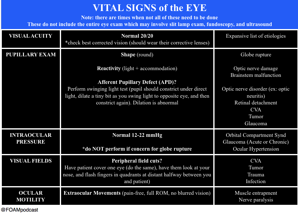
When calling ophthalmology, ensure you have the “Vital Signs of the Eye”

  • Vital Signs of the Eye - @FOAMpodcast
    Vital Signs of the Eye – @FOAMpodcast

Core Content

We delve into core content on eye trauma using Rosen’s (8th edition) Chapter 71 and Chapter 241 in Tintinalli (8th edition)

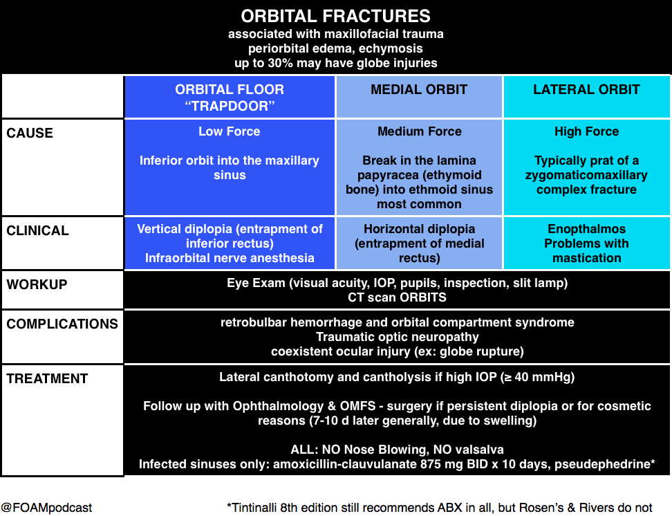
Orbital Fractures

Orbital Fractures

Note: entrapment is a clinical diagnosis.  A CT cannot comment on function, only structure.  Extraocular movements are a critical part of the fracture exam.

Pearl: young people are prone to “greenstick” fractures where the floor can actually pinch the muscle and trigger the oculocardiac reflex, which can be deadly.  They often have a “white eye” without too much impressive on exam but refuse to look in a certain direction due to nausea.

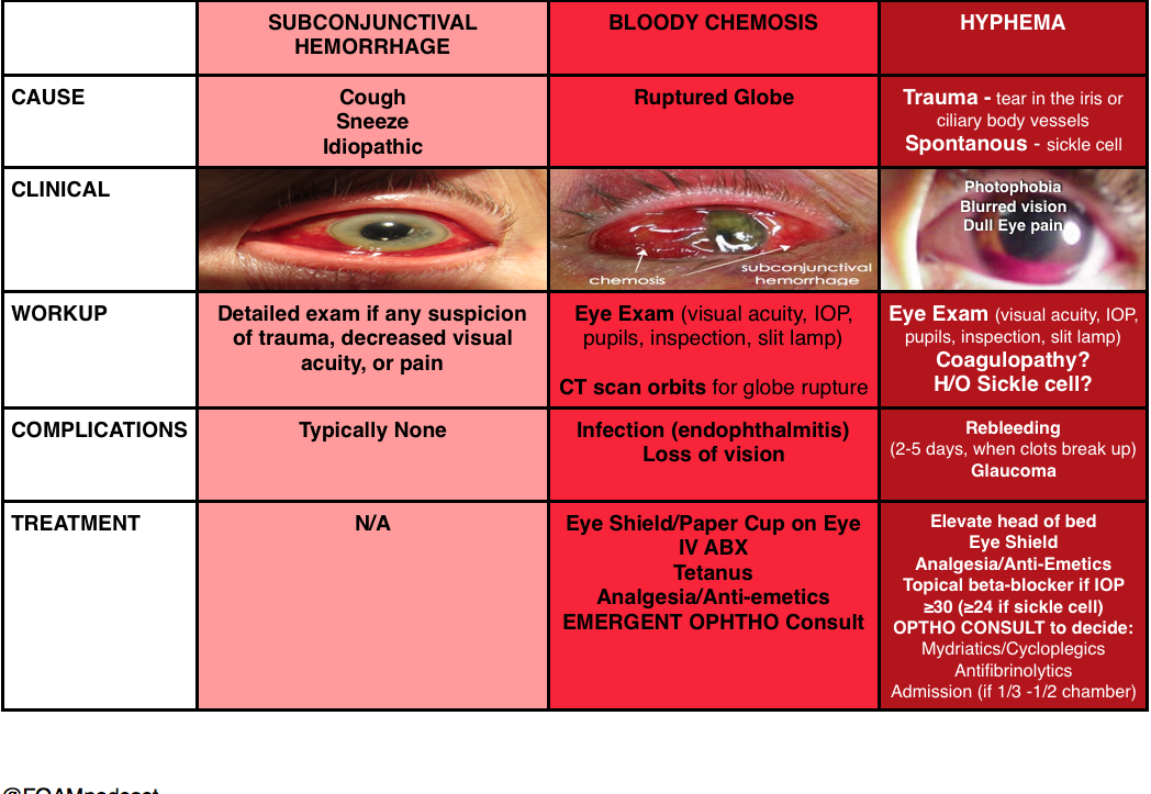
Hyphema

(and things confused with hyphema)

Hyphema

Burns

Ocular Burns

Pearl: An important thing to keep in mind is that a “white” eye after such an exposure is actually an ominous sign, as it implies ischemia of the limbal blood vessels, which portends a pretty bad prognosis.  People usually think that the more red an eye, the worse when, in truth, it’s often the other way around.

Generously Donated Rosh Review Questions

A 43-year-old construction worker presents with right eye pain. He states he was using a nail gun when he felt something hit his eye. Visual examination reveals a small nail penetrating the globe. What management should be pursued?

A. CT scan of the orbit and ophthalmology consultation

B. Measure intraocular pressure and consult ophthalmology

C. Perform lateral canthotomy and consult ophthalmology

D. Remove the foreign body, start topical antibiotics and send to ophthalmology for follow up

[accordion]
[toggle title=”Answer” state=”closed”]

This patient presents with a globe injury and should have a protective shield placed, intravenous antibiotics started, CT scan of the orbit performed and ophthalmology consulted emergently. Foreign body penetration of the globe is often associated with hammering, drilling, mechanical grinding or sanding. Any patient who presents with a foreign body sensation after one of these activities should increase suspicion for a penetrating injury of the globe. CT scan, MRI and ultrasound can all be used for diagnosis but MRI should be avoided if the suspected foreign body is metal containing. Many of these patients will require operative management and should be kept NPO. A protective shield should be placed to protect the eye but patching should be avoided as it may increase pressure on the eye. Emergent ophthalmologic consultation should be obtained

[/toggle]
[/accordion]

A 23-year-old man presents after a fight. His eye is seen below. Physical examination reveals intact extraocular movements, normal fluorescein staining, normal intraocular pressure, and normal visual acuity.

content_image-_medial_lower_lid_laceration

What management is indicated?

A. Delayed closure

B.  Laceration repair by the Emergency Physician

C. Ophthalmology consultation for repair

D. Tissue adhesive for repair

[accordion]
[toggle title=”Answer” state=”closed”]

C. Ophthalmology consultation for repair. This patient presents with a complex eyelid laceration possibly involving the canalicular system and should have a consultation with either ophthalmology or plastic surgery regarding repair. It is important to search for a penetrating globe injury in any patient with an eyelid laceration because of the proximity of structures. Simple horizontal and partial thickness lid lacerations can be repaired primarily by an Emergency Physician. However, more complicated lacerations should be considered for specialist repair because of the high likelihood of cosmetic or functional complications, or both. In general, lacerations through the orbital septum, lacerations with tissue loss, lacerations involving the lid margins, lacerations involving the levator or canthal tendons and those involving the canalicular system should be repaired by a skilled ophthalmologist or plastic surgeon. Injury to the canalicular system should be suspected in any laceration involving the medial lower eyelid. Tissue adhesive (D) is contraindicated this close to the eye and should not be used in an injury that requires precision alignment of tissue. Delayed closure (A) will likely lead to worse cosmetic outcomes. Primary repair by the Emergency Physician (B) is not recommended if the canalicular system may be involved.

[/toggle]
[/accordion]


Thanks to our peer reviewer, Michael Westafer, MD – Ophthalmologist and Glaucoma Fellow at Mayo Clinic

References

  1. “Eye Emergencies.”  Chapter 241.  Tintinalli’s Emergency Medicine: A Comprehensive Review. 8th edition.  
  2. Sharma R and Brunette D.  “Ophthalmology.”  Chapter 71.  Rosen’s Emergency Medicine.  8th edition, 909-930.