Olanzapine + Benzodiazepines – What is the FDA warning about?

ITUNES OR LISTEN HERE

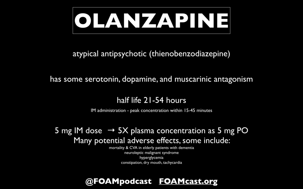
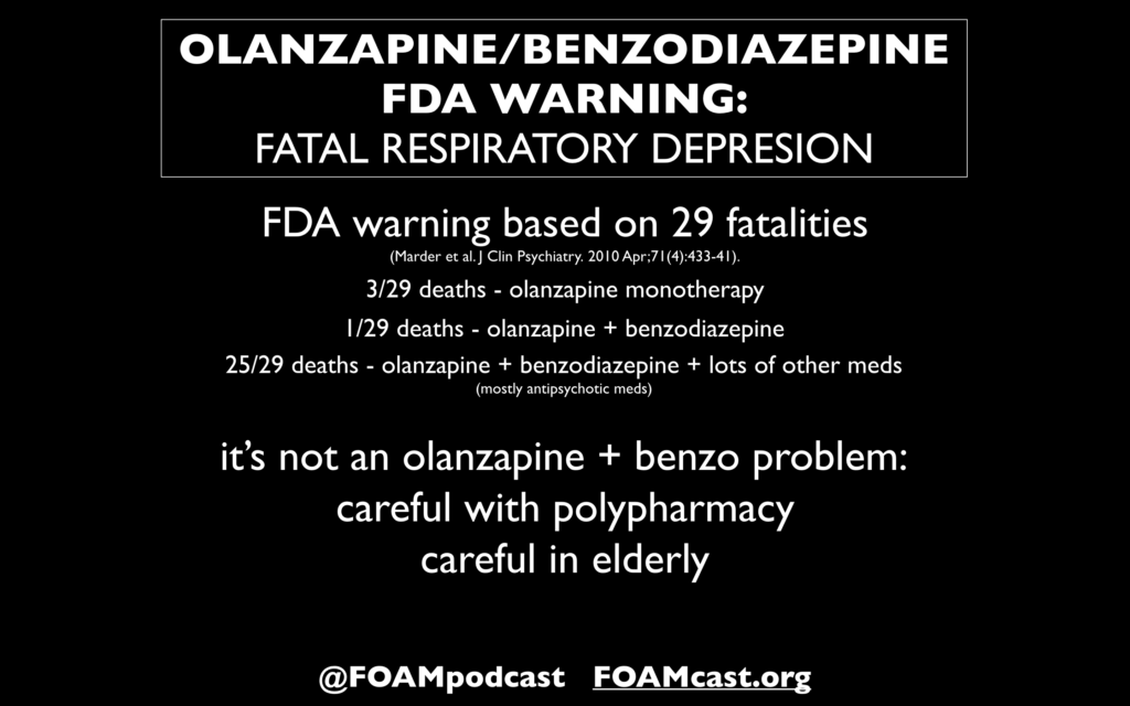
At ACEP 2019, a talk by Bryan Hayes (@PharmERToxGuy) was captivating. He addressed use of medications with black box warnings in the ED. One particular warning caught our eye – olanzapine + benzodiazepine.

The FDA has warned of the risk of potentially fatal respiratory depression with concomitant administration of antipsychotics and benzodiazepines, stating “Concomitant administration of intramuscular olanzapine and parenteral benzodiazepine has not been studied and is therefore not recommended.”

References:

  1. Marder SR, Sorsaburu S, Dunayevich E, et al. Case reports of postmarketing adverse event experiences with olanzapine intramuscular treatment in patients with agitation. J Clin Psychiatry. 2010;71(4):433-41.
  2. Cole JB, Moore JC, Dolan BJ, et al. A Prospective Observational Study of Patients Receiving Intravenous and Intramuscular Olanzapine in the Emergency Department. Ann Emerg Med. 2017;69(3):327-336.e2.

Top Literature of 2019 – Mid Year Review

iTunes or Listen Here

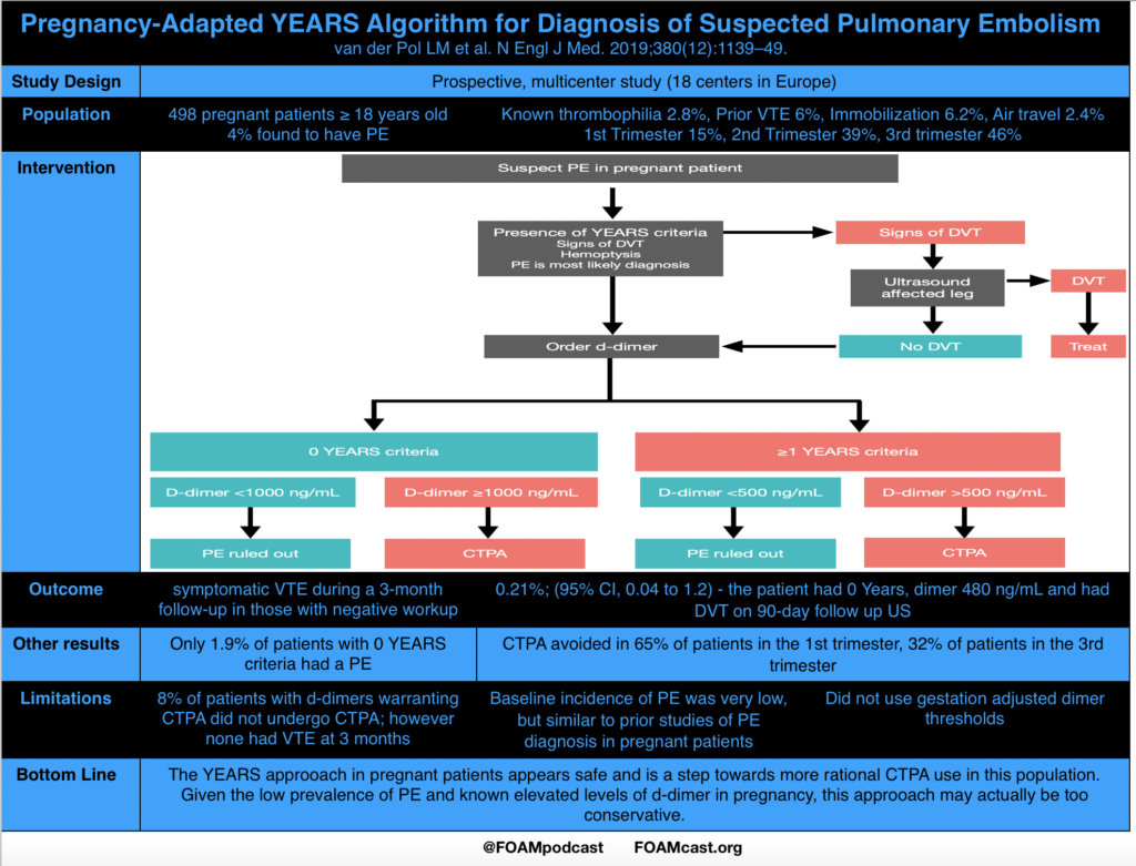
Risk Stratification and D-dimer in Pregnant Patients With Suspected Pulmonary Embolism (PE)

Van der pol LM, Tromeur C, Bistervels IM, et al. Pregnancy-Adapted YEARS Algorithm for Diagnosis of Suspected Pulmonary Embolism. N Engl J Med. 2019;380(12):1139-1149.

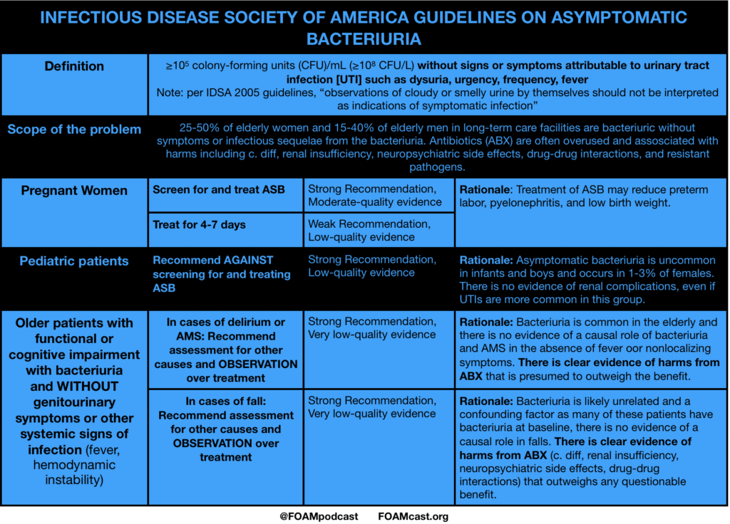
Infectious Disease Society of America (IDSA) Guidelines for Asymptomatic

Nicolle LE, Gupta K, Bradley SF, et al. Clinical Practice Guideline for the Management of Asymptomatic Bacteriuria: 2019 Update by the Infectious Diseases Society of America. Clin Infect Dis. 2019.

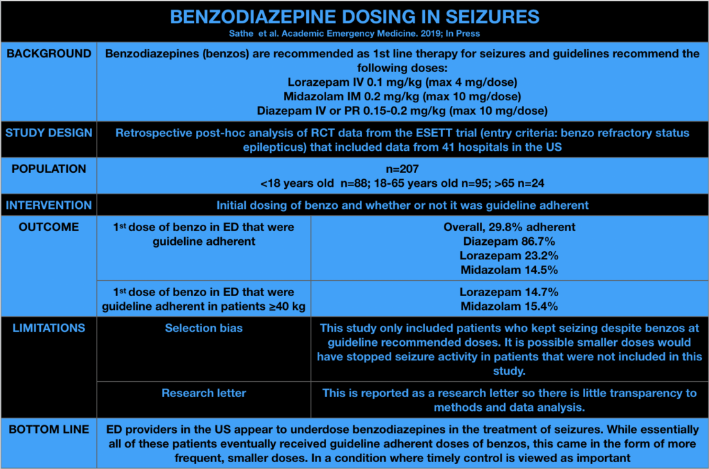
Benzodiazepine dosing for seizures

Sathe AG, Tillman C, Coles LD, et al. Underdosing of benzodiazepines in patients with status epilepticus enrolled in Established Status Epilepticus Treatment Trial. Acad Emerg Med. 2019 Jun 4.

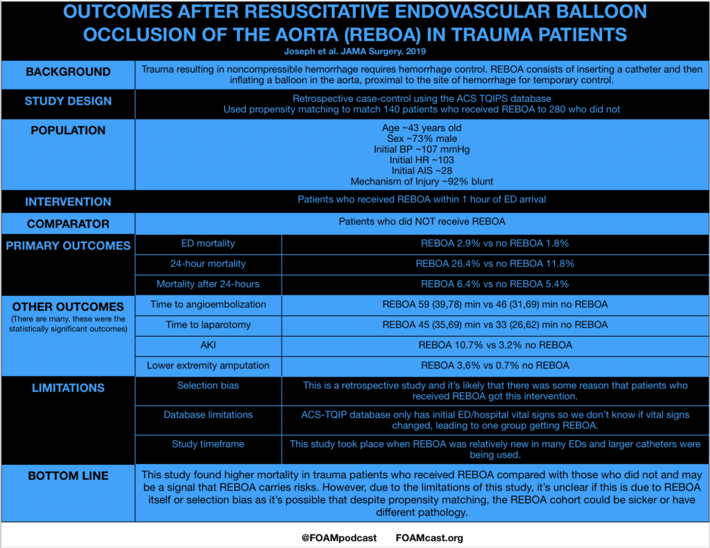
  • Outcomes after Resuscitative Endovascular Balloon Occlusion of the Aorta (REBOA) in Trauma Patients

Joseph B et al. Nationwide analysis of resuscitative endovascular balloon occlusion of the aorta in civilian trauma. JAMA Surg 2019. Mar 20.

Additional References:

  1. Brophy GM, Bell R, Claasen J, et al. Guidelines for the evaluation and management of status epilepticus. Neurocrit Care. 2012;17(1):3-13.
  2. Glauser T, Shinnar S, Gloss D, et al. American Epilepsy Society Guideline Evidence-Based Guideline: Treatment of convulsive status epilepticus in children and adults: report of the guideline committee of the American Epilepsy Society. Epilepsy Curr. 2016;16(1):48-61.

Medicated Assisted Therapy

iTunes or Listen Here

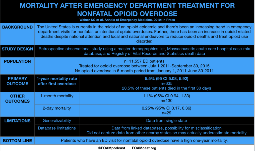
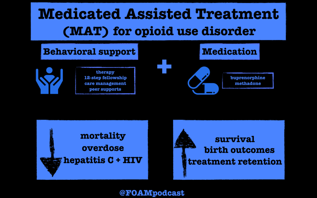
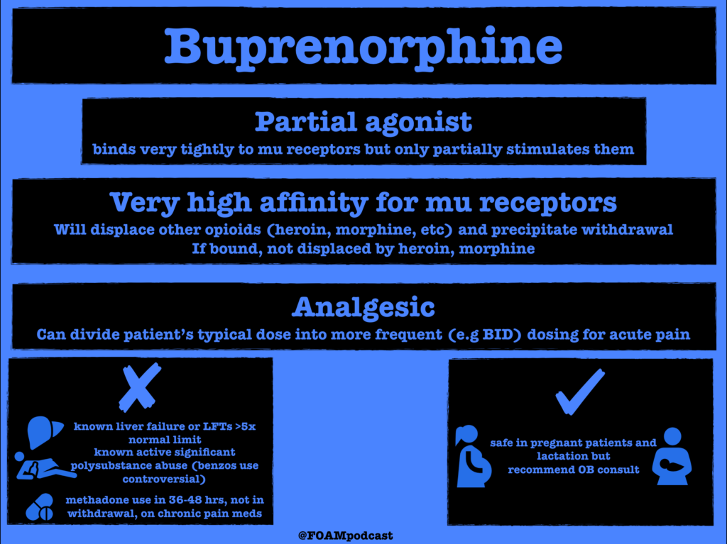
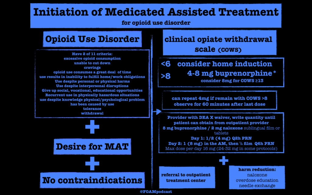
We discussed Medicated Assisted Treatment for opioid use disorder (OUD), focusing on buprenorphine initiated MAT from the ED. In the United States, providers must obtain a DEA X waiver in order to prescribe buprenorphine for OUD For $199, physicians can take a fully online training course, advanced practitioners require additional training. 

Protocol specifics vary by institution. Below is an example of a protocol. The clinical opiate withdrawal scale (COWS) can be found on MDCalcYale’s protocol, along with extensive resources for MAT from the ED can be found here.  Washington ACEP also has online resources .

Check out our sponsor for this show, Figure 1, a free open access application for providers using images and cases.

References:

  1. Berg ML, Idrees U, Ding R, Nesbit SA, Liang HK, and McCarthy ML. Evaluation of the use of buprenorphine for opioid withdrawal in an emergency department. Drug Alcohol Depend. 2007;86(2-3):239-44. doi:10.1016/j.drugalcdep.2006.06.014.
  2. Mattick RP, Breen C, Kimber J, Davoli M. Buprenorphine maintenance versus placebo or methadone maintenance for opioid dependence. Cochrane Database Syst Rev. 2014;(2):CD00220
  3. Pierce M, Bird SM, Hickman M, Marsden J, Dunn G, Jones A, and Millar T. Impact of treatment for opioid dependence on fatal drug-related poisoning: a national cohort study in England. Addiction. 2016;111(2):298-308. doi:10.1111/add.13193.
  4. Clark RE, Samnaliev M, Baxter JD, and Leung GY. The evidence doesn’t justify steps by state Medicaid programs to restrict opioid addiction treatment with buprenorphine. Health Aff (Millwood). 2011;30(8):1425-33. doi:10.1377/hlthaff.2010.0532
  5. Martin SA, Chiodo LM, Bosse JD, Wilson A. The Next Stage of buprenorphine Care for Opioid Use disorder. Annal of Internal Medicine

Episode 77 – Alcohol Withdrawal

iTunes or Listen Here

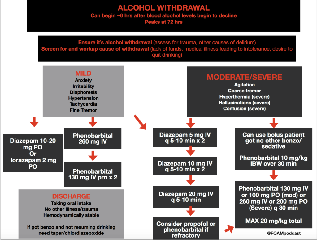
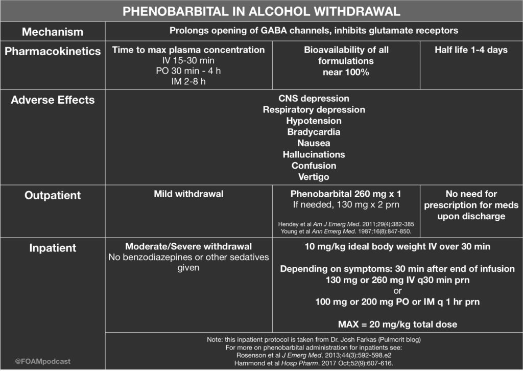
Over at the Pulmcrit blog, Dr. Josh Farkas has proposed the use of phenobarbital monotherapy for the treatment of ethanol withdrawal. He argues that phenobarbital has the following advantages:

  • Superior neurochemistry
  • Reliable – some patients will be resistant to benzos but this does not happen as much with phenobarbital
  • Predictable pharmacokinetics

Core Content

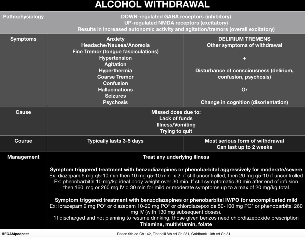
We cover alcohol withdrawal using Rosen’s Emergency Medicine (9th ed) Chapter 142 , Tintinalli’s Emergency Medicine (8th ed) Chapter 292, and Goldfrank’s Toxicologic Emergencies (10th ed) Chapter 81  as guides.


Rosh Review Emergency Board Review Questions

A 49-year-old man presents to the Emergency Department complaining of sweating and tremors. The patient drinks a bottle of liquor per day and stopped suddenly because of a pending court case. His last alcoholic drink was 3 days ago. On physical examination, his blood pressure is 168/105 mm Hg, pulse rate is 106/minute, respirations are 22/minute, and temperature is 99.3°F. The patient appears agitated and restless with a visible tremor of bilateral hands. The triage team ordered folic acid, thiamine, and a multivitamin. Which of the following is the most appropriate disposition?

A. Admit the patient and start diazepam

B.Admit the patient and start disulfiram

C.Discharge the patient with a prescription for diazepam

D.Discharge the patient with a prescription for disulfiram

[accordion]
[toggle title=”Answer” state=”closed”] A. Admit the patient and start diazepam is the correct disposition because this patient is suffering from alcohol withdrawal, which potentially can be fatal. Withdrawal symptoms occur when a patient has alcohol use disorder and has developed a tolerance to alcohol, where an increased amount of alcohol is needed to achieve the desired effect. When tolerance has developed, cessation leads to withdrawal. Early symptoms of alcohol withdrawal include anxiety, irritability, headache, tremor, tachycardia, hypertension, hyperthermia, and hyperactive reflexes. Seizures (usually grand mal) can develop between 12-24 hours after withdrawal starts. After 24-72 hours, life-threatening delirium tremens may occur, which manifests with signs of altered mental status, hallucinations and marked autonomic instability. Treatment of alcohol withdrawal involves giving a benzodiazepine (e.g. diazepam) until symptoms lessen and then tapering the dosage over days to weeks. Thiamine, folic acid, and vitamin B12 are also administered and any electrolyte abnormalities are corrected (typically low potassium and magnesium). Following withdrawal, the patient should be referred to support groups. Long term medication used to deter use of alcohol include naltrexone, disulfiram, and acamprosate.

Admit the patient and start disulfiram (A) is incorrect because the patient needs a benzodiazepine medication to prevent delirium tremens and potentially fatal consequences. Disulfiram is a medication used in some patients for long-term adherence to alcohol abstinence. Ingestion of alcohol while taking disulfiram causes copious vomiting and potentially more severe reactions. Discharge the patient with a prescription for diazepam (C) or disulfiram (D) is incorrect because alcohol withdrawal is potentially lethal and this patient should be admitted.

[/toggle]
[/accordion]

References:

  1. “Alcohol Related Diseases.” Rosen’s Emergency Medicine. 9th ed. Chapter 142, 1838-1851.e1
  2. “Substance Use Disorders.” Tintinalli’s Emergency Medicine: A Comprehensive Review. 9th ed. Chapter 281.
  3. “Ethanol Withdrawal.” Goldfrank’s Toxicologic Emergencies. 10th ed. Chapter 81.
  4. Hendey GW, Dery RA, Barnes RL, Snowden B, Mentler P. A prospective, randomized, trial of phenobarbital versus benzodiazepines for acute alcohol withdrawal. Am J Emerg Med. 2011;29(4):382-385.
  5. Young GP, Rores C, Murphy C, Dailey RH. Intravenous phenobarbital for alcohol withdrawal and convulsions. Ann Emerg Med. 1987;16(8):847-850.

Episode 74 – Physostigmine and Anticholinergic Toxidrome

iTunes or Listen Here

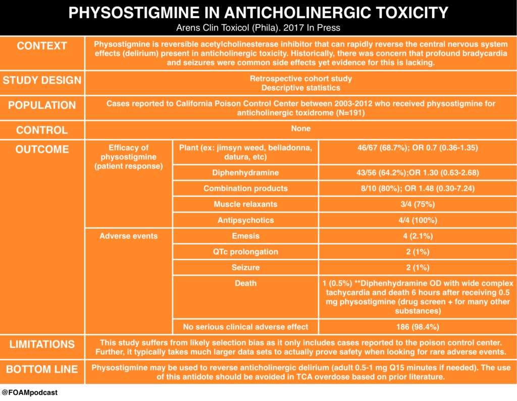
We review this blog post by Bryan Hayes, an ED pharmacist (@PharmERToxGuy), on the use of physostigmine in anticholinergic toxicity.
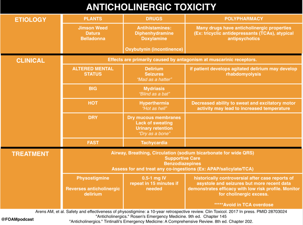
We review core content on anticholinergic toxicity using Rosen’s Emergency Medicine (9th ed), Tintinalli’s Emergency Medicine (8th ed) and Goldrank’s Toxicology as guides.
Rosh Review Emergency Board Review Questions

A 27-year-old man is brought to the ED by EMS after being found wandering in the street. His BP is 155/70, HR 115, T 37.5°C, RR 16, pulse ox 99% on room air, and finger stick glucose 98. On exam, the patient is confused with mumbling speech. His pupils are 7 mm and nonreactive. His face is flushed. Mucous membranes and skin are dry. Which of the following toxidromes is this patient exhibiting?

A. Anticholinergic

B. Cholinergic

C. Sedative-hypnotic

D. Sympathomimetic

[accordion]
[toggle title=”Answer” state=”closed”]

A. Anticholinergic Toxidrome refers to a constellation of physical findings that can provide important clues in a toxic ingestion. This patient exhibits an anticholinergic toxidrome. Anticholinergics are widely available as over-the-counter cold preparations and sleep aids (diphenhydramine, chlorpheniramine, doxylamine). They also are the basis of the toxicity of jimsonweed. Anticholinergics competitively inhibit acetylcholine in the central and peripheral nervous systems. This classic toxidrome is often described by the phrase “mad as a hatter (altered mental status), blind as a bat (mydriasis), red as a beet (flushed skin), hot as a hare (dry skin due to inability to sweat), dry as a bone (dry mucous membranes).” Other findings include tachycardia, delirium, seizures, and hallucinations. Treatment is mainly supportive with benzodiazepines for agitation and seizures, and cooling for temperature reduction. Gastric decontamination (whole-bowel irrigation or gastric lavage) is often recommended due to the delay in gastric emptying caused by anticholinergics. The use of physostigmine, a reversible cholinesterase inhibitor that will increase synaptic acetylcholine, is controversial and should be administered only in consultation with a toxicologist.

The cholinergic toxidrome (B) is characterized by the mnemonics SLUDGE (salivation, lacrimation, urination, defecation, GI upset, emesis) or DUMBBELS (defecation, urination, miosis, bronchospasm, bronchorrhea, emesis, lacrimation, salivation) and occurs after exposure to organophosphates or carbamates. The sedative-hypnotic toxidrome (C) is seen with benzodiazepines and barbiturates. Clinically, patients exhibit central nervous system and respiratory depression. The sympathomimetic toxidromes (D) can occur after ingestion of cocaine, amphetamines, or decongestants. It typically presents with delirium, paranoia, tachycardia, hypertension, hyperpyrexia, diaphoresis, mydriasis, seizures, and hyperactive bowel sounds. Sympathomimetic and anticholinergic toxidromes are frequently difficult to distinguish. The key difference is that sympathomimetics are associated with diaphoresis, whereas anticholinergics cause dry skin.

[/toggle]
[/accordion]

A 22-year-old man is brought into the Emergency Department by Emergency Medical Services with altered mental status. He is an agitated man and mumbling incoherent words. His pupils are 6 mm and reactive to light. His axillae are dry. His heart rate is 115 beats/minute and temperature is 101.6 oF. Which of the following is a complication of the antidote used in the treatment of this condition?

A. Asystole

B. Bladder retention

C. QT-interval prolongation

D. Respiratory depression

[accordion]
[toggle title=”Answer” state=”closed”]

Asystole. The mnemonic for the anticholinergic toxidrome is blind as a bat (mydriasis), red as a beet (vasodilation leading to flushing), hot as a hare (hyperthermia), dry as a bone (dry skin and mucosa), mad as a hatter (visual or auditory hallucinations, agitation, and mumbling or incoherent speech), bloated as a toad (ileus and urinary retention), and the heart runs alone (tachycardia). Additionally, anticholinergic toxicity can cause orthostatic hypotension (especially in the elderly), picking behavior, and can lead to seizures, coma, and even death. There are multiple anticholinergic medications, including tricyclic antidepressants, antihistamines, antiparkinson medications, antipsychotics, antispasmodics, belladonna alkaloids, skeletal muscle relaxants, and mydriatics. Antihistamine overdose is the most common cause of anticholinergic toxicity. Treatment is supportive with IV hydration. If the ingestion occurred within one hour, GI decontamination with activated charcoal is reasonable. Benzodiazepines are used for significant agitation and seizures. Good temperature control should be achieved with antipyretics and active cooling is essential. For wide complex tachyarrhythmias, treatment is with sodium bicarbonate. The antidote is physostigmine, a reversible acetylcholinesterase inhibitor. There is, however, a risk of profound bradycardia, seizures, and asystole in patients with widened QRS complexes. This risk is minimal if there is a pure anticholinergic syndrome without concomitant sodium channel blockade, but this is unfortunately not always known. Since the most common cause of anticholinergic toxicity is from diphenhydramine overdose (which causes sodium channel blockade), physostigmine should only be used with the assistance of a medical toxicologist.  

[/toggle]
[/accordion]

References:

  1. Arens AM, et al. Safety and effectiveness of physostigmine: a 10-year retrospective review. Clin Toxicol. 2017 Jul 13:1-7. [Epub ahead of print] PMID 28703024
  2. “Anticholinergics.” Rosen’s Emergency Medicine. 9th ed.  Chapter 145
  3. “Anticholinergics.” Tintinalli’s Emergency Medicine: A Comprehensive Review. 8th ed. Chapter 202.
Episode 71 – Opioids

ITUNES OR LISTEN HERE

We cover Free Open Access Medical Education on opioids.

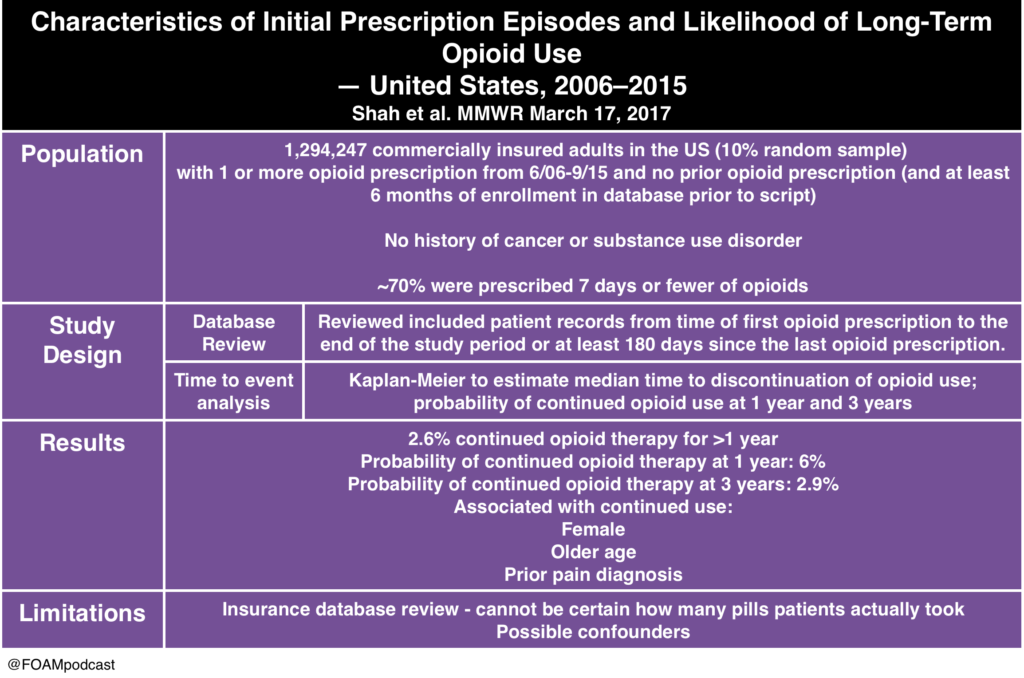
The Poison Review podcast reviews papers on the opioid epidemic, including a paper by Rudd et al demonstrating an increase in opioid-related deaths over the past five years as well as the following paper by Shah and colleagues.

Long term opioid use

The Dantastic Mr. Tox and Howard, a new podcast on the scene, also covers multiple topics on opioids, including common misconceptions of tramadol.


The American College of Medical Toxicology recently released a statement on fentanyl exposure, stating how absolutely unlikely it is that accidental dermal exposure would result in toxicity.  ” If bilateral palmar surfaces were covered with fentanyl patches, it would take approximately 14 minutes to receive 100 mcg of fentanyl…This extreme example illustrates that even a high dose of fentanyl prepared for transdermal administration cannot rapidly deliver a high dose.”

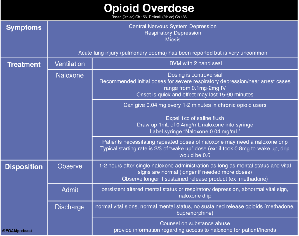
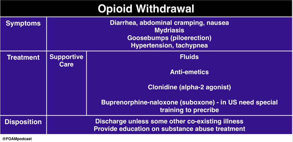
We cover core content on opioid overdose and withdrawal using Rosen’s Emergency Medicine (9th ed) Chapter 156 and Tintinalli’s Emergency Medicine (8th ed), Chapter 186 as a guide.

opioid withdrawal

Episode 57 – Lithium Toxicity & Rhabdomyolysis

The Free Open Access Medical Education (FOAM) 

Listen Here or iTunes

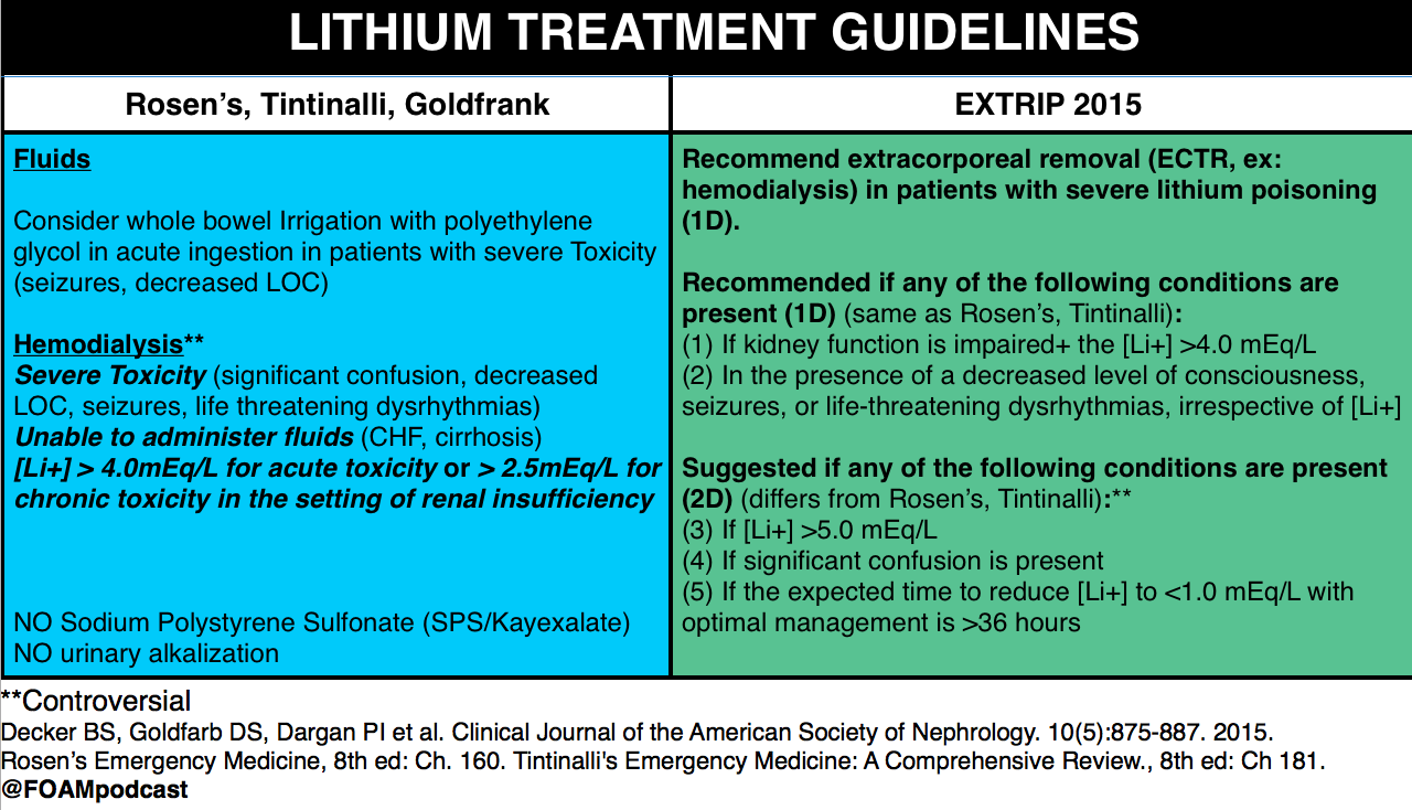
We cover a post from Dr. Josh Farkas on PULMcrit on lithium toxicity. The key message from the post is: a single serum lithium level doesn’t necessitate dialysis, despite a recommendation from the EXTRIP working group  to initiate dialysis in patients with a lithium level > 5 mEq/L [1].  Dr. Farkas advocates for aggressive management in asymptomatic patients with chronic lithium toxicity and patients without impaired renal function.

Lithium neurotoxicity does not correlate with serum lithium levels; it depends on the concentration in the CNS.

  • The argument is that the neurologic effects, among the most worrisome sequelae of lithium toxicity, results from lithium crossing the blood-brain-barrier (BBB) into the central nervous system (CNS). Thus, in an acute ingestion, patients may have a higher serum lithium level, but given the acuity, the lithium may not cross into the CNS.  Conversely, a patient with chronic toxicity may have a mildly elevated serum lithium level but the lithium has had time to cross the BBB into the CNS so more neurotoxicity can result from lower serum lithium concentrations.

Differentiating between acute, chronic, and acute on chronic toxicity is important in management.

  • Acute – expect the levels will rise rapidly as the bolus of lithium is absorbed.
  • Chronic – expect the levels will not rise rapidly given a lack of lithium bolus.
  • Acute on chronic – again, expect levels to rise rapidly and may see more neurotoxicity as some lithium may have passed through the BBB into the CNS.

screen-shot-2016-09-30-at-6-19-25-pm

screen-shot-2016-09-30-at-6-02-28-pm

Core Content

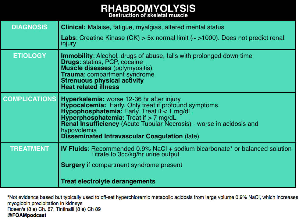
We review rhabdomyolysis using Rosen’s Emergency Medicine (8e)  Chapter 160 and Tintinalli’s Emergency Medicine (8e), Chapter 181.

screen-shot-2016-10-01-at-12-05-27-pm

Generously Donated Rosh Review Questions

A 17-year-old man presents to the ED from a correctional facility complaining of general malaise with nausea and vomiting one day after a weightlifting competition. Vital signs are T 37.2°C, BP 100/65 mm Hg, HR 125, and RR 22. Physical exam reveals an uncomfortable, fatigued male who has diffuse muscle soreness. Urinalysis shows 3+ blood without red blood cells. What is the most important next test to direct the acute management of this patient?

A. Creatine kinase assay

B. Electrocardiogram

C. Electrolyte panel with blood urea nitrogen and creatinine

D.Microscopic urinalysis

[accordion]
[toggle title=”Answer” state=”closed”]

B. Electrocardiogram. The patient presents with exercised-induced rhabdomyolysis. Although the causes of rhabdomyolysis are manifold, potential complications are independent of etiology. Of all the complications, hyperkalemia is the most concerning and, undiagnosed, can lead to sudden cardiac death. ECG changes indicative of hyperkalemia are thus critical to identify early in the course of management. Hyperkalemia results from impaired calcium transport with increased intracellular calcium accumulation, cellular necrosis, and expulsion of intracellular contents (including potassium) into the bloodstream. Rhabdomyolysis can also lead to acute tubular necrosis and kidney failure, which will exacerbate developing hyperkalemia by decreasing renal potassium clearance. Hyperkalemia is an immediate life-threatening condition that develops shortly after muscle injury. Absent point-of-care electrolyte analyzers, the most rapid way to screen for hyperkalemia is through an electrocardiogram.

Rhabdomyolysis is not defined by a specific creatine kinase (A) level. But, in general, a serum CK >5 times the upper limit of normal (a threshold that may differ by lab) is considered indicative of rhabdomyolysis. Although kidney injury can occur at any level, higher CKs correlate with an increased likelihood for the development of acute renal failure. In the absence of cerebral or myocardial infarction, a CK >5 times is diagnostic for serious muscle injury. Urinalysis (D) typically shows brownish discoloration with “large” blood on dipstick but few, if any, red blood cells on microscopic evaluation. This occurs because most dipstick tests cannot distinguish myoglobinuria from hematuria or hemoglobinuria. Protein, brown casts, and renal tubular epithelial cells may also be present. Measures of renal function and an electrolyte panel (C) should be obtained in all patients with suspected rhabdomyolysis. But, as mentioned, waiting for results may lead to a delay in the identification of life-threatening complications. In addition to hyperkalemia, hyperphosphatemia and hypocalcemia may also be seen. Additional (though less worrisome) laboratory abnormalities include elevated uric acid and low albumin.

[/toggle]
[/accordion]

One Step Further Question: What is the late complication of rhabdomyolysis associated with thrombocytopenia, hypofibrinogenemia, and an elevated D-dimer?

[accordion]
[toggle title=”Answer” state=”closed”]

Disseminated intravascular coagulopathy may occur and is a result of muscle necrosis with liberation of activating substances from injured cells.

[/toggle]
[/accordion]


  • References:
  • 1. Decker BS, Goldfarb DS, Dargan PI et al. Extracorporeal Treatment for Lithium Poisoning: Systematic Review and Recommendations from the EXTRIP Workgroup. Clinical Journal of the American Society of Nephrology. 10(5):875-887. 2015.
  • 2. “Chapter 160. Lithium.”  Rosen’s Emergency Medicine.  8th edition.
  • 3. “Chapter 181. Lithium.” Tintinalli’s Emergency Medicine: A Comprehensive Review. 8th edition.
  • 4. Grellar H. “Chapter 72. Lithium.” Goldfrank’s Toxicology. 10th edition.
  • 5.  “Chapter 127. Rhabdomyolysis. Rosen’s Emergency Medicine.  8th edition.
  • 6. “Chapter 89. Rhabdomyolysis.” Tintinalli’s Emergency Medicine: A Comprehensive Review. 8th edition.

Episode 48 – Urine Drug Screen, Cocaine, and PCP

(ITUNES OR LISTEN HERE)

The Free Open Access Medical Education (FOAM)

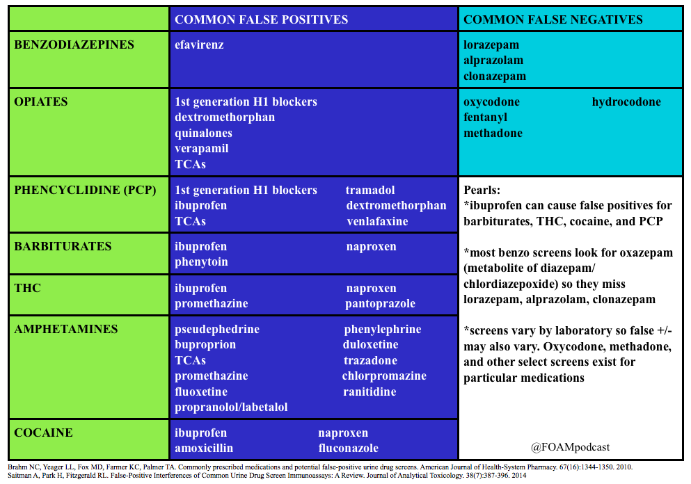
We review a post by Dr. Seth Trueger (@MDaware) on false positives of common medications in the urine drug screen.  We delve into posts by Dr. Bryan Hayes (@PharmERToxGuy) on false negatives for benzodiazepines and opioids in the urine drug screen.

Screen Shot 2016-04-25 at 8.29.32 PM

Core Content

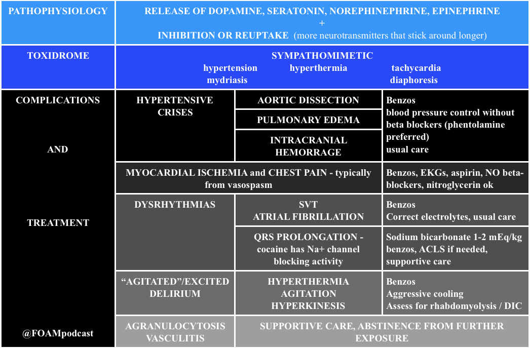
We delve into core content on cocaine and phencyclidine (PCP) using Rosen’s (8th edition), Chapter 154 and Tintinalli (8th edition)

Cocaine

Cocaine
Cocaine

Note: Beta-blockers are contra-indicated in cocaine induced hypertension and chest pain.  Much of the ischemia induced by cocaine is thought to be due to vasospasm, predominantly from alpha-1 receptor effects.  Beta-blockers block the relaxation provided by beta-2 stimulus on muscles, leaving alpha-1 constricting vessels, “unopposed.”  This is largely theoretical/based on canine literature as there are only two human studies on this, (a) 10 humans given propranolol + cocaine with vasospasm and (b) 9 humans given labetalol + cocaine without vasospasm [4,5]. However, recent papers attempting to dispel this teaching don’t quite prove the point. One retrospective chart review looked at cocaine positive urine screens in patients with chest pain and found no worsened troponins. Cocaine stays positive in the urine for 3 days so it is not clear that these were patients presenting with cocaine associated chest pain [6].

Generously Donated Rosh Review Questions

  1. An 18-year-old man is brought to the ED by the police after being found running around a parking lot, screaming at bystanders. He reportedly smoked phencyclidine (PCP) earlier that day. His vital signs are notable for a heart rate of 130 beats per minute and a blood pressure of 150/86 mm Hg. On physical exam, he is diaphoretic, with vertical nystagmus and equal pupils and appears acutely agitated.

A. Administer haloperidol for acute agitation

B. Administer IV fluids for presumed rhabdomyolysis and benzodiazepines for agitation

C. Apply physical restraints; this is adequate treatment for most cases

D. Monitor for traumatic injuries; they are the most common cause of morbidity and mortality with PCP use

E. Urinary acidification to increase PCP elimination

[accordion]
[toggle title=”Answer” state=”closed”]

The most common cause of morbidity and mortality in patients with phencyclidine (PCP) intoxication is rhabdomyolysis. Fluid hydration with normal saline is the initial therapy for rhabdomyolysis. Creatinine kinase (CK) should be obtained in patients with PCP intoxication. If abnormal, serial levels should be obtained until a downward trend is noted. Serum creatinine should also be monitored for evidence of renal insufficiency. PCP is a dissociative anesthetic drug that works on the NMDA glutamate receptor. People who abuse PCP can have sensations of euphoria initially, but this can be followed by an emergence reaction that is characterized by dysphoria and psychosis. If initial calming measures such as placing the patient in a calm environment fail and chemical restraints are needed, benzodiazepines are the treatment of choice. Benzodiazepines are also the preferred medication for acute agitation. Haloperidol (A) may cause dysrhythmias and hypotension. It also lowers the seizure threshold and may precipitate PCP-induced movement disorders. PCP is a weak base and its excretion is increased in acidic urine. Physical restraints (C) may be required for patients with PCP intoxication, but they should be used in conjunction with chemical restraints (preferably benzodiazepines). An agitated patient fighting against physical restraints is at risk for worsening rhabdomyolysis, which, if unrecognized, can precipitate cardiac arrest. Traumatic injuries (D)frequently occur in PCP-intoxicated patients; however, rhabdomyolysis with subsequent renal insufficiency is the most common cause of associated morbidity and mortality. Patients with PCP intoxication should be evaluated for suspected trauma, and any injuries should be stabilized and treated. Acidification of the urine (E) can cause a metabolic acidosis, which is potentially harmful and has not demonstrated improved outcomes. For these reasons, urinary acidification is not routinely recommended.

[/toggle]
[/accordion]

2. A 39-year-old woman presents with chest pain and difficulty breathing that began shortly after smoking crack cocaine. Vital signs are BP 190/85 mm Hg, HR 105, RR 18, T 99.1℉. The ECG demonstrates ST segment depression and T wave inversions in the lateral leads.

A. Diazepam

B. Metoprolol

C. Morphine

D. Phenylephrine

[accordion]
[toggle title=”Answer” state=”closed”]

Diazepam, a benzodiazepine, should be administered to this patient who is hyperadrenergic from cocaine intoxication. Signs and symptoms of cocaine intoxication include, dilated pupils, diaphoresis, tachycardia, hypertension and hyperthermia. Many patients experience euphoria, though some will develop acute psychosis. Benzodiazepines decrease the cocaine-induced hyperadrenergic state. Reduction of sympathetic tone induces coronary and peripheral vasodilation. Coronary artery dilation directly improves myocardial blood flow. Peripheral vasodilation reduces preload and afterload. Reductions in preload and afterload decrease blood pressure and improve myocardial oxygen demand. Several factors, including, excess sympathetic stimulation, dehydration, hyperthermia, and cocaine-induced cardiac sodium channel blockade, may cause patients with cocaine intoxication to develop dysrhythmias. These contributing factors should be treated with benzodiazepines, IV fluid resuscitation and temperature management. In some patients, cocaine-induced cardiac sodium channel blockade may cause wide complex tachycardia that should be treated with sodium bicarbonate. Hyperthermia should be managed aggressively with a target temperature of less than or equal to 102.0℉. Severe agitation, aggression or psychosis should be initially managed with benzodiazepines. Most antipsychotic agents have pronounced anticholinergic side effects. This may worsen dysrhythmias.and decrease sweating, further complicating temperature management

[/toggle]
[/accordion]

  1. Rao R, Hoffman RS.  Cocaine and other Sympathomimetics. Rosen’s Emergency Medicine (8e). Chapter 154, 1999-2006.e2
  2. “Cocaine and Amphetamines.” Tintinalli’s Emergency Medicine: A Comprehensive Study Guide (8e). Chapter 187
  3. “Prison Medicine.” Tintinalli’s Emergency Medicine: A Comprehensive Study Guide (8e). Chapter 301
  4. Lange RA, Cigarroa RG, Flores ED, et al. Potentiation of cocaine, induced coronary vasoconstriction by beta adrenergic blockade. Ann Intern Med. 1990;112:897–90
  5. Boehrer JD, Moliterno DJ, Willard JE, Hillis LD, Lange RA. Influence of labetalol on cocaine-induced coronary vasoconstriction in humans. Am J Med. 1993;94(6):608–610
  6. Ibrahim M, Maselli DJ, Hasan R, Hamilton A. Safety of β-blockers in the acute management of cocaine-associated chest pain. The American Journal of Emergency Medicine. 31(3):613-616.


Episode 43 – Alcohols

(ITUNES OR LISTEN HERE)

The Free Open Access Medical Education (FOAM)

The Skeptic’s Guide to Emergency Medicine Episode 144, “That Smell of Isopropyl Alcohol for Nausea in the Emergency Department.” This podcast reviews an article by Beadle et al, an RCT on the use of inhalational isopropyl alcohol for nausea.

Population – Adults in the Emergency Department

Intervention – Nasal inhalation of an isopropyl alcohol pad for ~ 60 seconds (time 0 and, if still nauseated, at 2 minutes and 4 minutes)

Comparison – Nasal inhalation of an identical pad soaked in saline.

Outcome -Nausea score at 10 minutes post treatment using an 11-point verbal numeric response scale (0:“no nausea”; 10:“worst nausea imaginable”)

  • Lower in the isopropyl group (Median score of 3 vs. 6 on an 11 point scale, p<0.001). This gave an effect size of 3 (95% CI 2 to 4).

Limitations – single center, convenience sample, unclear blinding as isopropyl likely has a particular smell to it, outcome nausea score, not vomiting

Eagerton-Warburton et al conducted an RCT of intravenous ondansetron 4mg, metoclopramide 20mg, or saline (placebo) of adult patients with nausea and found  no significant difference in nausea scores on a visual analog scale between groups (n=270).

Core Content

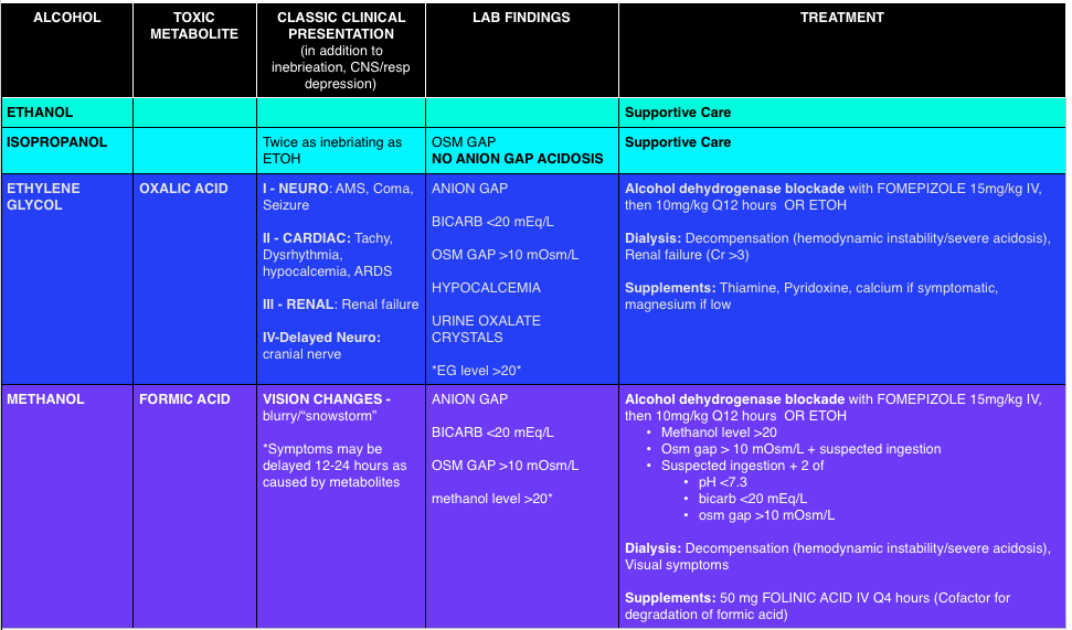
We delve into core content on vertigo using Rosen’s Medicine (8e),  Chapter 155 “Toxic Alcohols”  and Tintinalli’s Emergency Medicine: A Comprehensive Study Guide  (7e) Chapter 179“Alcohols.”

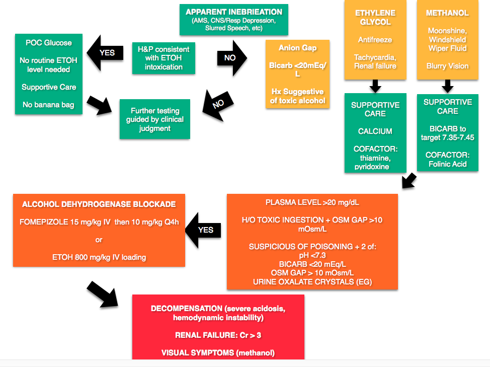
Any alcohol can be “toxic,” the ramifications depend on the dose. Toxic alcohols typically refer to ethylene glycol (EG) and methanol.  In these cases, the parent compound (EG or methanol) is inebriating but not particularly toxic. These compounds are metabolized, like any alcohol, but unfortunately the metabolites (oxalic acid – EG, formic acid – methanol) have unique toxic properties. For example, the oxalic acid produced from EG metabolism combines with calcium.  These deposit into tissues causing renal failure and neurologic sympotms. The formic acid has a predilection for the retina, causing visual symptoms.

Screen Shot 2016-02-19 at 7.01.02 PM

Toxic Alcohols

Osmolal Gap (calculator) – One of the touted features of toxic alcohols is the elevated anion gap, that is, the difference between the measured serum osmolality and the calculated osmolarity.

Calculated Osmolarity: 2[Sodium] + [Glucose]/18 + [BUN]/2.8 + [ETOH]/4.6    (Note: Some references use ETOH/3.7)

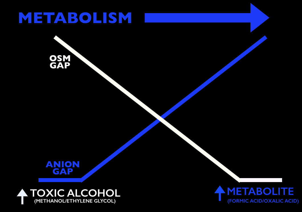
  • Problems:  A normal osm gap does not exclude the presence of toxic alcohols as the osm gap decreases as the alcohol is metabolized.  Also, other disease processes can increase the osm gap, including shock, diabetic ketoacidosis, etc [2,3].
Osm Gap vs Anion Gap As Toxic Alcohols Metabolize

A common pearl exists that urine can be placed under a Wood’s lamp for fluorescence due to the fluorescein in ethylene glycol antifreeze (to detect leaks). Unfortunately this is neither sensitive nor specific [4,5].

Treatment

Screen Shot 2016-02-17 at 9.04.49 PM

Rosh Review Questions

Question 1. [polldaddy poll=9311166]

Question 2. [polldaddy poll=9311159]

Answers

1. A. Hypocalcemia. Most laboratories do not have the capability to measure serum levels of toxic alcohols in a timely fashion. As a result, physicians must use other clinical markers to determine the presence of these unmeasured toxins. The classic laboratory abnormality is an elevated serum osmolality with an osmolar gap since the actual toxic alcohol is not measured in its own quantity but contributes to the osmolality of the blood. When calculating the osmolar gap, it is important to have a measured serum ethanol level as this is a large contributor to the final osmolality.Ethylene glycol ingestion leads to the formation of calcium oxalate crystals. As a result, serum calcium levels will decrease and hypocalcemia is a surrogate marker for ethylene glycol toxicity.Hypoglycemia (B) is not characteristic of a toxic alcohol ingestion. Hypoglycemia is often seen in chronic alcoholics due to poor nutritional intake. In children, ethanol may induce hypoglycemia. Other toxic ingestions which may lead to hypoglycemia include beta-blockers, sulfonylureas and insulin. Hypokalemia (C) is not associated with any toxic alcohol ingestions. As patients become more acidemic from alcohol ingestions, hyperkalemia may develop. Hyponatremia (D) is not associated with toxic alcohol ingestions. Evidence of hyponatremia on laboratory analysis should prompt further investigation for other underlying causes.

2.C.  Glycolic acid level greater than 8 mmol/L. A glycolic acid level greater than 8 mmol/L is an indication for emergent hemodialysis following an acute ethylene glycol overdose. Ethylene glycol is found in automobile coolants, antifreeze, hydraulic brake fluid, de-icing agents and industrial solvents. Symptoms progress through 4 stages: inebriation and intoxication, cardiopulmonary symptoms (tachycardia, hypertension, pulmonary edema), renal symptoms (acute renal failure), and delayed neurologic symptoms (cranial neuropathy). Treatment includes 1.) Alcohol dehydrogenase (ADH) enzyme blockade with fomepizole or ethanol to prevent toxic and acidic metabolite production, 2.) Correction of the metabolic acidosis with sodium bicarbonate, and 3.) Hemodialysis for removal of the parent compound and toxic metabolites. Although somewhat controversial with the advent of fomepizole other indications for hemodialysis following ethylene glycol ingestion include severe metabolic acidosis, renal failure, deterioration despite intensive care, and electrolyte disturbances.Other indications for emergent hemodialysis following an acute ethylene glycol overdose include: ethylene glycol level greater than 50 mg/dL (B), anion gap greater than 20 mmol/L (A)and initial pH less than 7.3 (D).

References

  1.  Beadle KL, Helbling AR, Love SL, April MD, Hunter CJ. Isopropyl Alcohol Nasal Inhalation for Nausea in the Emergency Department: A Randomized Controlled Trial. Annals of emergency medicine. 2015. [pubmed]
  2. Chapter 155.  “Toxic Alcohols. ” In: Marx JA, Hockberger RS, Walls RM eds.  Rosen’s Emergency Medicine, 8th e.
  3. Chapter 179. Alcohols. In: Tintinalli JE, Stapczynski J, Ma O, Cline DM, Cydulka RK, Meckler GD, T. eds. Tintinalli’s Emergency Medicine: A Comprehensive Study Guide, 7e.New York, NY: McGraw-Hill; 2011.
  4. Tobe TJM, Braam GB, Meulenbelt J, et al: Ethylene glycol poisoning mimicking Snow White. Lancet 2002; 359: pp. 444
  5. Wallace KL, Suchard JR, Curry SC, et al: Diagnostic use of physicians’ detection of urine fluorescence in a simulated ingestion of sodium fluorescein–containing antifreeze. Ann Emerg Med 2001; 38: pp. 49