Episode 56 – Altitude

iTunes or Listen Here

The Free Open Access Medical Education (FOAM)

We review a FOAM post by Dr. Matthew MacPartlin on Rollcage Medic on flying after a pneumothorax.

Increases in altitude are accompanied by decreases in pressure that cause the volume of gas to expand (Boyle’s law – pressure and gas volume are inversely associated). As such, gas filled spaces, such as a pneumothorax, may expand during ascent, particularly many thousand meters, as in is the case of aircraft.

  • British Thoracic Society (2011) – No commercial flights until full resolution of pneumothorax, confirmed by chest x-ray. They ideally recommend waiting 7 days after resolution of spontaneous pneumothorax and 14 days after resolution of traumatic pneumothorax. The risk of pneumothorax recurrence drops after one year [1].
    • An observational paper by Sacco and colleagues report the experience of patients flying after chest tube removal, but before the 7-14 day waiting period and found, in their experience, this was safe [2]

Core Content

Tintinalli (8e) Ch 221, Rosen’s Emergency Medicine (8e) Ch 144.

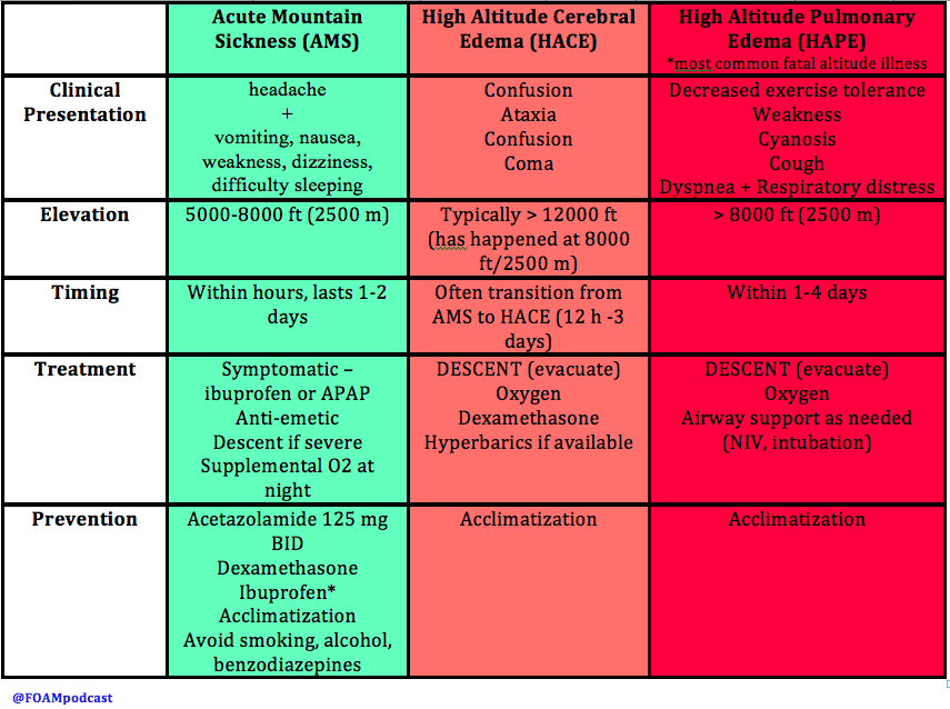
screen-shot-2016-09-09-at-6-22-50-am

Generously Donated Rosh Review Questions

A 20-year-old woman is climbing Mt. Kilimanjaro when she begins developing a headache followed by vomiting. As you begin to assess her, she has a grand mal seizure. Which of the following treatments should immediately be started?

A. Descent and acetazolamide

B. Ibuprofen and dexamethasone

C. Supplemental oxygen and acetazolamide

D. Supplemental oxygen, descent, and dexamethasone

[accordion]
[toggle title=”Answer” state=”closed”]

This patient has developed high-altitude cerebral edema (HACE), a rare but potentially life-threatening form of high-altitude illness. Most cases of HACE are described in ascension past 12 000 feet although it may happen at altitudes as low as 8 200 feet. In HACE, mild altered mental status can rapidly progress to coma in as little as 12 hours. HACE is characterized by global cerebral dysfunction evidenced by headache, fatigue, vomiting, ataxia, confusion, generalized seizures, slurred speech, and focal neurologic deficits. It is a clinical diagnosis although imaging will show cerebral edema. Treatment should start with high-flow oxygen, dexamethasone, and immediate descent. Additionally, if hyperbaric treatment is available, it should be initiated. Ibuprofen (B) and other NSAIDs can be used for prophylaxis prior to ascent as can acetazolamide (A and C).

[/toggle]
[/accordion]

A 32-year-old man complains of dyspnea on exertion and a cough with clear, watery sputum. He has been climbing Mt. Kilimanjaro for 2 days. Other than descent, what treatment can be started immediately?

A. Acetazolamide

B. Albuterol

C. Furosemide

D. Portable hyperbaric chamber therapy

[accordion]
[toggle title=”Answer” state=”closed”]

The patient is suffering from high-altitude pulmonary edema (HAPE) and should be treated with descent from altitude and hyperbaric oxygen therapy. HAPE is the most common fatal manifestation of high-altitude illness. It typically does not develop until the climber has passed 10,000 feet of elevation but it can happen at lower altitudes with heavy activity. The symptoms of HAPE usually begin 2-4 days after arrival at high altitude. Typically, patients experience marked dyspnea on exertion, fatigue with minimal effort, dry cough and difficulty with recovering from exertion. As HAPE progresses, patients will have a cough productive of copious clear secretions and have rales on examination. In severe cases, hemoptysis can develop. Although symptoms may mimic pneumonia or acute cardiogenic pulmonary edema, HAPE should be suspected in the correct clinical scenario. Rapid identification and management is central to preventing morbidity and mortality. The first and most important step in management is descent of the patient. Moderate decreases in altitude (1500 – 3000 feet) can rapidly resolve symptoms. If a hyperbaric chamber is available, it should be employed as simulated descent is just as effective and may be more logistically feasible.

Furosemide (C) is a loop diuretic, which decreases intravascular volume. There is a delay in onset of action and in recent years it has been replaced by pulmonary vasodilators like nifedipine. Additionally, the dehydration associated with furosemide makes it potentially dangerous in these patients. Moreover, HAPE is caused by hypoxia-induced pulmonary vasoconstriction and not from contractility problems of the heart. Acetazolamide (A) is a carbonic anhydrase inhibitor and can be used to treat mild altitude related symptoms like acute mountain sickness (AMS) or to prevent the development of high-altitude illnesses but it is not an appropriate treatment in HAPE. Albuterol (B) plays no role in the treatment of HAPE as bronchospasm is not the problem

[/toggle]
[/accordion]

References

  1. Ahmedzai S, Balfour-Lynn IM, Bewick T et al. Managing passengers with stable respiratory disease planning air travel: British Thoracic Society recommendations. Thorax. 66(Suppl 1):i1-i30. 2011. [article]
  2. Sacco F, Calero KR. Safety of early air travel after treatment of traumatic pneumothorax.  Int J Circumpolar Health. 2014; 73.
  3. Cheatham ML, Safcsak K. Air travel following traumatic pneumothorax: when is it safe? Am Surg 1999; 65:1160–1164
  4. Yaron M, Paterson RD, Davis CB. “High Altitude Medicine.” Chapter 144. Rosen’s Emergency Medicine (8e).
  5. Hackett PH, Davis CB. “High Altitude Disorders.” Chapter 221. Tintinalli’s Emergency Medicine: A Comprehensive Study Guide.
Episode 19 – Environment: Mushrooms and Hypothermia

(ITUNES OR LISTEN HERE)

The Free Open Access Medical Education (FOAM)

We review the Tox Talk podcast, Episode 23 – Mushrooms.  Our favorite pearls:

Clitocybe, Inocybe – contain muscarine which stimulates muscarinic receptors (acetylcholine/parasympathetic), causing a cholinergic toxidrome. Think SLUDGE (salivation, lacrimation, urination, defecation, gastric emptying/emesis) and the Killer B’s (bradycardia, bronchorrhea, bronchospasm) or DUMBELLS (diarrhea/diaphoresis, urination, miosis, bradycardia, emesis, lacrimation, lethargy, salivation). Basically, cholinergic toxidrome: SMALL, WET, SLOW.

  • Memory aid: these mushrooms end in -yBE, akin to the “killer B’s” that make cholinergic toxicity deadly.

Gyromitra – (false morel) contains gyromitrin which can cause seizures, in addition to gastrointestinal upset and liver failure.  Treatment: pyridoxine (B6).

  • Memory aid: gyromitra named because they look like the gyri of the brain and, conveniently, make the brain seize through depletion of GABA.

Amanita phalloides – contains amatoxins which cause delayed gastrointestinal symptoms and liver failure., echoing acetaminophen toxicity.

  • Caution: this is different than the amanita muscaria ‘mushroom, which is tricky because that amanita muscaria has neither muscarinic properties nor the toxicity of amanita phalloides.

Bonus pearl: Coprinus species can cause a disulfiram like reaction.

FOAM article on mushrooms by Jo et al

The Bread and Butter

We summarize some key topics from the following readings, Tintinalli (7e) Chapter  ; Rosen’s 8(e) Chapter  – but, the point isn’t to just take our word for it.  Go enrich your fundamental understanding yourself!

Hypothermia starts at 35°C and then is categorized based on severity.

Pearls:

  • Ethanol + hypothermia = bad news.  Ethanol is the most common cause of excessive heat loss in urban areas as people tend to not take warming measures, may be homeless or without heat, and have impaired thermoregulation.  Hypothermia also slows alcohol metabolism, making people drunker for longer.
  • Elderly patients are more susceptible to hypothermia, particularly as they may not sense the cooler temperatures.  Some may also have impaired thermoregulation.
  • Have a low threshold

Diagnostics:

  • Get a temperature, on all patients.  This applies to patient’s “found down” as well as the chronic alcoholic who just seems really drunk.
  • If patients aren’t rewarming 1°C/hr and they’re above 32°C, consider: sepsis, cortisol deficiency, myxedema, ethanol.
  • The J wave or “Osborn” wave is found in many cases of hypothermia, often quoted at ~80%.  However, it is not pathognomonic for hypothermia.
From the Rosh Review

Treatment: Warm the patient.  Don’t call the patient dead until they’re warm and dead, which means their temp is above 30-32°C.

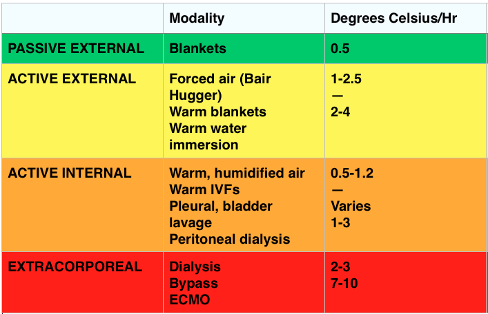
Screen Shot 2014-11-22 at 9.30.13 PM

Passive Rewarming – effective when the patient can still shiver (33-35°C).

  • Generates ~1.5°C of heat/hr

Active Rewarming – direct transfer of heat to the patient.

  • Indications: Cardiovascular instability, temp ≤30-32° C, inadequate rate of rewarming or failure to rewarm, endocrine problem, trauma, tox, secondary hypothermia impairing thermoregulation
  • Can be external or internal (which can be minimally invasive like IV fluids or quite invasive with things like bypass or pleural lavage).

Outcomes

  • Unlikely survival with a potassium > 12 mmol/L and recommendations are to terminate resuscitation for potassium >12 mmol/L and consider cessation for potassium between 10-12 mmol/L

FOAM Resources:

EBM Gone Wild on Prognostication

ScanCrit on ECMO in Accidental Hypothermia

EMCrit on Severe Accidental Hypothermia

Generously Donated Rosh Review Questions (Scroll for Answers)

Question 1.  A 40-year-old man with a history of substance abuse is brought in by EMS after being found unconscious outside of a nightclub in the middle of winter. It is unclear how long he was outside. He is unresponsive http://www.mindanews.com/buy-topamax/ with a GCS of 3.[polldaddy poll=8469565]

Question 2.  What is the most common cause of death in hypothermic patients after successful resuscitation?

Question 3.

[polldaddy poll=8473489]

Question 4. What abnormal rhythm is common with temperatures below 32°C?

References:

Danzl DF, Zafren K. Accidental Hypothermia, in Marx JA, Hockberger RS, Walls RM, et al (eds): Rosen’s Emergency Medicine: Concepts and Clinical Practice, ed 7. St. Louis, Mosby, Inc., 2013, (Ch) 140: pp 1883-1885.

Brown D JA, Brugger H, et al. Accidental Hypothermia. N Engl J Med 2012;367:1930-1938.

Mair P, Kornberger E, et al. Prognostic markers in patients with severe accidental hypothermia and cardiocirculatory arrest. Resuscitation 1994;27:47-54.

Answers:

1. D. When the serum potassium is greater than 12 mmol/L resuscitative efforts should be halted as the patient is unlikely to survive and further efforts constitute futile care. Accidental hypothermia is not an uncommon occurrence particularly in colder climates. It may occur in conjunction with substance abuse when an individual becomes impaired and is subsequently exposed to the outdoors. It can also occur as a result of drowning, avalanche and other trauma. Bio-makers other than potassium have been studied including serum lactate (B), pH (C) and clotting time. None have been proven prognostically reliable and therefore should not be used as a guide to determine if resuscitation should be continued. Hypothermic patients that present in cardiac arrest should be warmed to a minimum of 32°C (A) preferably via ECMO or cardiopulmonary bypass. However, if a hypothermic patient is warmed to 32°C and remains in asystole, recovery is unlikely and resuscitative efforts should be terminated. Other indications to cease resuscitative efforts include: obvious signs of irreversible death (e.g. major trauma), valid DNR order, conditions that are unsafe for the rescuer or provider, and an avalanche burial > 35 minutes in which the airway is packed with snow and the patient is asystolic.

2. Pulmonary edema.

2. C. Hypothermia. The ECG demonstrates the presence of J waves or Osborn waves which are seen in hypothermia. One of the first cardiac effects of hypothermia is bradycardia secondary to decreased firing of the cardiac pacemaker cells in cold temperatures. Osborn waves may appear at any temperature below 32°C. The waves are an upward deflection at the terminal portion of the QRS complex. They may represent abnormal ion flux in cold temperatures along with delayed depolarization and early repolarization of the left ventricular wall. As temperatures continue to drop, the ECG will demonstrate prolonged intervals: PR, followed by QRS and then QTc. Both diabetic ketoacidosis (A) and digoxin toxicity (B) may lead to hyperkalemia. In diabetic ketoacidosis, hyperkalemia develops as a result of the acidic pH in the blood and the transport of hydrogen ions intracellularly in exchange for a potassium ion. Digoxin toxicity poisons the cellular Na+/K+ ATPase resulting in elevated extracellular levels of potassium. The ECG manifestations of hyperkalemia begin with peaked T waves. Multiple other findings eventually develop including a shortened QT interval, ST depression, bundle branch blocks, widened QRS, prolonged PR interval, flattened T wave and ultimately a sine wave. Hyperparathyroidism (D) may lead to hypercalcemia. In hypercalcemia, the ECG shows a shortened QT interval, flattened T waves and QRS widening at very high levels.

4.  Atrial fibrillation.