Stop Giving Lidocaine in “GI Cocktails”

Apple Podcasts or Listen Here

We are trialing a new series of quick literature reviews. Let us know what you think

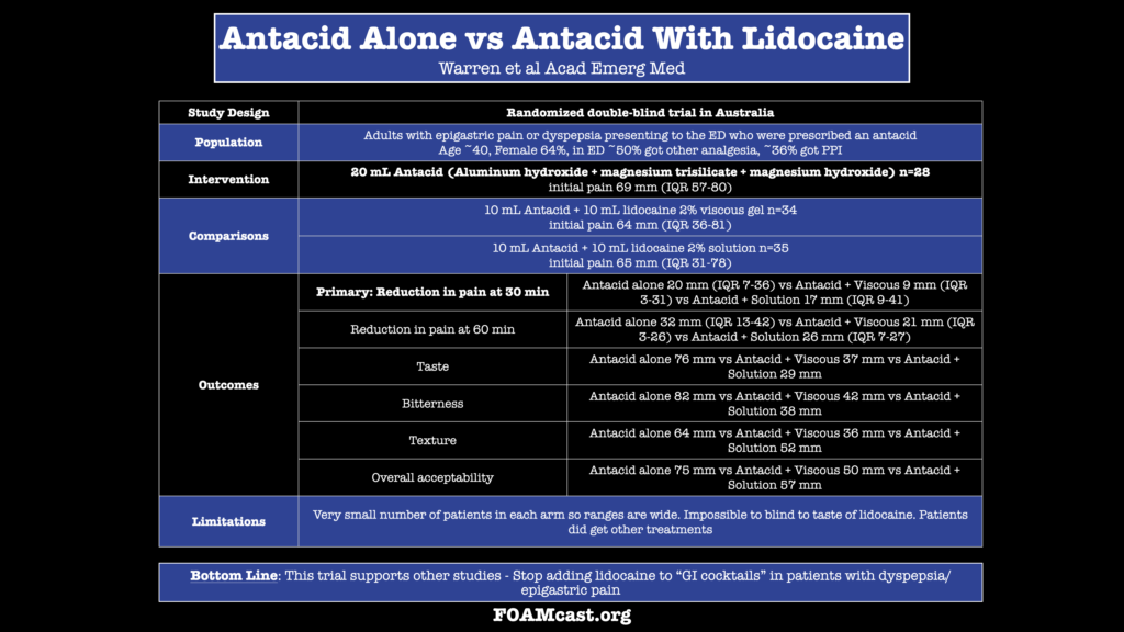
“GI cocktails” are commonly used in the Emergency Department for epigastric discomfort, but the evidence behind “cocktail” components is lacking and while lidocaine is commonly used, a trial by Berman et al found no benefit (versus a prior RCT in 1990 with high risk of bias).

In this episode we review a new trial by Warren et al on antacids versus antacids with lidocaine for epigastric pain and dyspepsia

Mechanical CPR, Balloon Tamponade, and Advocacy

iTunes or Listen Here

We are at #SMACC in Sydney, Australia, thanks to the Rosh Review, delivering updates from the conference to your earbuds.

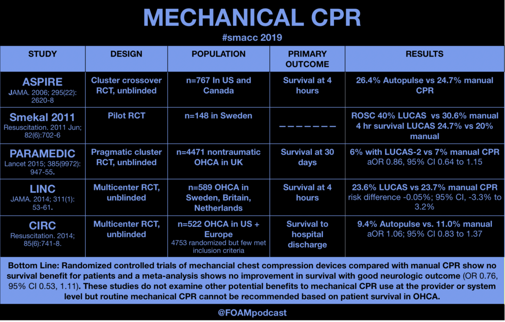
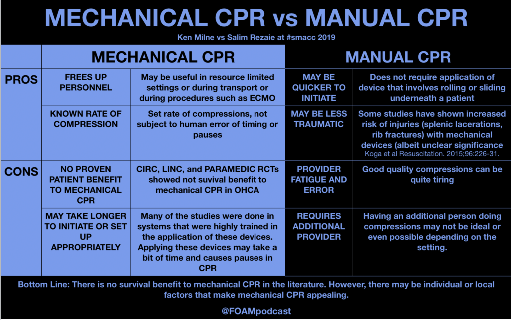
Mechanical CPR vs Manual CPR – Ken Milne vs Salim Rezaie

  1. Perkins GD, Lall R, Quinn T, et al. Mechanical versus manual chest compression for out-of-hospital cardiac arrest (PARAMEDIC): a pragmatic, cluster randomised controlled trial. Lancet (London, England). 2015; 385(9972):947-55. [pubmed]
  2. Rubertsson S, Lindgren E, Smekal D, et al. Mechanical chest compressions and simultaneous defibrillation vs conventional cardiopulmonary resuscitation in out-of-hospital cardiac arrest: the LINC randomized trial. JAMA. 2014; 311(1):53-61. [pubmed]
  3. Wik L, Olsen JA, Persse D. Manual vs. integrated automatic load-distributing band CPR with equal survival after out of hospital cardiac arrest. The randomized CIRC trial. Resuscitation. 2014; 85(6):741-8. [pubmed]
  4. Gates S, Quinn T, Deakin CD, Blair L, Couper K, Perkins GD. Mechanical chest compression for out of hospital cardiac arrest: Systematic review and meta-analysis. Resuscitation. 2015;94:91-7.  [pubmed]

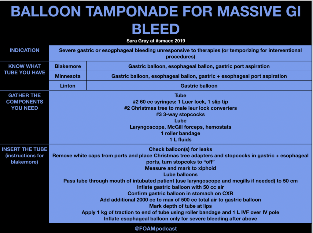
Massive GI Bleed and Balloon Tamponade – Dr. Sara Gray

Blakemore, Linton, and Minnesota Tube Review Video

Blakemore Placement

Advocacy in Emergency Medicine – Esther Choo and Hugh Montgomery

Dr. Choo spoke about how she became an advocate and helped start TIMES UP HEALTHCARE.  Check out her NEJM perspective article.

Hugh Montgomery spoke on ways to motivate individuals to change behavior by appealing to emotional aspects.

Episode 76 – Pneumoperitoneum, Gastritis, & PUD

iTunes or Listen Here

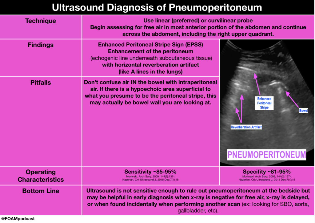
5MinSono has a great 5 minute videocast on the ultrasound diagnosis of pneumoperitoneum.
Core Content

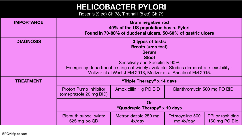
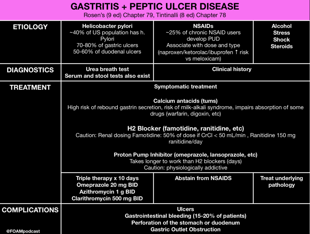
We cover gastropathies and peptic ulcer disease (PUD) using Rosen’s Emergency Medicine (9th ed) Chapter 79 and Tintinalli’s Emergency Medicine (8th ed) Chapter 78  as guides.
Rosh Review Emergency Board Review Questions

A 67-year old man with chronic osteoarthritis complains of gnawing and burning in the epigastric area that is occasionally accompanied by nausea and vomiting. His current BMI is 26 and he is physically active. What is the most probable cause for these symptoms?

A. Cholelithiasis

B. Gastric carcinoma

C. Nonsteroidal anti-inflammatory drug induced gastritis

D. Peptic ulcer disease

[accordion]
[toggle title=”Answer” state=”closed”] C. 
Nonsteroidal anti-inflammatory drug (NSAID) therapy is the first line treatment in osteoarthritis, however chronic NSAID use can often destroy the gastric mucosa leading to hemorrhage, erosions and ulcers. NSAIDs such as naproxen and ibuprofen are the most common agents associated with acute erosive gastritis. A long-term prospective study found that patients with arthritis who were older than 65 years and regularly took low-dose aspirin were at increased risk for dyspepsia severe enough to necessitate the discontinuation of NSAIDs. This suggests that better management of NSAID use should be discussed with older patients in order to reduce NSAID-associated upper GI events. COX -2 inhibitors, such as celecoxib, are an alternate therapy to NSAIDs. Other common agents that cause gastritis include alcohol and Helicobacter pylori. The mainstay treatment of erosive gastritis is to refrain from the offending agent. The gold standard for diagnoses of gastritis is an upper GI endoscopy.

Symptomatic cholelithiasis (A) is often seen in populations who have risk factors for gallstones, which include persons with diabetes mellitus, persons who are obese, women, rapid weight cyclers, and patients on hormone therapy or taking oral contraceptives. This patient does not portray the colicy pain associated with gallstones. Patients with gastric cancer (B) often present with weight loss, dysphagia, postprandial fullness and loss of appetite. Gastric cancer is multifactorial involving both inherited predisposition and environmental factors. Environmental factors implicated in he development of gastric cancer include diet, Helicobacter pylori, previous gastric surgery, pernicious anemia, chronic atrophic gastritis and radiation exposure. Smoking and smoked meats also have a high correlation with gastric cancer. Peptic ulcer disease (D) is a complication of chronic gastritis and can present in a similar manner as gastritis. Peptic ulcers include both gastric and duodenal ulcers. Peptic ulcers present with gnawing or burning sensation that occur after meals. Common risk factors include H. pylori infection and ingestion of NSAIDs. An upper GI endoscopy must be performed to visualize the ulcers. Biopsy is indicated if ulcers are seen on endoscopy in order to rule out Helicobacter pylori. Active ulcers associated with NSAID use are treated with an appropriate course of proton pump inhibitor (PPI) therapy and the cessation of NSAIDs. For patients with a known history of ulcer and in whom NSAID use is unavoidable, the lowest possible dose and duration of NSAID and co-therapy with a PPI is recommended.

[/toggle]
[/accordion]

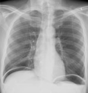
A 55-year-old man presents with severe abdominal pain and tenderness on examination that began acutely approximately 12 hours prior to arrival. His X-ray is shown below. What is the most appropriate next step?
A. Computed tomography scan of the abdomen and pelvis

B. Nasogastric tube insertion

C. Observation and serial abdominal exams

D. Surgical consultation

[accordion]
[toggle title=”Answer” state=”closed”]

D. The X-ray demonstrates free air under the diaphragm representing a perforated viscus within the intraabdominal cavity. The presence of free air is an indication for an emergent surgical consultation for repair. The emergency provider should administer broad-spectrum antibiotics covering aerobic and anaerobic organisms along with intravenous fluid resuscitation.  

A CT scan of the abdomen and pelvis (A) is indicated if the X-ray does not reveal evidence of free air and the patient has ongoing pain and tenderness requiring a diagnosis. Some perforations will not show on plain films, and as time progresses, the area of perforation may wall off and not show on X-ray. A nasogastric tube (B) is not indicated in the management of a patient with a perforated viscus. Observation and serial abdominal exams (C) are not sufficient for a patient with a perforation.  

A patient presents with hematemesis. What test is most likely to determine the etiology of the bleeding?

A. CT scan of the abdomen and pelvis

B. Nasogastric tube lavage

C. Right upper quadrant ultrasound

D. Upper endoscopy

[/toggle]
[/accordion]

[accordion]
[toggle title=”Answer” state=”closed”]

Upper endoscopy is the modality that is most likely to identify the culprit lesion in a patient with upper gastrointestinal bleeding (UGIB). UGIB is a common presentation caused by a variety of pathologies including gastritis, esophageal varices, peptic ulcer disease, Mallory-Weiss tears, arteriovenous malformations and Boerhaave’s syndrome. Of these causes, peptic ulcer disease is the most common. Regardless of the etiology, endoscopy represents the best modality for diagnosis. It allows direct visualization of the esophagus, stomach and first two sections of the duodenum. Additionally, it allows for interventions to be performed if active bleeding or stigmata of recent bleeding are found.

CT scan of the abdomen and pelvis (A) is limited in its ability to give a diagnosis. Nasogastric tube lavage (B) may show the presence of blood in the upper GI tract but cannot differentiate between causes. Right upper quadrant ultrasound (C) may give information about the patient including the presence of cirrhosis but cannot give a specific diagnosis as the cause.

[/toggle]
[/accordion]

References

  1. Nazerian P, Tozzetti C, Vanni S. Accuracy of abdominal ultrasound for the diagnosis of pneumoperitoneum in patients with acute abdominal pain: a pilot study. Critical ultrasound journal. 7(1):15. 2015. [pubmed]
Episode 73 – Gastroparesis & Biliary Pathology

iTunes or Listen Here

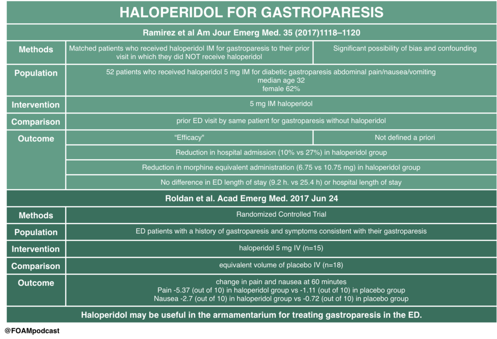
Dr. Salim Rezzaie of RebelEM wrote a blog post on HUGS – Haloperidol for Gastroparesis.  This study had significant methodologic limitations but provides some interesting insight into a practice that many providers have adopted – using haloperidol for gastroparesis. A tiny RCT by Roldan and colleagues also looked at this practice and found promising results, although the study was very small.  Haloperidol has also been used in cannabinoid hyperemesis but literature on this is limited to case reports/series. 

Core Content

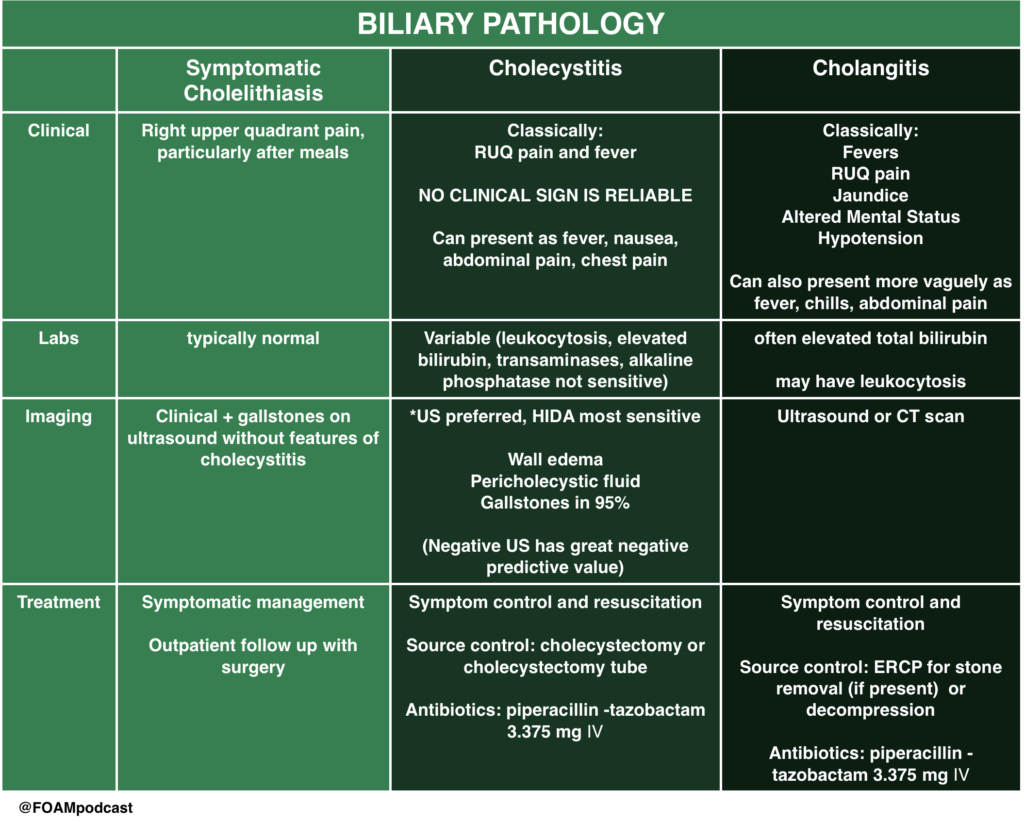
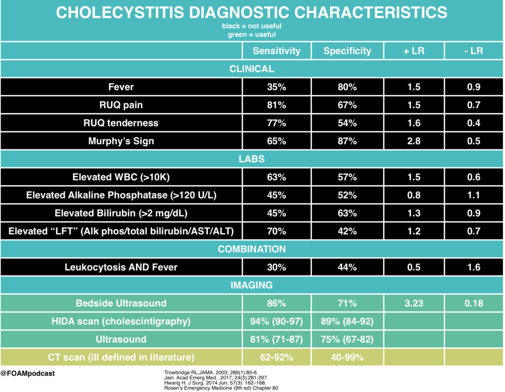
We review biliary colic, cholecystitis, and cholangitis using Tintinalli (8th ed) Chapter 79 and Rosen’s Emergency Medicine (9th ed) Chapter 80 as guides.

Rosh Review Emergency Board Review Questions

A 60-year-old woman presents with two days of right upper quadrant abdominal pain constant in nature and associated with subjective fever, nausea, and vomiting. Vital signs are temperature of 38.1°C, heart rate 87, blood pressure 140/80 mm Hg, respiratory rate 14, and oxygen saturation of 99% on room air. Her abdomen is soft with right upper quadrant tenderness and a positive Murphy’s sign. Which of the following tests is most sensitive and specific in diagnosing this patient’s condition?

A. Computed tomography scan with intravenous contrast

B. Hepatobiliary iminodiacetic acid (HIDA) scan

C. MRI with gadolinium

D. Ultrasound

[accordion]
[toggle title=”Answer” state=”closed”]

This patient has suspected acute cholecystitis and requires an imaging study for confirmation. Hepatobiliary iminodiacetic acid (HIDA) scanning is considered the most sensitive and specific test for diagnosing acute cholecystitis. IDA is administered intravenously, taken up by hepatocytes, and excreted into the bile canaliculi. Failure to obtain an outline of the gallbladder within one hour proves cystic duct obstruction and, in the appropriate clinical setting, confirms the diagnosis of acute cholecystitis. Visualization of the gallbladder and common duct within one hour has a high negative predictive value. A HIDA scan is usually obtained when the ultrasound study is equivocal.

Computed tomography scan with intravenous contrast (A) can identify cholecystitis with a reported sensitivity of 92% and specificity of 99%. It is most useful in cases of emphysematous and hemorrhagic cholecystitis. MRI with gadolinium (C) provides similar diagnostic yield to CT scan. Ultrasound (D) is most useful in the ED setting because it is a quick, noninvasive test. Its sensitivity and specificity, however, are lower than a HIDA scan’s for pathology-confirmed cholecystitis. Ultrasound findings and a clinical exam consistent with acute cholecystitis are highly predictive, and many such patients will undergo cholecystectomy without further diagnostic testing.

[/toggle]
[/accordion]


References:

  1. Ramirez R, Stalcup P, Croft B, Darracq MA. Haloperidol undermining gastroparesis symptoms (HUGS) in the emergency department. Am J Emerg Med. 2017; 35(8):1118-1120. [pubmed]
  2. Roldan CJ, Chambers KA, Paniagua L, Patel S, Cardenas-Turanzas M, Chathampally Y. Randomized Controlled Double-blind Trial Comparing Haloperidol Combined With Conventional Therapy to Conventional Therapy Alone in Patients With Symptomatic Gastroparesis. Acad Emerg Med. 2017; In Press[pubmed]
  3. Trowbridge RL, Rutkowski NK, Shojania KG. Does this patient have acute cholecystitis? JAMA. 2003; 289(1):80-6. [pubmed]
  4. Jain A, Mehta N, Secko M. History, Physical Examination, Laboratory Testing, and Emergency Department Ultrasonography for the Diagnosis of Acute Cholecystitis. Acad Emerg Med.. 2017; 24(3):281-297. [pubmed]
  5. Hwang H, Marsh I, Doyle J. Does ultrasonography accurately diagnose acute cholecystitis? Improving diagnostic accuracy based on a review at a regional hospital.   J Surg. 2014 Jun; 57(3): 162–168. [pubmed]

Episode 52 – The Esophagus

ITUNES OR LISTEN HERE

The Free Open Access Medical Education (FOAM)

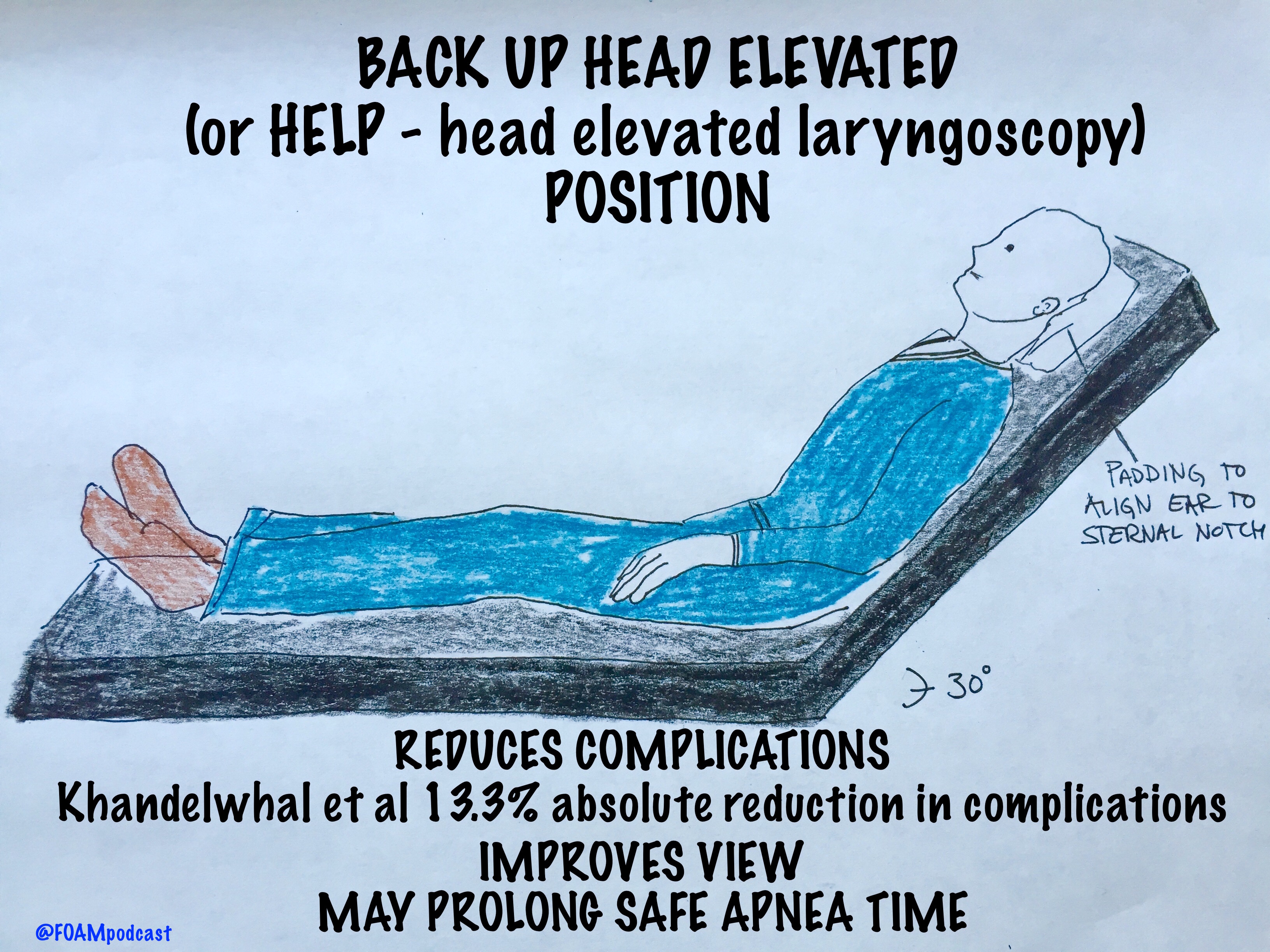
We cover a Scancrit post on the Back Up Head Elevated (BUHE) intubation position.  This post details a multicenter retrospective observational study by Khandelwal et al in Anesthesia & Analgesia

FullSizeRender (4)

Population: 528 adults undergoing emergent intubation

Intervention: Head up at least 30 degrees during intubation

Control: Supine intubation

Primary Outcome: Occurrence of intubation related complication (difficult intubation: >3 attempts or prolonged intubation, hypoxemia, esophageal intubation, or pulmonary aspiration) – 22.6% in supine group vs 9.3% in the head elevated position.  Absolute difference of 13.3%

Limitations: Did not look at emergency department intubation.  More experienced intubators used the BUHE positioning, which could confound the reduction in intubation related complications [1].

BUHE or Head Elevated Laryngoscopy Position (HELP) has also been found to

  • Improve laryngeal view [2,3]
  • Prolong safe apnea time [4]

Note: in patients with possible spinal injuries, one may use reverse trendelenberg (or forego the back up head elevated position)

Core Content

We delve into core content on the esophagus using Rosen’s (8th ed) Chapter 71 and Chapter 77 in Tintinalli (8th ed)

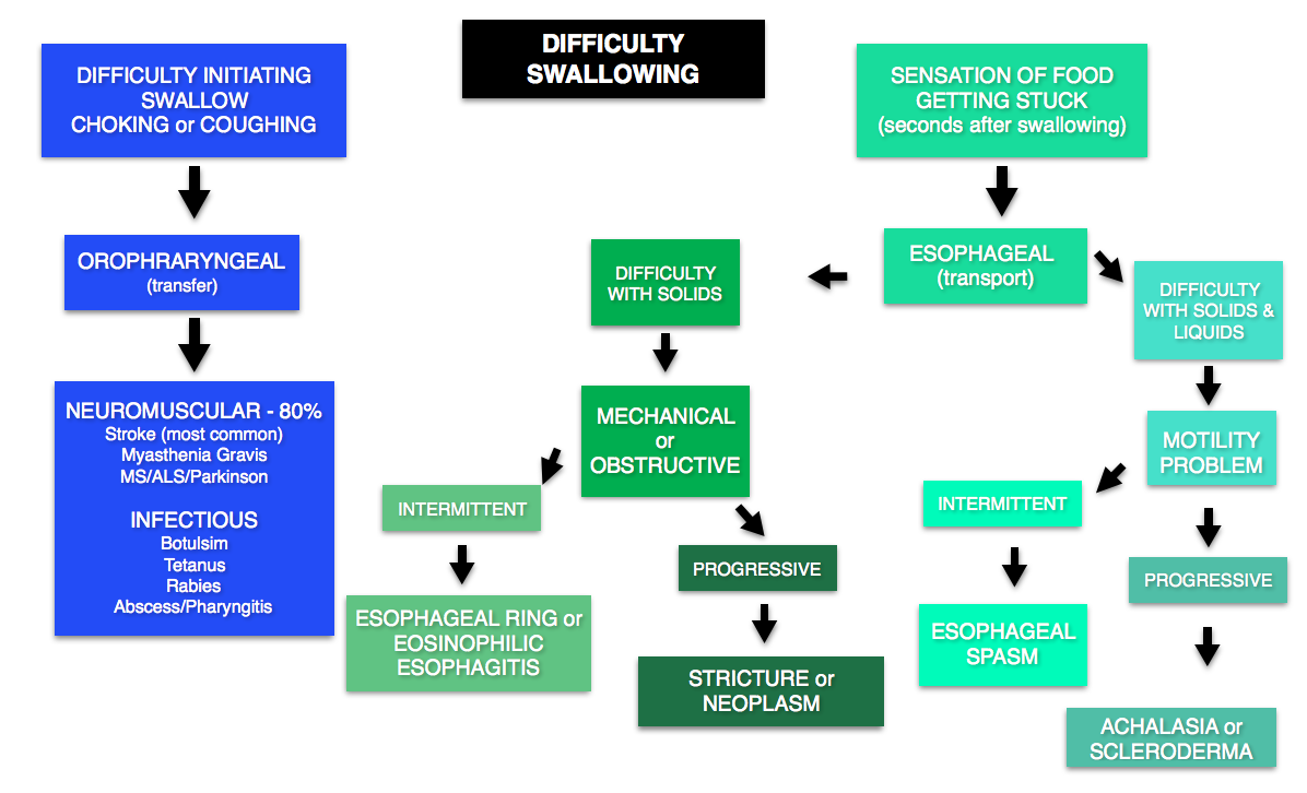
DYSPHAGIA

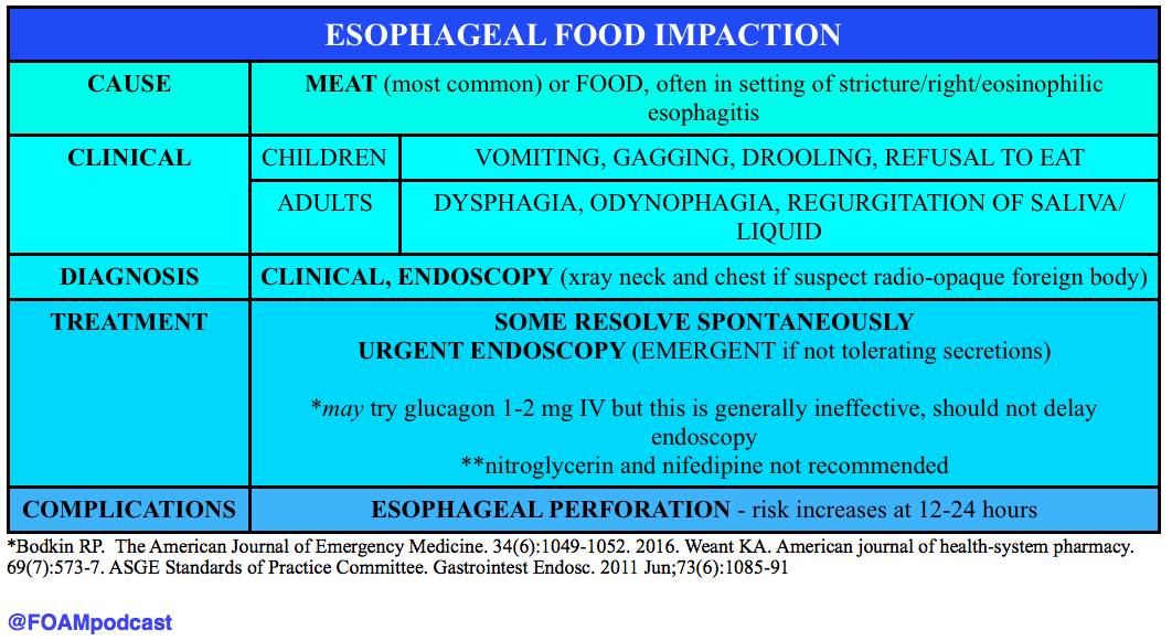
Emergent conditions may include stroke (most common cause), myasthenia/botulism/or other neuromuscular problems (may also have concomitant respiratory failure). Many causes do not need emergent workup.

@FOAMpodcast
@FOAMpodcast

ESOPHAGEAL FOOD BOLUS

Screen Shot 2016-06-27 at 5.29.03 AM

Generously Donated Rosh Review Questions

An 87-year-old woman presents to the ED after her caregiver witnessed the patient having difficulty swallowing over the past 2 days. The patient is having difficulty with both solids and liquids. She requires multiple swallowing attempts and occasionally has a mild choking episode. She has no other complaints. Your exam is unremarkable. Which of the following is the most likely cause of her condition?

A. Achalasia

B. Cerebrovascular accident

C. Esophageal neoplasm

D. Foreign body

[accordion]
[toggle title=”Answer” state=”closed”]

B. Cerebrovascular accident.  Dysphagia can be divided into two categories: transfer and transport. Transfer dysphagia occurs early in swallowing and is often described by the patient as difficulty with initiation of swallowing. Transport dysphagia occurs due to impaired movement of the bolus down the esophagus and through the lower sphincter. This patient is experiencing a transfer dysphagia. This condition is most commonly due to neuromuscular disorders that result in misdirection of the food bolus and requires repeated swallowing attempts. A cerebrovascular accident (stroke)that causes muscle weakness of the oropharyngeal muscles is frequently the underlying cause. Achalasia (A) is the most common motility disorder producing dysphagia. It is typically seen in patients between 20 and 40 years of age and is associated with esophageal spasm, chest pain, and odynophagia. Esophageal neoplasm (C) usually leads to dysphagia over a period of months and progresses from symptoms with solids to liquids. It is also associated with weight loss and bleeding. Foreign bodies (D) such as a food bolus can lead to dysphagia, but patients are typically unable to tolerate secretions and are often observed drooling. These patients do not have difficulty in initiating swallowing.

[/toggle]
[/accordion]

A 33-year-old man presents with dysphagia to both solids and liquids, with solids being worse than liquids. He describes a sensation of the food getting stuck in his chest. Occasionally, he needs to raise his arms above his head to help food pass into his stomach. His primary care doctor has been treating him for GERD over the previous six months, but his symptoms are getting worse. Which of the following is the most likely diagnosis?

A. Achalasia

B. Diffuse esophageal spasm

C. Schatzki ring

D. Zenker’s diverticulum

[accordion]

[toggle title=”Answer” state=”closed”]

Achalasia This patient most likely has achalasia, which is an esophageal dysmotility disorder due to failure of the lower esophageal sphincter to relax. Dysphagia is the most common symptom. While all patients have dysphagia to solids, only two-thirds have liquid dysphagia. By standing after eating, straightening one’s back, or raising the arms above the head, the esophageal pressure increases, which can help emptying into the stomach. Symptoms usually begin with mild dysphagia in patients who are 20 to 40 years old; symptoms are usually progressive. Diffuse esophageal spasm (B) is a hypermotility disorder causing strong, uncoordinated peristaltic contractions that do not propel food effectively to the stomach. Dysphagia, regurgitation of food, and chest pain are commonly present. The dysphagia is intermittent and does not progress over time. A Schatzki ring (C) is a fibrous band-like structure in the distal esophagus that is the most common cause of dysphagia with solids. Patients typically do not experience difficulty with liquids. Zenker’s diverticulum (D) is an acquired disease that is due to an out-pouching in the mucosa of the pharynx. It typically occurs in individuals older than 50 years and can cause regurgitation, cough, and halitosis from food that becomes stuck in the diverticulum.

[/toggle]
[/accordion]

[accordion]
[toggle title=”What are three common treatments for achalasia?” state=”Closed”]

Nitroglycerin to reduce lower esophageal sphincter tone, endoscopic injection of botulinum toxin into the muscle of the sphincter, and surgical myotomy.  Diffuse esophageal spasm (B) is a hypermotility disorder causing strong, uncoordinated peristaltic contractions that do not propel food effectively to the stomach. Dysphagia, regurgitation of food, and chest pain are commonly present. The dysphagia is intermittent and does not progress over time. A Schatzki ring (C) is a fibrous band-like structure in the distal esophagus that is the most common cause of dysphagia with solids. Patients typically do not experience difficulty with liquids. Zenker’s diverticulum (D) is an acquired disease that is due to an out-pouching in the mucosa of the pharynx. It typically occurs in individuals older than 50 years and can cause regurgitation, cough, and halitosis from food that becomes stuck in the diverticulum.

[/toggle]
[/accordion]

References

  1. Khandelwal et al. Head-elevated Patient Positioning Decreases Complications of Emergent Tracheal Intubation in the Ward and Intensive Care Unit. Anesthesia and Analgesia. Apr 2016.
  2. Lee et al. Laryngeal exposure during laryngoscopy is better in the 25 degree back-up position than in the supine position. British Journal of Anaesthesia. July 2007.
  3. Levitan RM, Mechem CC, Ochroch EA, Shofer FS, Hollander JE. Head-elevated laryngoscopy position: improving laryngeal exposure during laryngoscopy by increasing head elevation. Annals of emergency medicine. 41(3):322-30. 2003.
  4. Ramkumar V, Umesh G, Philip FA. Preoxygenation with 20º head-up tilt provides longer duration of non-hypoxic apnea than conventional preoxygenation in non-obese healthy adults. Journal of anesthesia. 25(2):189-94. 2011. [pubmed]
  5. Bodkin RP, Weant KA, Baker Justice S, Spencer MT, Acquisto NM. Effectiveness of glucagon in relieving esophageal foreign body impaction: a multicenter study. The American Journal of Emergency Medicine. 34(6):1049-1052. 2016.
  6. Weant KA, Weant MP. Safety and efficacy of glucagon for the relief of acute esophageal food impaction. American journal of health-system pharmacy : AJHP : official journal of the American Society of Health-System Pharmacists. 69(7):573-7. 2012
  7. ASGE Standards of Practice Committee, Ikenberry SO, Jue TL, Anderson MA, Appalaneni V, Banerjee S, Ben-Menachem T, Decker GA, Fanelli RD, Fisher LR, Fukami N, Harrison ME, et al. Management of ingested foreign bodies and food impactions. Gastrointest Endosc. 2011 Jun;73(6):1085-91

Episode 45 – Diverticulitis

(ITUNES OR LISTEN HERE)

The Free Open Access Medical Education (FOAM)

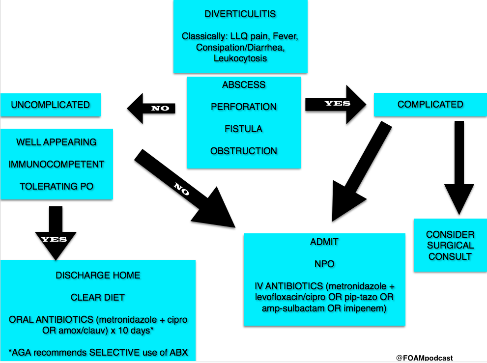
Life in the Fast Lane Research and Reviews (LITFL R&R) #121  featured a section on the new American Gastroenterology Association (AGA) guidelines on diverticulitis. The game changer?  Antibiotics aren’t a requirement in select patients with uncomplicated acute diverticulitis [1].

The guidelines based this recommendation on two studies, previously covered by Dr. Ryan Radecki on Emergency Medicine literature of note over the past 3 years. This post details a prospective observational study on antibiotics for acute diverticulitis [2].  In another post, Dr. Radecki discusses an RCT of antibiotics (ABX) vs IV fluids only.

  • 623 patients with an episode with a short history and with clinical signs of diverticulitis, with fever (>38 Celsius) and inflammatory parameters, verified by computed tomography (CT), and without any sign of complications (fistula, perforation, abscess) or signs of sepsis
  • Randomized to IVF only or IVF + antibiotics
  • Primary Outcome – 6 patients (1.9%) developed complications in the no ABX arm vs 3 patients (1.0%) in the ABX arm (not statistically significant). Overall study complication rate was 1.4% [3].

Of note, since 2012, the Cochrane Review suggests that antibiotics may not be necessary in uncomplicated appendicitis [4].

A note on LITFL R&R – every week this blog post features 5-10 high yield articles, culled from contributors across the globe from all kinds of literature – pediatrics, critical care, emergency medicine, etc. It is difficult to keep up with the literature and some have estimated that the number needed to read (NNR) to of 20-200, depending on the journal [5].  Those looking for high yield articles may find their time well spent focused on this cherry picked selection of articles.

Core Content

We delve into core content on diverticula and clostridium difficile using Rosen’s Medicine (8e),  Chapters 31, 173 and Tintinalli’s Emergency Medicine: A Comprehensive Study Guide  (7e) Chapters 76, 85.

Diverticulosis

Diverticula are small herniations through the wall of the colon (small outpouchings). Often this is asymptomatic, identified incidentally on imaging or colonoscopy. Most common cause of lower gastrointestinal bleeding (LGIB) in adults in the U.S.

Diverticulitis

Diverticulitis Algorithm

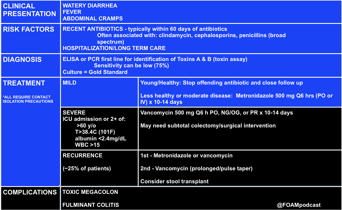
Clostridium Difficile (c. diff)

C. Difficile


Note on testing – asymptomatic carriage rates of c.diff vary based on the population but may be between 3-50%.  Textbooks quote a 3% carriage rate in newborns and rates of 20%-50% in hospitals and long term care facilities, respectively [10,11].

C. diff historically has a unique odor, refrains of “it smells like c. diff” echo in the halls.  Yet this does not perform very well, essentially a coin flip based on a 2013 study by  Rao and colleagues.  They  had 18 nurses smell 10 stool samples (5 c. diff positive and 5 c. diff neg) and found the median percent correct identification of c. diff positive vs negative was 45% [6]. 

Rosh Review Questions

Question 1. [polldaddy poll=9330955]

Question 2.A 75-year-old woman presents with several days of voluminous watery stools. She was discharged from the hospital one week ago following treatment for pneumonia. Stool studies reveal C. difficile toxin. [polldaddy poll=9333580]

Answers:

  1. C. Patients who present with uncomplicated diverticulitis should be treated with oral antibiotics for 7-10 days. Diverticulitis is an inflammation of the diverticulum in the large intestine. In uncomplicated cases of diverticulitis, patients present with abdominal pain typically in the left lower quadrant with tenderness to palpation in the same area. Patients should not have peritoneal signs or masses on examination. Complicated diverticulitis is defined as the presence of either extensive inflammation or complications such as abscess, peritonitis or obstruction. Patients with uncomplicated diverticulitis can be empirically treated with antibiotics (typically as an outpatient) for 7-10 days. Patients with uncomplicated diverticulitis typically do not require CT imaging (A). Patients with complicated diverticulitis should be treated with intravenous antibiotics (B) and admitted to the hospital. Ultrasound (D) has shown promise in diagnosing diverticulitis but CT is the imaging modality of choice.
  2. C.C. difficile infection is caused by a spore-forming obligate anaerobic bacillus that causes a spectrum of disease ranging from diarrhea to pseudomembranous colitis. C. difficile is the most common cause of infectious diarrhea in hospitalized patients in the United States. Risk factors for infection include broad-spectrum antibiotic use, particularly clindamycin, though other antibiotics have also been implicated. Additional risk factors include prolonged hospitalization, advanced age, and underlying comorbidities. The spectrum of clinical manifestations includes frequent watery stools to a more toxic clinical presentation with profuse stools (up to 20-30 per day), crampy abdominal pain, fever, leukocytosis, and hypovolemia. C. difficile colitis should be suspected in patients who develop diarrhea while taking or after recent cessation of antibiotics, or among recently discharged patients who develop diarrhea. Diagnosis is confirmed by identification of C.difficile toxin in the stool. Colonoscopy, while not usually necessary for diagnosis, reveals characteristic yellowish plaques in the intestinal lumen, confirming pseudomembranous colitis. Treatment for C. difficile infection depends on disease severity. Previously healthy patients with very mild symptoms may be managed by cessation of the offending antibiotic and close clinical monitoring. Oral metronidazole, 500 mg po every 6 hours for 10-14 days is the treatment for moderately severe colitis. Severely ill patients should be hospitalized and treated with oral vancomycin, 125 mg po every 6 hours for 10-14 days.

References:

  1. Stollman N, Smalley W, Hirano I, AGA Institute Clinical Guidelines Committee. American Gastroenterological Association Institute Guideline on the Management of Acute Diverticulitis. Gastroenterology. 2015;149(7):1944–9. doi:10.1053/j.gastro.2015.10.003.
  2. Isacson D, Thorisson A, Andreasson K, Nikberg M, Smedh K, Chabok A. Outpatient, non-antibiotic management in acute uncomplicated diverticulitis: a prospective study. Int J Colorectal Dis. 2015;30(9):1229–1234. doi:10.1007/s00384-015-2258-y.
  3. Chabok A, Phlman L, Hjern F, Haapaniemi S, Smedh K. Randomized clinical trial of antibiotics in acute uncomplicated diverticulitis. Br J Surg. 2012;99(4):532–539. doi:10.1002/bjs.8688.
  4. Shabanzadeh DM1, Wille-Jørgensen P.Antibiotics for uncomplicated diverticulitis.  Cochrane Database Syst Rev. 2012 Nov 14;11:CD009092. doi: 10.1002/14651858.CD009092.pub2. 
  5. McKibbon KA, Wilczynski NL, Haynes RB. What do evidence-based secondary journals tell us about the publication of clinically important articles in primary care journals? BMC Med. 2004;2:33.
  6. Rao K, Berland D, Young C, Walk ST, Newton DW. The Nose Knows Not: Poor Predictive Value of Stool Sample Odor for Detection of Clostridium difficile. Clinical Infectious Diseases. 56(4):615-616. 2012.
  7. “Chapter 85: Diverticulitis.” Tintinalli’s Emergency Medicine: A Comprehensive Study Guide, 7e.New York, NY: McGraw-Hill; 2011. p 578-581.
  8. “Disorders of the Large Intestine.” Rosen’s Emergency Medicine, 8th e. p 1261-1275.
  9. “Gastrointestinal Bleeding.”  Rosen’s Emergency Medicine, 8th e. p 248-253.
  10.  “Infectious Diarrheal Disease and Dehydration.” Rosen’s Emergency Medicine, 8th ep 2188-2204.
  11. “Chapter 76: Disorders Presenting Primarily with Diarrhea.” Tintinalli’s Emergency Medicine: A Comprehensive Study Guide, 7e.New York, NY: McGraw-Hill; 2011. p 534-535

Episode 23 – SBO and Mesenteric Ischemia

(ITUNES OR LISTEN HERE)

The Free Open Access Medical Education (FOAM)

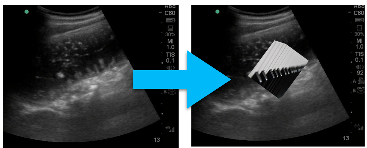
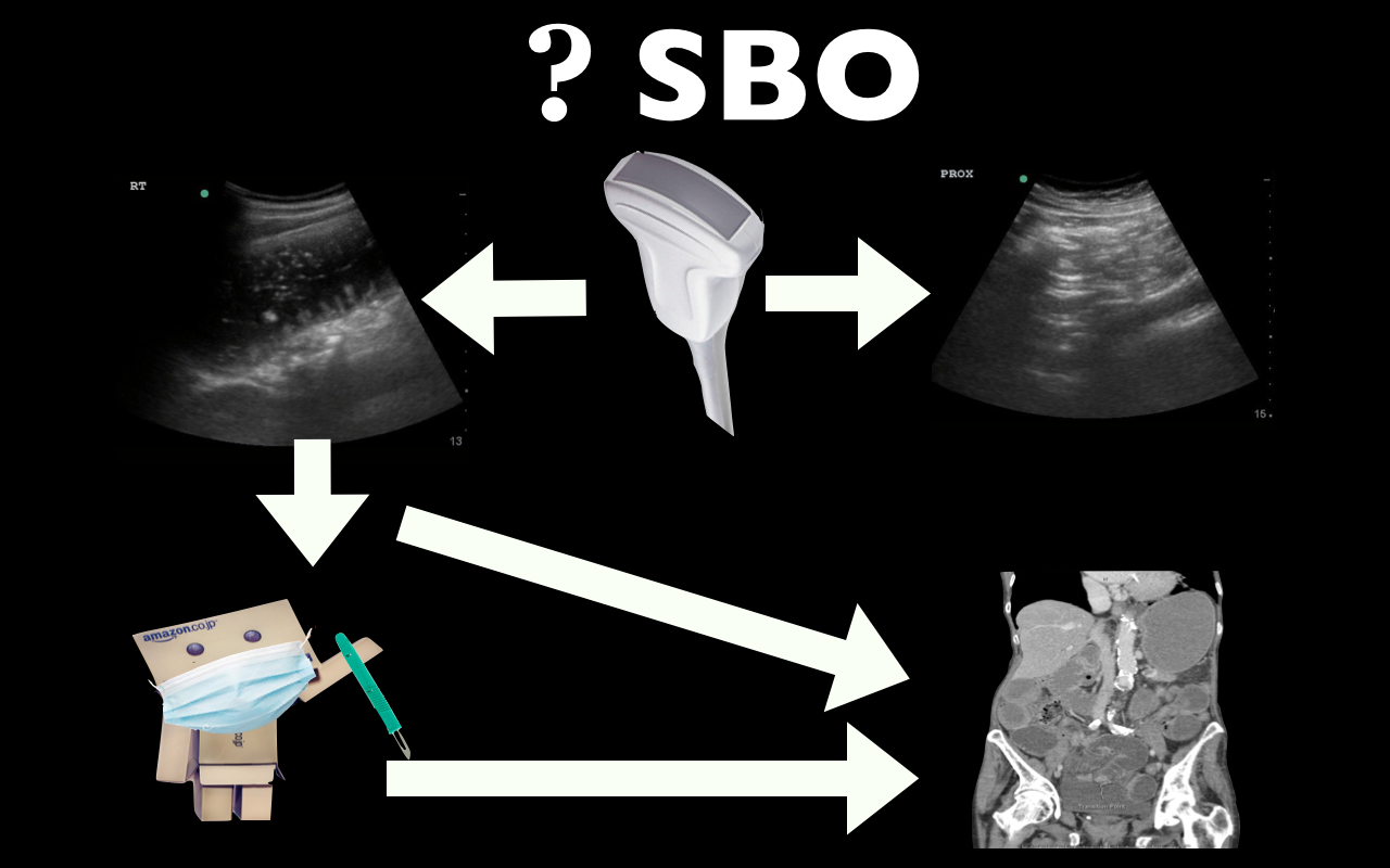
This week we’re covering Dr. Jacob Avila’s post on ultrasound for small bowel obstruction (SBO)  located at Ultrasound of the week.  He has an accompanying video on 5minSono.

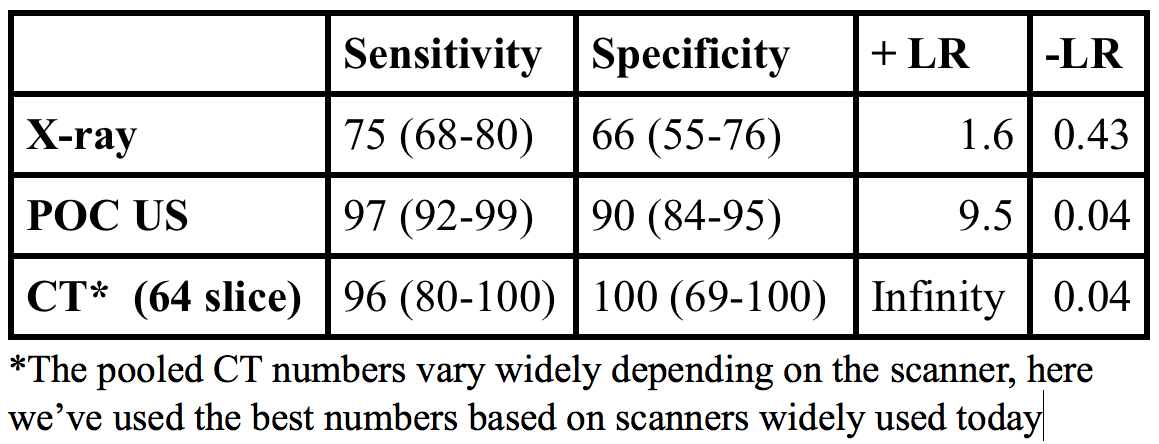
Point of care ultrasound has good operating characteristics for diagnosis of SBO with a LR+ 9.5, LR- 0.04, far better than abdominal x-ray [1].

Screen Shot 2015-01-25 at 10.20.44 AM

What to look for:

  • Dilated loops of bowel > 2.5 cm in diameter
  • Additional clues:  “To and fro” peristalsis
  • The piano key sign, Tanga sign

Piano Key Sign
Piano Key Sign

Problems with abdominal x-ray:

  • Rosen’s: Abdominal x-rays are “diagnostic in approximately 50 to 60% of cases of SBO, equivocal in 20 to 30%, and normal, nonspecific, or misleading in 10 to 20%” [2].
  • American College of Radiology: they can “prolong the evaluation period … while often not obviating the need for additional examinations, particularly CT” [3].

Limitations:

  • While ultrasound can diagnose SBO, there is little evidence to suggest that we can identify transition points or strangulation/necrosis.  As such, there can still be a role for CT scan, particularly in first time SBO to identify a transition point.
  • The EAST guidelines acknowledge the utility of ultrasound yet this practice is far from accepted in the surgical community.  Surgical colleagues will likely still want concrete imaging such as an x-ray or CT; however, ultrasound performed concurrent with the history and physical may speed up patient’s disposition to definitive care/imaging.

Possible algorithm for use of US in SBO

More FOAM on the topic:

The Bread and Butter

We cover key points on SBO and Acute Mesenteric Ischemia from Rosenalli, that’s Tintinalli (7e) Chapter 86; Rosen’s (8e) Chapter 92.  But, don’t just take our word for it.  Go enrich your fundamental understanding yourself.

Small Bowel Obstruction

Etiology of intestinal obstruction: “HANG IV.” Hernia, Adhesions (most common cause), Neoplasm, Gallstone ileus, Intussusception, Volvulus

Treatment

  • Intravenous fluids – resuscitate the patient!
  • Antiemetics.  If a patient is compromising their airway, an aspiration risk, or vomiting despite antiemetics, consider the use of a nasogastric tube.  Shockingly, “use of nasogastric decompression is considered dogma by many emergency physicians and surgeons, its effect in decreasing the duration of SBO has scant support in the medical literature” [2, 5].  This point demonstrates that SBO is not a monolithic disease entity but a spectrum of pathology with variable treatments depending on patient’s sickness.
  • Antibiotics that cover gram-negative and anaerobic organisms
  • Admit. Most of these patients will likely go to the surgical service; however,

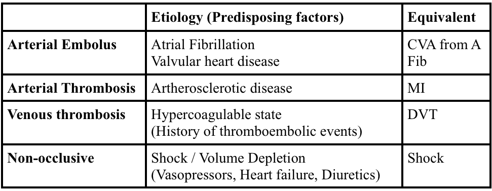
Acute Mesenteric Ischemia

Screen Shot 2015-01-24 at 7.18.02 PM

 Generously Donated Rosh Review Questions (scroll for answers)

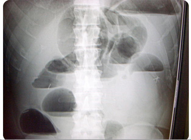
Question 1. A 73-year-old man presents with vomiting and abdominal pain for 2 days. The patient has a remote history of cholecystectomy and appendectomy. Examination reveals a markedly distended abdomen and absent bowel sounds. Lab studies show an elevated WBC count and a lactate of 4.3 mmol/L. An abdominal radiograph is obtained that is shown below. [polldaddy poll=8607202]

Screen Shot 2015-01-26 at 8.40.19 AM

Question 2. An 87-year-old woman presents with worsening abdominal pain over the last 24 hours. She has minimal tenderness on examination but an elevated lactic acid. An abdominal CT Scan demonstrates mesenteric ischemia. [polldaddy poll=8607198]

References:

1. Taylor MR, Lalani N. Adult small bowel obstruction. Acad Emerg Med. 2013;20(6):528–44.

2. Roline CE, Reardon RF.  “Disorders of the Small Intestine.  Rosen’s Emergency Medicine.  8th ed. pp 1216-1224.e2.

3. Ros PR, Huprich JE. ACR Appropriateness Criteria on suspected small-bowel obstruction. J Am Coll Radiol. 2006;3:(11)838-41.

4. Vicario SJ, Price TG.  “Bowel Obstruction and Volvulus.” Tintinalli’s Emergency Medicine: A Comprehensive Study Guide.  7th ed. pp 581-583.

5.  Fonseca AL, Schuster KM, Maung AA, Kaplan LJ, Davis KA. Routine nasogastric decompression in small bowel obstruction: is it really necessary? Am Surg. 2013;79:(4)422-8.

Answers.

1. D. This patient presents with a high-grade small bowel obstruction (SBO) with evidence of bowel ischemia (elevated lactate). Mortality has fallen in the last century with aggressive surgical treatment (from 60% to 5%). The abdominal radiograph above shows multiple air-fluid levels consistent with an SBO. Radiographs are abnormal in 50-60% of cases and are more likely to demonstrate abnormality when the obstruction is high-grade versus partial. Two views (upright and supine or supine and decubitus) should be obtained. Mechanical obstruction refers to the presence of a physical barrier to the flow of intestinal contents. In a simple obstruction, the intestinal lumen is partially or completely obstructed causing intestinal distension proximally but does not cause compromise of the vascular supply. In a closed-loop obstruction, a segment of bowel is obstructed at two sequential sites usually by twisting on a hernia opening or adhesive band leading to compromise of blood flow eventually resulting in bowel ischemia. Ischemia may only be seen on CT scan or occasionally, on laparoscopy or laparotomy. However, an elevated lactate in the setting of an SBO is highly suggestive of intestinal ischemia. The presence of blood in stool (either gross blood or guaiac positive stools) also suggests the presence of ischemia or infarction. When compromise of the vascular supply is suspected, the patient should have an emergent surgical consultation for operative management. Immediate management should also include placement of a nasogastric tube for decompression of the proximal parts of the intestines, intravascular volume resuscitation and intravenous antibiotics when vascular compromise is suspected or confirmed. CT scan of the abdomen and pelvis (A) is considered complimentary to plain films and is more sensitive and specific. Additionally, CT scan can reveal the site and cause of obstruction. However, surgical evaluation of a high-grade SBO should not be delayed for advanced imaging. Colonoscopy (B) is not indicated in small bowel obstruction. There is an increased risk of perforation. An enema and polyethylene glycol (C) is the treatment for constipation, and may worsen the outcome in patients with high-grade bowel obstruction.

2. Arterial emboli account for more than 50% of cases of mesenteric ischemia. The classic presentation of mesenteric ischemia is abdominal pain out of proportion to examination. Most commonly, thrombi develop in the left ventricle or atrium and embolize into the aorta. From the aorta, the emboli pass into one of the branches supplying the circulation to the gut. Thesuperior mesenteric artery is the most common site of embolization because of its large diameter and narrow angle of takeoff from the aorta. Mesenteric ischemia usually involves the small intestine and sometimes the right colon. The large intestine has significantly more collateral flow and is not as susceptible to ischemia. Aortic dissection (A) may lead to mesenteric ischemia depending on the location of the dissection. It is also possible to have a primary dissection of the mesenteric blood supply (e.g. SMA).Primary arterial thrombosis (C) of the mesentery is much less common and arises from progression of underlying atherosclerotic disease. Patients will often have a history of intestinal “angina” or chronic mesenteric ischemia during which symptoms occur after eating when the gut requires additional blood supply which is limited by the atherosclerotic changes. Venous thrombosis (D) is the least common etiology of mesenteric ischemia and most commonly affects the superior mesenteric vein.