Episode 61 – The Elbow

iTunes or Listen Here

We review a podcast from Dr. Tim Horeczko’s Pediatric Emergency Playbook on elbow injuries.

critoe

Core Content

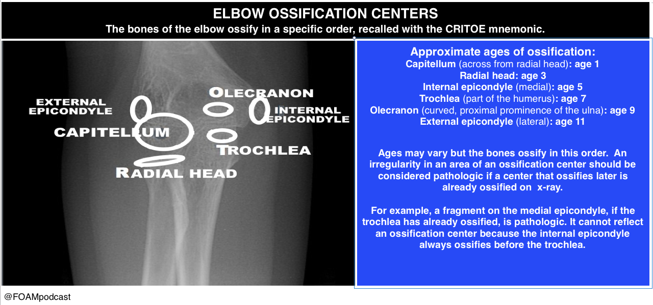
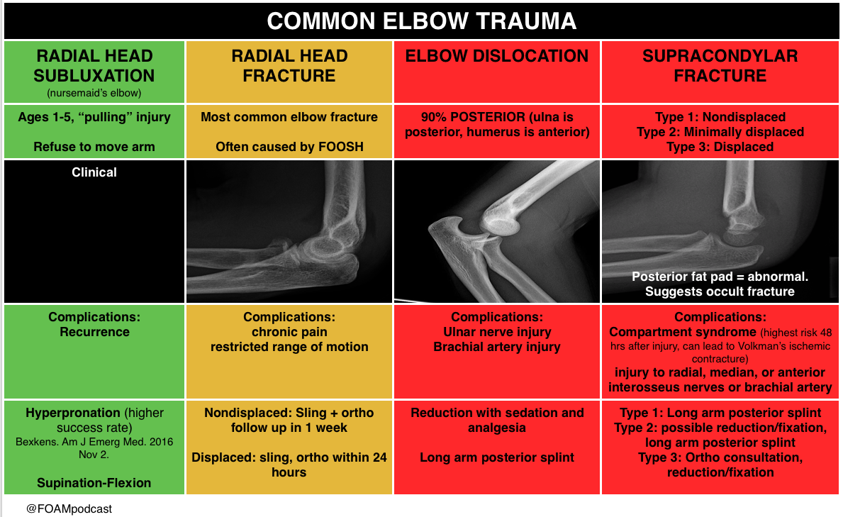
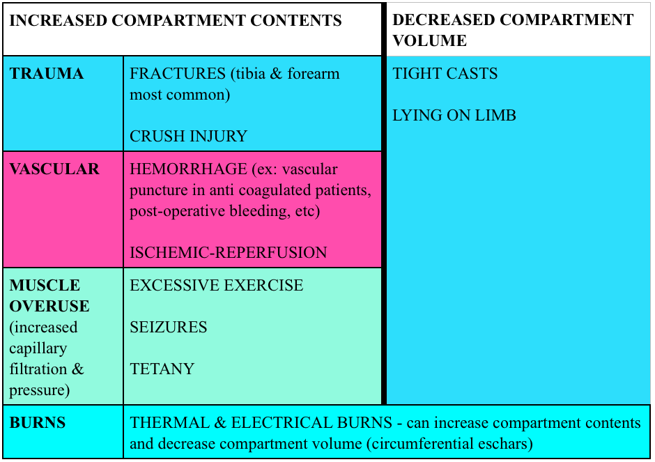
We delve into core content on other elbow adjacent injuries using Rosen’s Emergency Medicine (8th edition) Chapter  and Tintinalli’s Emergency Medicine (8th edition) Chapter  as a guide.

Elbow Trauma

olecranon bursitis

 Rosh Review Emergency Board Review Questions

A 63-year-old man presents with left arm pain after a fall. His X-ray is shown below. What structure is commonly injured with this fracture?

A. Axillary nerve

B. Median nerve

C. Radial nerve

D. Ulnar nerve

[accordion]
[toggle title=”Answer” state=”closed”]

C. Radial nerve injury is the most common nerve injury seen after humeral shaft fractures. These fractures usually occur from a direct blow to the arm and can be seen in falls and motor vehicle collisions. Patients present with severe pain, arm swelling and decreased range of motion. The arm can be shortened or rotated in a complete fracture depending on the location of the fracture. A complete neurovascular exam should be performed as with all fractures and dislocations. The radial nerve may be injured during humeral fracture in up to 20% of patients. The injury is usually a neuropraxia and resolves spontaneously in most patients. However, this recovery can take months. Humeral fractures rarely need specific reduction maneuvers for treatment. They should be placed in a sugar tong splint and placed in a sling. Gravity alone is typically successful in fracture reduction. The axillary nerve (A) may be injured during glenohumeral dislocations. The median nerve (B) may be injured during posterior elbow dislocations. Anterior elbow dislocations can be associated with ulnar nerve injury (D).

[/toggle]
[/accordion]


A 3-year-old girl was walking on the sidewalk with her mom when she fell onto the street. In a panicked state, her mom picked up the little girl by her arm. Immediately after, the little girl refused to move her right arm complaining that it hurt. In the emergency room, the girl is holding her right arm in a flexed, pronated, and adducted position. There is no crepitus, swelling or point tenderness along the entire right arm or clavicle. Which of the following is the next step in management of this patient?

A. Actively supinate and flex the elbow while applying pressure over the radial head

B. Consult orthopedics for casting

C. Obtain an ultrasound

D. Perform a skeletal survey

[accordion]
[toggle title=”Answer” state=”closed”]

A. This child has “nursemaid’s elbow” that is due to subluxation of the annular ligament rather than dislocation of the radial head. The etiology is slippage of the head of the radius under the annular ligament. The distal attachment of the annular ligament covering the radial head is weaker in children than in adults, allowing it to be more easily torn. It occurs in toddlers due to traction via pulling on a pronated and extended arm. The child immediately refuses to the move the arm and often cradles the affected arm. Parents often give a history of a young child with no history of trauma who suddenly refuses to use an arm. The diagnosis is clinical and imaging studies are generally not needed. If reduction is unsuccessful after 2–3 attempts then imaging studies may be warranted. Treatment is manual reduction via supination and flexion or hyperpronation. A palpable click may be felt and the child usually regains immediate movement of the arm and relief of discomfort. A skeletal survey (D) should be obtained in all cases of suspected child abuse to assess for fractures in multiple stages of healing. Child abuse should be on the differential in all pediatric orthopedic cases. Consulting orthopedics for casting (B) is not necessary as this is a dislocation injury. Ultrasonography (C) has been used as a noninvasive modality to assess for annular ligamentous injury and displacement of the radial head from the capitellum. It has also been used to assess progress of treatment for patients with recurrent subluxations, however it is not the first-line diagnostic nor treatment option.

[/toggle]
[/accordion]

Episode 40 – Femoral Nerve Blocks & Compartment Syndrome

(ITUNES OR LISTEN HERE)

The Free Open Access Medical Education (FOAM)

We cover one of Dr. Ken Milne’s podcasts on The Skeptic’s Guide to Emergency Medicine on regional anesthesia for hip fractures.  They cover a systematic review by Ritcey et al that found a reduction in pain scores in adult patients with femoral neck or hip fractures receiving one of the following means of regional anesthesia:

Why may this be important?

  • Better pain control for uncomfortable patients. Many patients with hip and femoral neck fractures are elderly. As such, opioids may be underdosed or pain medications may be used sparingly in these patients. Further, patients may still have pain with transfers.
  • Reduction in opioids. Opioids relieve pain but often have deleterious side effects. In addition to hypotension and allergic reactions, opioids may cause delirium.

Why don’t we do it? As a knowledge translation project, Dr. Milne’s podcast aims to propagate the best available, clinically relevant information to practitioners to mitigate the knowledge translation gap as much of the other world provides these blocks routinely to suspected fractures. A study by Haslam and colleagues from Canada suggests that while emergency providers know regional anesthesia is good in this scenario there are other barriers to adoption in North America [2].  Systemic barriers exist and include consultant worries about compartment syndrome, which is largely unfounded [3].  See this post for a more in-depth exploration.

Core Content

Compartment Syndrome – Tintinalli (7e) Chapter 275, “Compartment Syndrome.” Rosen’s  (8e) Chapter 49, “General Principles of Orthopedic Injuries.”

Compartment syndrome is typically caused by too much pressure in a confined space. Compartments are often surrounded by fascia and tissue with limited ability to stretch.  When the volume in the compartment increases or external forces compresses the compartment, blood flow in and out of the compartment are compromised.  Certain areas of the body are more predisposed to compartment syndrome (classics are the lower leg in tibia fractures and the forearm compartments).

Causes: Most cases of compartment syndrome are caused by fractures (~75%) but certainly not all.

Screen Shot 2015-12-23 at 8.04.41 AM

Diagnosis:

  • Clinical.  Classically this was taught as the 5 P’s (pain, parethesias, pallor, pulselessness, poikilothermia).  Yet, many of these perform poorly in real life.
  • Compartment pressures.  Many commercial devices exist to measure compartment pressures, yet these readings have poor specificity for compartment syndrome.
    • Historically, compartment pressures >30 mmHg or a delta pressure (Diastolic pressure minus the compartment pressure) <20-30 mmHg has indicated compartment syndrome.
    • Studies have measured compartment pressures on long bone fracture patients NOT suspected of having compartment syndrome and found that acting based on pressures would have resulted in 24-35% overdiagnosis (depending on a delta pressure cut-off of 20 mmHg and 30 mmHg respectively) [4-5]

Treatment:

  • Fasciotomy. Suspect compartment syndrome? Call surgery. In the the meantime, remove constrictive bands or clothing, give analgesia, place the limb in a dependent position, assess for rhabdomyolysis.

More FOAM on compartment syndrome: CoreEM

Generously Donated Rosh Review Questions 

1. A 24-year-old long distance athlete was brought to the emergency room complaining of severe leg pain in his left anterior lower leg. The pain became worse while running his ultra marathon yesterday and subsided after he finished. However he went for a run today and the pain returned. He describes the pain as a burning, tight pain that is 10/10. On physical exam he is in exquisite discomfort. There are no signs of trauma or broken bones. The pain is worsened on passive stretching of the leg. On palpation of his legs there is a firm wooden feeling. Distal pulses are palpated however his left leg does appear very pale. He has diminished 2-point sensory discrimination in his left leg compared to his right leg. [polldaddy poll=9241018]

2. You obtain a radiograph of a patient who was in a MVC. His GCS is 15. While being observed in the ED, the patient requests increasing doses of pain medication and is complaining of a deep, burning, unrelenting pain to his left lower extremity. He also states that he now feels tingling in his calf. [polldaddy poll=9241033]

Answers.

  1. Compartment syndrome is a serious emergency complication that should be considered whenever pain and paresthesias occur in an extremity after a fracture within an enclosed osseofascial space. It is caused by increased pressure within the compartment space that prevents adequate tissue perfusion. Compartment syndrome is most commonly associated with closed long-bone fractures of the tibia, but it can occur with isolated soft tissue trauma and even in open fractures. It has been described in a variety of situations such as prolonged procedures in the lithotomy position, the tuck position (knees tucked to chest for lumbar surgery), bedridden patients, from a spontaneous hemorrhage, and even the application of excessive traction in the reduction of a fracture. Compression dressings (A) and external wrappings should be avoided; increased compression will worsen perfusion. Elevating the limb (B) results in reduction in the local arteriovenous gradient and may be counterproductive and exacerbate compartment syndrome. It is best to keep the extremity level or slightly elevated (<10 degrees). Pressures <30 mmg Hg (C) generally do not produce compartment syndrome. The best measure of adequate limb perfusion, however, is not absolute compartment pressure but rather the differential between diastolic blood pressure and absolute compartment pressure. A pressure differential <30 mm Hg is considered by most to be an indication for emergent fasciotomy. However, with strong clinical suspicion, a fasciotomy may be required at any pressure differential as this is largely a clinical diagnosis. The initial presentation of compartment syndrome (E) usually begins with pain with passive stretching of the muscle groups, paresthesias with decreased sensation, and pain that is out of proportion to exam. Pallor and the loss of pulses are late and ominous findings.
  2. Compartment syndrome is a serious emergency complication that should be considered whenever pain and paresthesias occur in an extremity after a fracture within an enclosed osseofascial space. It is caused by increased pressure within the compartment space that prevents adequate tissue perfusion. Compartment syndrome is most commonly associated with closed long-bone fractures of the tibia, but it can occur with isolated soft tissue trauma and even in open fractures. It has been described in a variety of situations such as prolonged procedures in the lithotomy position, the tuck position (knees tucked to chest for lumbar surgery), bedridden patients, from a spontaneous hemorrhage, and even the application of excessive traction in the reduction of a fracture. Compression dressings (A) and external wrappings should be avoided; increased compression will worsen perfusion. Elevating the limb (B) results in reduction in the local arteriovenous gradient and may be counterproductive and exacerbate compartment syndrome. It is best to keep the extremity level or slightly elevated (<10 degrees). Pressures <30 mmg Hg (C) generally do not produce compartment syndrome. The best measure of adequate limb perfusion, however, is not absolute compartment pressure but rather the differential between diastolic blood pressure and absolute compartment pressure. A pressure differential <30 mm Hg is considered by most to be an indication for emergent fasciotomy. However, with strong clinical suspicion, a fasciotomy may be required at any pressure differential as this is largely a clinical diagnosis. The initial presentation of compartment syndrome (E) usually begins with pain with passive stretching of the muscle groups, paresthesias with decreased sensation, and pain that is out of proportion to exam. Pallor and the loss of pulses are late and ominous findings.

References:

  1. “Hip and Femur Injuries.” Chapter 270. Tintinalli’s Emergency Medicine: A Comprehensive Study Guide.
  2. Haslam L, Lansdown A, Lee J, et al. Survey of Current Practices: Peripheral Nerve Block Utilization by ED Physicians for Treatment of Pain in the Hip Fracture Patient Population. Canadian geriatrics journal : CGJ. 2013;16(1):16–21.
  3.  Karagiannis G, Hardern R. Best evidence topic report. No evidence found that a femoral nerve block in cases of femoral shaft fractures can delay the diagnosis of compartment syndrome of the thigh. Emerg Med J. 2005 Nov;22(11):814.Bhalla MC, Dick-Perez R. Exercise Induced Rhabdomyolysis with Compartment Syndrome and Renal Failure. Case Reports in Emergency Medicine. 2014:1-3. 2014.
  4. Whitney A, O’Toole RV, Hui E. Do one-time intracompartmental pressure measurements have a high false-positive rate in diagnosing compartment syndrome? The journal of trauma and acute care surgery. 76(2):479-83. 2014.
  5. Nelson JA. Compartment pressure measurements have poor specificity for compartment syndrome in the traumatized limb. The Journal of emergency medicine. 44(5):1039-44. 2013.

Episode 22 – The Knee

(ITUNES OR LISTEN HERE)

The Free Open Access Medical Education (FOAM)

This week we’re covering a post from the incredible pediatric resource, Don’t Forget the Bubbles, “Knee X-ray Interpretation” by Dr. Tessa Davis.  We use a systematic approach to assessing chest x-rays, so why not knee x-rays?

  •  Know the anatomy
  •  Look at:
    • Effusion
    • Main bones
    • Tibiofemoral alignment
    • Tibial plateaus
    • Intercondylar eminence
    • Patellar tendon disruption
    • Patellar fracture

The Bread and Butter

We summarize some key topics from Rosenalli, that’s Tintinalli (7e) Chapter s271, 281; Rosen’s (8e) Chapters 57, 136.  But, don’t just take our word for it.  Go enrich your fundamental understanding yourself.

Knee Dislocation

  • Anterior is most common (40%), posterior (33%)
  • Approximately 50% of knee dislocations may be relocated upon presentation to the hospital (this does not reduce risk of badness)
  • Most worrisome sequelae = popliteal artery disruption.  Of patients with popliteal disruption, the amputation rate rises to 90% 8 hours after the injury without surgical intervention.
  • Workup may depend on your institution (ex: angiogram vs. CT angio vs. ultrasound) but all patients will need an ABI + 24 hour of pulse checks per current standards.
Screen Shot 2015-01-08 at 1.10.48 PM
Algorithm (adopted from Rosen’s)

Septic Arthritis

  • Most Common Organisms: S. aureus, N. gonorrhea
  • Hematogenous spread
  • Most Common Location: knee, hip

Risk factors such as immunocompromised hosts and use of steroids are risk factors for septic arthritis but the ones with the highest likelihood ratio (LR+ >10 is ideal):

  • Skin infection overlying prosthetic joint (LR+ 15)
  • Joint surgery within the preceding 3 months (LR+ 6.9)
  • Age > 80 (LR+ 3.5)

Diagnosis:  In the red, hot, swollen, painful joint, think septic arthritis.  Clinical and laboratory indicators aren’t great. Synovial fluid analysis, particularly the culture exists as the gold standard.  Arthrocentesis Trick of the Trade from ALiEM. Here are the operating characteristics from Margaretten et al:

  • Fever: Sensitivity 57%
  • Lab tests: White Blood Cell count (WBC), sedimentation rate (ESR), and c-reactive protein don’t perform well
    • WBC LR+  1.4 (1.1-1.8); LR- 0.28 (0.07-1.10)
    • Erythrocyte sedimentation rate 1.3 (1.1-1.8); LR- 0.17 (0.20-1.30)
    • C-reactive protein  1.6 (1.1-2.5); LR- 0.44 (0.24-0.82)
  • Synovial fluid gram stain and culture is the “gold standard.”

Treatment: Intravenous antibiotics and washout of the joint by orthopedics in the operating room

 Generously Donated Rosh Review Questions 

Question 1. A 67-year-old man with a history of gout presents with atraumatic left knee pain. Physical examination reveals an effusion with overlying warmth and erythema. There is pain with passive range of motion. He reports a history of gout in this joint in the past. [polldaddy poll=8568492]

Question 2.  A 27-year-old woman presents with severe left knee pain after an MVC where she was the front passenger. She states her knee hit the dashboard. An X-ray of the patient’s knee is shown below. After reduction, the physical examination reveals swelling of the knee and an Ankle-Brachial Index (ABI) of 0.8. [polldaddy poll=8569540]

Screen Shot 2015-01-12 at 5.13.06 PM

Answers.

1. D. Septic arthritis is a bacterial or fungal infection of a joint typically spread hematogenously unless there is direct bacterial http://www.mindanews.com/buy-imitrex/ contamination. The synovium is highly vascular and lacks a basement membrane making it susceptible to bacterial seeding. Certain conditions predispose individuals to septic arthritis including diabetes, sickle cell disease, immunocompromise, alcoholism or pre-existing joint disease like rheumatoid arthritis or gout. Fever is present in less than half of cases of septic arthritis so with clinical suspicion an arthrocentesis is indicated. The knee is the most common joint affected and patients have pain (especially on passive range of motion) and decreased range of motion often accompanied by warmth, erythema and fever. This patient may have an acute gouty flare, but the clinician must exclude an infection. On joint fluid analysis, the white blood cell count of a septic joint is typically > 50,000. Indomethacin (B) is a non-steroidal anti-inflammatory agent commonly used in the treatment of acute gout. Gout is an arthritis caused by deposition of monosodium urate monohydrate crystals in the joint space. Acute flares involve a monoarticular arthritis with a red, hot, swollen and tender joint. Acute episodes of gout result from overproduction or decreased secretion of uric acid. However, measurement of serum uric acid (C) does not correlate with the presence of absence of an acute flare. A radiograph of the knee (D) may show chronic degenerative changes associated with gout but will not help to differentiate a gouty arthritis versus septic arthritis.

2. C. Obtain Angiography. This patient presents with a knee dislocation and signs of a popliteal artery injury requiring angiography for diagnosis. A knee dislocation refers to a dislocation of the tibia in relation to the femur and not a patellofemoral dislocation. A tibiofemoral dislocation is a limb-threatening emergency due to the high rate of popliteal artery injury. The neurovascular bundle (popliteal artery, popliteal vein and common peroneal nerve) runs posteriorly in the popliteal fossa. The popliteal artery is tethered to the femur and tibia by a fibrous tunnel and is inherently immobile making it susceptible to injury during dislocation. Knee dislocations typically occur in major trauma. An MVC where the knee strikes the dashboard is a common scenario. The dislocation is usually clinically obvious and should be emergently reduced regardless of the presence of confirmatory X-rays. The leg should rapidly be assessed for any “hard” signs of vascular injury including an absence of pulse, limb ischemia, rapidly expanding hematoma, the presence of a bruit or thrill and pulsatile bleeding. Neurologic status should also be assessed prior to and after reduction. After reduction, all patients should have ankle-brachial index (ABI) performed. A normal ABI is > 0.9. Any patient with an ABI less than this should be further investigated for a popliteal injury with angiography. Splint and elevation (D) may be appropriate once a vascular injury is ruled out. The patient should not be discharged home (A) with an abnormal ABI. Observation and repeat ABI (B) is indicated if the initial ABI is normal.

Episode 22 – The Knee

(ITUNES OR LISTEN HERE)

The Free Open Access Medical Education (FOAM)

This week we’re covering a post from the incredible pediatric resource, Don’t Forget the Bubbles, “Knee X-ray Interpretation” by Dr. Tessa Davis.  We use a systematic approach to assessing chest x-rays, so why not knee x-rays?

  •  Know the anatomy
  •  Look at:
    • Effusion
    • Main bones
    • Tibiofemoral alignment
    • Tibial plateaus
    • Intercondylar eminence
    • Patellar tendon disruption
    • Patellar fracture

The Bread and Butter

We summarize some key topics from Rosenalli, that’s Tintinalli (7e) Chapter s271, 281; Rosen’s (8e) Chapters 57, 136.  But, don’t just take our word for it.  Go enrich your fundamental understanding yourself.

Knee Dislocation

  • Anterior is most common (40%), posterior (33%)
  • Approximately 50% of knee dislocations may be relocated upon presentation to the hospital (this does not reduce risk of badness)
  • Most worrisome sequelae = popliteal artery disruption.  Of patients with popliteal disruption, the amputation rate rises to 90% 8 hours after the injury without surgical intervention.
  • Workup may depend on your institution (ex: angiogram vs. CT angio vs. ultrasound) but all patients will need an ABI + 24 hour of pulse checks per current standards.
Screen Shot 2015-01-08 at 1.10.48 PM
Algorithm (adopted from Rosen’s)

Septic Arthritis

  • Most Common Organisms: S. aureus, N. gonorrhea
  • Hematogenous spread
  • Most Common Location: knee, hip

Risk factors such as immunocompromised hosts and use of steroids are risk factors for septic arthritis but the ones with the highest likelihood ratio (LR+ >10 is ideal):

  • Skin infection overlying prosthetic joint (LR+ 15)
  • Joint surgery within the preceding 3 months (LR+ 6.9)
  • Age > 80 (LR+ 3.5)

Diagnosis:  In the red, hot, swollen, painful joint, think septic arthritis.  Clinical and laboratory indicators aren’t great. Synovial fluid analysis, particularly the culture exists as the gold standard.  Arthrocentesis Trick of the Trade from ALiEM. Here are the operating characteristics from Margaretten et al:

  • Fever: Sensitivity 57%
  • Lab tests: White Blood Cell count (WBC), sedimentation rate (ESR), and c-reactive protein don’t perform well
    • WBC LR+  1.4 (1.1-1.8); LR- 0.28 (0.07-1.10)
    • Erythrocyte sedimentation rate 1.3 (1.1-1.8); LR- 0.17 (0.20-1.30)
    • C-reactive protein  1.6 (1.1-2.5); LR- 0.44 (0.24-0.82)
  • Synovial fluid gram stain and culture is the “gold standard.”

Treatment: Intravenous antibiotics and washout of the joint by orthopedics in the operating room

 Generously Donated Rosh Review Questions 

Question 1. A 67-year-old man with a history of gout presents with atraumatic left knee pain. Physical examination reveals an effusion with overlying warmth and erythema. There is pain with passive range of motion. He reports a history of gout in this joint in the past. [polldaddy poll=8568492]

Question 2.  A 27-year-old woman presents with severe left knee pain after an MVC where she was the front passenger. She states her knee hit the dashboard. An X-ray of the patient’s knee is shown below. After reduction, the physical examination reveals swelling of the knee and an Ankle-Brachial Index (ABI) of 0.8. [polldaddy poll=8569540]

Screen Shot 2015-01-12 at 5.13.06 PM

Answers.

1. D. Septic arthritis is a bacterial or fungal infection of a joint typically spread hematogenously unless there is direct bacterial contamination. The synovium is highly vascular and lacks a basement membrane making it susceptible to bacterial seeding. Certain conditions predispose individuals to septic arthritis including diabetes, sickle cell disease, immunocompromise, alcoholism or pre-existing joint disease like rheumatoid arthritis or gout. Fever is present in less than half of cases of septic arthritis so with clinical suspicion an arthrocentesis is indicated. The knee is the most common joint affected and patients have pain (especially on passive range of motion) and decreased range of motion often accompanied by warmth, erythema and fever. This patient may have an acute gouty flare, but the clinician must exclude an infection. On joint fluid analysis, the white blood cell count of a septic joint is typically > 50,000. Indomethacin (B) is a non-steroidal anti-inflammatory agent commonly used in the treatment of acute gout. Gout is an arthritis caused by deposition of monosodium urate monohydrate crystals in the joint space. Acute flares involve a monoarticular arthritis with a red, hot, swollen and tender joint. Acute episodes of gout result from overproduction or decreased secretion of uric acid. However, measurement of serum uric acid (C) does not correlate with the presence of absence of an acute flare. A radiograph of the knee (D) may show chronic degenerative changes associated with gout but will not help to differentiate a gouty arthritis versus septic arthritis.

2. C. Obtain Angiography. This patient presents with a knee dislocation and signs of a popliteal artery injury requiring angiography for diagnosis. A knee dislocation refers to a dislocation of the tibia in relation to the femur and not a patellofemoral dislocation. A tibiofemoral dislocation is a limb-threatening emergency due to the high rate of popliteal artery injury. The neurovascular bundle (popliteal artery, popliteal vein and common peroneal nerve) runs posteriorly in the popliteal fossa. The popliteal artery is tethered to the femur and tibia by a fibrous tunnel and is inherently immobile making it susceptible to injury during dislocation. Knee dislocations typically occur in major trauma. An MVC where the knee strikes the dashboard is a common scenario. The dislocation is usually clinically obvious and should be emergently reduced regardless of the presence of confirmatory X-rays. The leg should rapidly be assessed for any “hard” signs of vascular injury including an absence of pulse, limb ischemia, rapidly expanding hematoma, the presence of a bruit or thrill and pulsatile bleeding. Neurologic status should also be assessed prior to and after reduction. After reduction, all patients should have ankle-brachial index (ABI) performed. A normal ABI is > 0.9. Any patient with an ABI less than this should be further investigated for a popliteal injury with angiography. Splint and elevation (D) may be appropriate once a vascular injury is ruled out. The patient should not be discharged home (A) with an abnormal ABI. Observation and repeat ABI (B) is indicated if the initial ABI is normal.