#dasSMACC – Pulmonary Embolism, Pulmonary Edema, TEE, and Pediatric Cardiology

ITUNES OR LISTEN HERE

We are in Berlin for #dasSMACC and have lots of pearls to share from the speakers at this amazing conference.  Talks will be released for free on the SMACC podcast over the next year, but this podcast holds some pearls that we thought couldn’t wait.

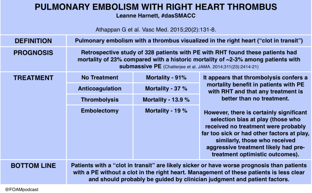
Dr. Leanne Harnett on Pulmonary Embolism with Right Heart Thrombus (PE with RHT)

das smacc pe

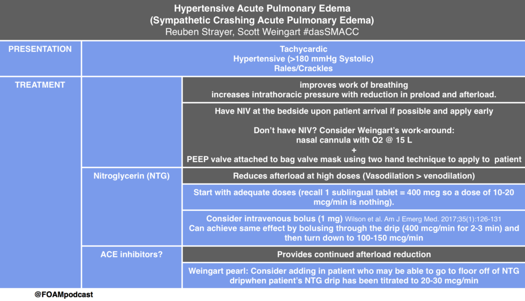
Drs. Reuben Strayer (@emupdates) and Scott Weingart (@emcrit) on Acute Hypertensive Pulmonary Edema

Screen Shot 2017-06-29 at 11.39.15 AM

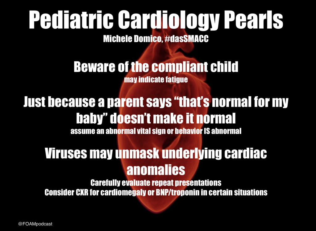
Dr. Michele Domico on Pediatric Cardiology Emergencies

Pediatric Cardiology dasSMACC

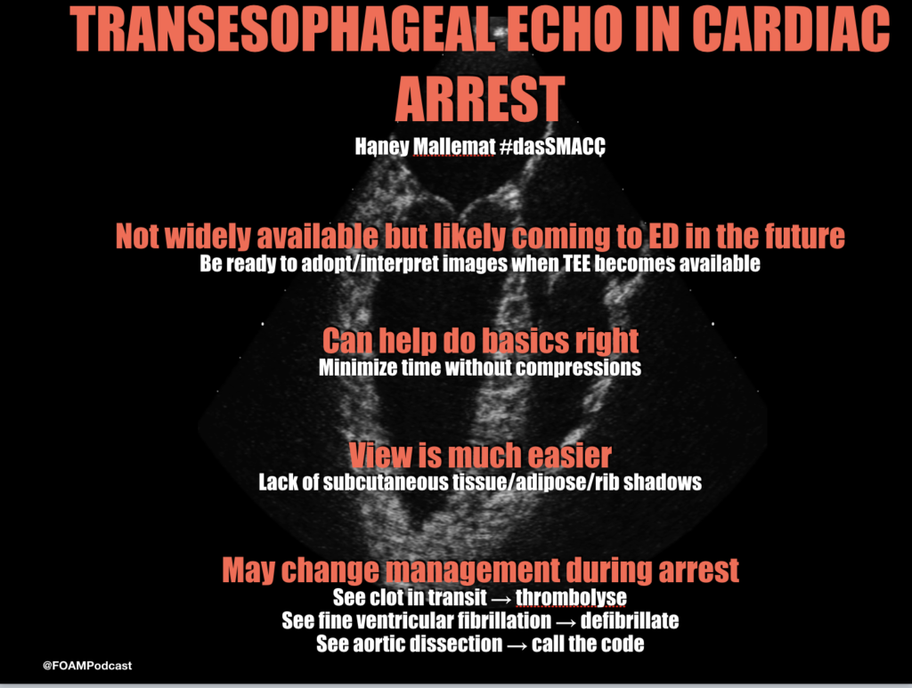
Dr. Haney Mallemat (@CriticalCareNow) on Transesophageal Echocardiography (TEE) during Cardiac Arrest

TEE

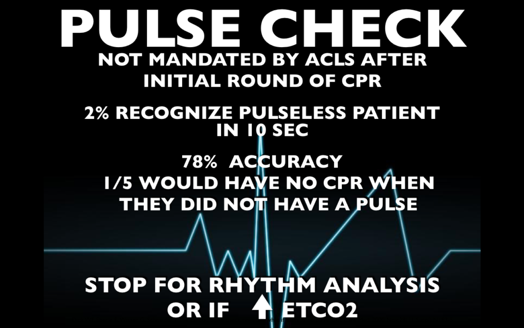
pulsecheck2

For more on pulse checks, see this blog post.

References:

  1. Athappan G, Sengodan P, Chacko P, Gandhi S. Comparative efficacy of different modalities for treatment of right heart thrombi in transit: a pooled analysis. Vasc Med. 2015;20(2):131-8.
  2. Wilson SS, Kwiatkowski GM, Millis SR, Purakal JD, Mahajan AP, Levy PD. Use of nitroglycerin by bolus prevents intensive care unit admission in patients with acute hypertensive heart failure. Am J Emerg Med. 2017;35(1):126-131.

Episode 67 – Serious Pediatric Fever

iTunes or Listen Here

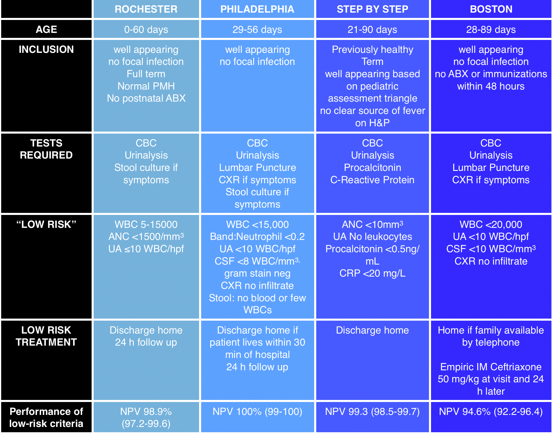
We cover an episode of The Skeptic’s Guide to Emergency Medicine that covers a validation study of the Step by Step approach to pediatric fever.  This approach to infants with a fever <3 months old is alluring as it does not necessitate a lumbar puncture.  This algorithm had a better sensitivity and negative predictive value than the Rochester criteria.   The approach did miss some infants with a serious bacterial infection and these tended to be those between 21 and 28 days old and those with fever onset <2 hours prior to arrival.
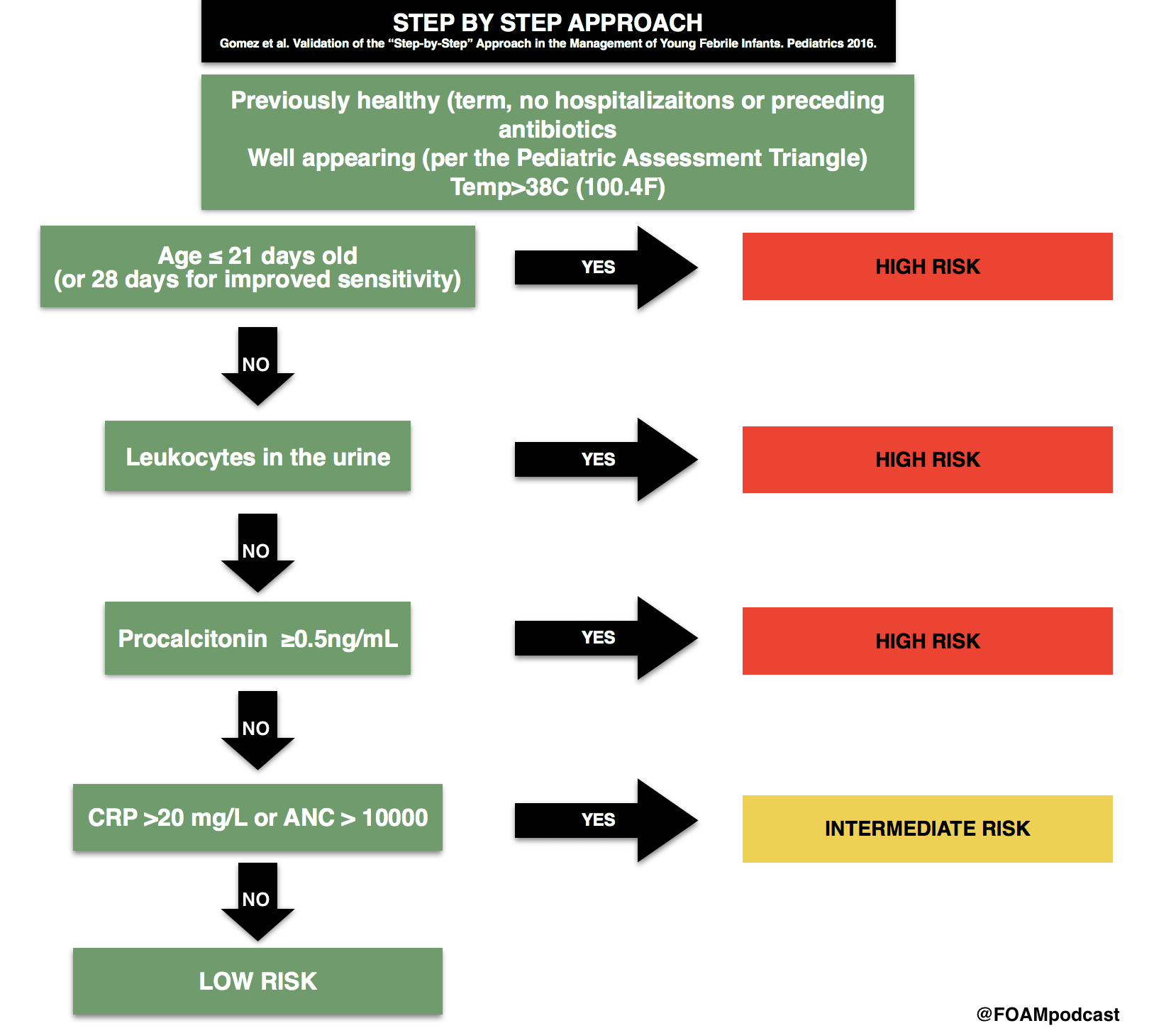
Step by Step for Pedi Fever

 Core Content

We cover Chapter 116 in Tintinalli’s Emergency Medicine (8th ed) and Rosen’s on pediatric fever.

Infants <3 months old with fever algorithms
Screen Shot 2017-03-25 at 3.06.45 PM

Rosh Review Emergency Board Review Questions

A 25-day-old female presents with fever and cough. Mom denies any symptoms at home. The patient’s 2-year-old brother had a cough and rhinorrhea 1 week prior. On exam, the patient’s temperature is 38.7°C with clear lungs, a benign abdomen, and normal tympanic membranes bilaterally. What is the appropriate workup for this patient?

  1. CBC, chest X-ray
  2. CBC, chest X-ray, urinalysis
  3. CBC, chest X-ray, urinalysis, blood cultures
  4. CBC, chest X-ray, urinalysis, blood cultures, lumbar puncture

[accordion]
[toggle title=”Answer” state=”closed”]

4.  Neonates with fever aged 28 days or younger may have few clues on history and physical examination to guide therapy. Therefore, a high index of suspicion is necessary to detect the febrile neonate with a serious bacterial infection. Obtaining the pertinent medical history from the mother regarding the pregnancy, delivery, and early neonatal life of the febrile neonate is essential. Typically, infections in the 1st week of life are secondary to vertical transmission, and those infections after the 1st week are usually community acquired or hospital acquired. Bacterial meningitis is more common in the 1st month of life than at any other time. An estimated 5%–10% of neonates with early onset group B streptococcal (GBS) sepsis have concurrent meningitis. Therefore, febrile infants (temperature >38°C) younger than 28 days should receive a full sepsis workupCBC, chest X-ray (A), urinalysis (B), and blood cultures (C) are a partial workup for neonatal fever.

[/toggle]
[/accordion]

A two-day-old boy presents to the ED with fever for the past four hours. His birth history includes a normal spontaneous vaginal delivery at term. Parents report noticing that the child “felt warm,” and that he was having copious nasal secretions while feeding. On physical examination, the child appears lethargic, has mottled extremities, and is hot to the touch. Breath sounds are clear bilaterally, and there are no rashes. His vital signs are T 102.9°F, BP 74/48 mm Hg, HR 170 beats per minute, and RR 40 breaths per minute. Which of the following groupings of organisms should your antibiotic choices cover when treating this febrile neonate?

  1. Listeria monocytogenes, Group B streptococcus, Escherichia coli
  2. Mycoplasma pneumoniae, Neisseria meningitidis, Streptococcus pneumoniae
  3. Neisseria meningitidis, Listeria monocytogenes, Streptococcus pneumoniae
  4. Streptococcus pneumoniae, Neisseria meningitidis, Haemophilus influenzae
    [accordion]
    [toggle title=”Answer” state=”closed”]

Answer 1. The febrile neonate is a child 28 days and younger who presents with a fever. These children are at very high risk of serious bacterial infections, including urinary tract infection, pneumonia, meningitis, and bacteremia. Risk factors for serious bacterial infection in a neonate include prematurity, low birth weight, premature or prolonged rupture of membranes, meconium aspiration, or maternal group B streptococcus infection. The evaluation of a neonate with a fever includes CBC, urinalysis, blood culture, urine culture, and a lumbar puncture in order to obtain CSF for cell count, Gram stain, and culture. If the child has respiratory symptoms, a chest X-ray should be performed. If the child has diarrhea, stool testing should also be performed. The most common pathogens involved in serious bacterial infections, including meningitis and bacteremia, in neonates are Listeria monocytogenes, Group B streptococcus, and Escherichia coli. These children can become critically ill very rapidly; therefore, initial management should include a fluid bolus of 20 mL/kg and broad-spectrum antibiotics to cover the most common pathogens in this age group. The most appropriate antibiotics to use in neonates with a fever are ampicillin and cefotaxime. Ampicillin will cover Listeria monocytogenes while cefotaxime will cover Group B streptococcus and Escherichia coli. If there is a history of maternal infection with herpes simplex virus, acyclovir should be added to the empiric broad-spectrum treatment. These patients universally need to be admitted to the hospital for IV antibiotics and observation until all cultures have returned.  Mycoplasma pneumoniae, Neisseria meningitidis, Streptococcus pneumoniae (B) are common pathogens seen in adolescents and young adults. Mycoplasma pneumoniae is a common cause of atypical pneumonia in this age group. Streptococcus pneumoniae is a common bacterial cause of pneumonia, bacteremia, and meningitis while Neisseria meningitidis is primarily a cause of meningitis. Neisseria meningitidis, Listeria monocytogenes, Streptococcus pneumoniae (C) are the primary pathogens causing serious bacterial infections in adults over the age of 65. Listeria monocytogenes is a pathogen that is seen in infants and then later reemerges as a prominent pathogen in older adults. Streptococcus pneumoniae, Neisseria meningitidis, Haemophilus influenzae (D) are the most common pathogens causing serious bacterial infections in children ages one to five years. There has been a significant decline in the incidence of Haemophilus influenzae type B in recent years due to childhood vaccination programs.

[/toggle]
[/accordion]
Episode 61 – The Elbow

iTunes or Listen Here

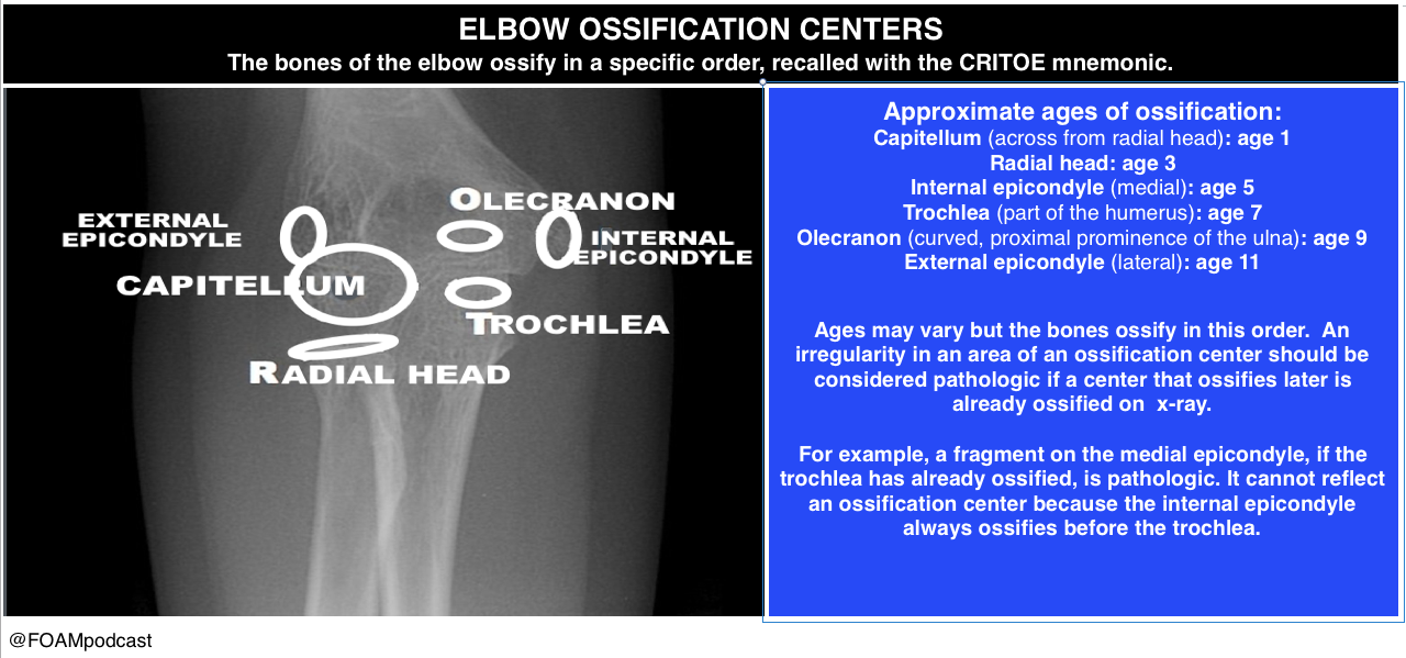
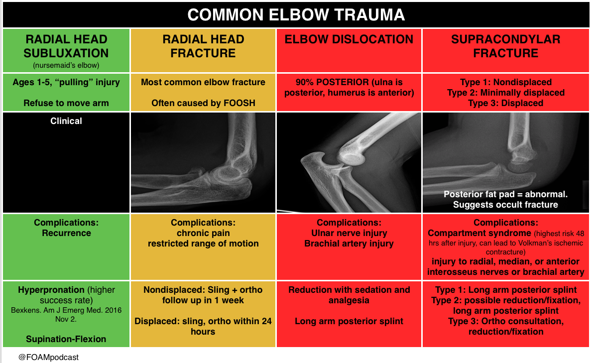
We review a podcast from Dr. Tim Horeczko’s Pediatric Emergency Playbook on elbow injuries.

critoe

Core Content

We delve into core content on other elbow adjacent injuries using Rosen’s Emergency Medicine (8th edition) Chapter  and Tintinalli’s Emergency Medicine (8th edition) Chapter  as a guide.

Elbow Trauma

olecranon bursitis

 Rosh Review Emergency Board Review Questions

A 63-year-old man presents with left arm pain after a fall. His X-ray is shown below. What structure is commonly injured with this fracture?

A. Axillary nerve

B. Median nerve

C. Radial nerve

D. Ulnar nerve

[accordion]
[toggle title=”Answer” state=”closed”]

C. Radial nerve injury is the most common nerve injury seen after humeral shaft fractures. These fractures usually occur from a direct blow to the arm and can be seen in falls and motor vehicle collisions. Patients present with severe pain, arm swelling and decreased range of motion. The arm can be shortened or rotated in a complete fracture depending on the location of the fracture. A complete neurovascular exam should be performed as with all fractures and dislocations. The radial nerve may be injured during humeral fracture in up to 20% of patients. The injury is usually a neuropraxia and resolves spontaneously in most patients. However, this recovery can take months. Humeral fractures rarely need specific reduction maneuvers for treatment. They should be placed in a sugar tong splint and placed in a sling. Gravity alone is typically successful in fracture reduction. The axillary nerve (A) may be injured during glenohumeral dislocations. The median nerve (B) may be injured during posterior elbow dislocations. Anterior elbow dislocations can be associated with ulnar nerve injury (D).

[/toggle]
[/accordion]


A 3-year-old girl was walking on the sidewalk with her mom when she fell onto the street. In a panicked state, her mom picked up the little girl by her arm. Immediately after, the little girl refused to move her right arm complaining that it hurt. In the emergency room, the girl is holding her right arm in a flexed, pronated, and adducted position. There is no crepitus, swelling or point tenderness along the entire right arm or clavicle. Which of the following is the next step in management of this patient?

A. Actively supinate and flex the elbow while applying pressure over the radial head

B. Consult orthopedics for casting

C. Obtain an ultrasound

D. Perform a skeletal survey

[accordion]
[toggle title=”Answer” state=”closed”]

A. This child has “nursemaid’s elbow” that is due to subluxation of the annular ligament rather than dislocation of the radial head. The etiology is slippage of the head of the radius under the annular ligament. The distal attachment of the annular ligament covering the radial head is weaker in children than in adults, allowing it to be more easily torn. It occurs in toddlers due to traction via pulling on a pronated and extended arm. The child immediately refuses to the move the arm and often cradles the affected arm. Parents often give a history of a young child with no history of trauma who suddenly refuses to use an arm. The diagnosis is clinical and imaging studies are generally not needed. If reduction is unsuccessful after 2–3 attempts then imaging studies may be warranted. Treatment is manual reduction via supination and flexion or hyperpronation. A palpable click may be felt and the child usually regains immediate movement of the arm and relief of discomfort. A skeletal survey (D) should be obtained in all cases of suspected child abuse to assess for fractures in multiple stages of healing. Child abuse should be on the differential in all pediatric orthopedic cases. Consulting orthopedics for casting (B) is not necessary as this is a dislocation injury. Ultrasonography (C) has been used as a noninvasive modality to assess for annular ligamentous injury and displacement of the radial head from the capitellum. It has also been used to assess progress of treatment for patients with recurrent subluxations, however it is not the first-line diagnostic nor treatment option.

[/toggle]
[/accordion]

Episode 49 – The AAP BRUE (formerly ALTE) Guidelines

(ITUNES OR LISTEN HERE)

The Free Open Access Medical Education (FOAM)

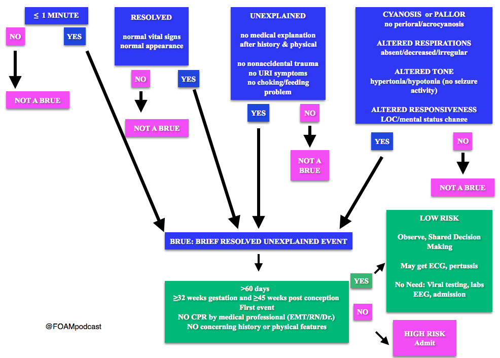
We review the American Academy of Pediatrics guideline on Brief Resolved Unexplained Events (Formerly Apparent Life-Threatening Events) and Evaluation of Lower-Risk Infants.

Screen Shot 2016-05-04 at 9.33.37 AM

Note: Some have voiced concerns that these guidelines potentially downplay the event. The concern is that the yield of these workups and admissions may be low, but possibly worthwhile.  At FOAMcast, we are not qualified to critique these guidelines but there are helpful tables and charts in them to realize that these recommendations really are only for specific events and children AFTER thorough history and physical.

Generously Donated Rosh Review Question

A 6-week-old boy is brought to the emergency room because of cyanosis. He was sleeping comfortably in a supine position right after a feeding when he suddenly choked, became limp and lips turned blue. The mother witnessed the event and blew to the face of the boy. The whole episode lasted for about two minutes. EMS was called and upon arrival at the house, the boy was back to his usual self. At the ER, he boy has normal vital signs with normal physical examination findings.

A. Admit for cardiorespiratory monitoring

B. Discharge after reassuring the parents

C. Observe for four hours in the ER

D. Request for complete blood count

[accordion]
[toggle title=”Answer” state=”closed”]

A. Admit for cardiorespiratory monitoring. The boy in the vignette had an apparent life-threatening event (ALTE) which is not a specific diagnosis but a description of an acute, unexpected episode that is frightening to the caretaker. ALTE includes one or more of the following features: apnea, color change (may be cyanotic, pallid, erythematous or plethoric), marked change in muscle tone (limpness or rigidity) and choking or gagging. A specific cause for ALTE can be identified in over one-half of patients after a careful history, physical examination, and appropriate laboratory evaluation. The remaining cases are considered idiopathic if no cause can be identified after a thorough assessment. Common etiologies for ALTE include gastroesophageal reflux, neurologic problems (such as seizures), and respiratory infection. The history of an ALTE must be taken seriously, even if the infant appears entirely well by the time he or she is evaluated. In-hospital observation with cardiorespiratory monitoring is indicated for infants whose initial evaluation suggests physiologic compromise. Hospital admission may provide important clinical information where additional episodes may be witnessed by medical personnel during the observation period. In addition, serious underlying medical conditions may become apparent.  Discharging the patient after reassuring the parents (B) and observing the patient for four hours in the ER (C) are not appropriate management strategies for the infant in the vignette who needs admission for cardiorespiratory monitoring. Requesting for complete blood count (D) is not routinely done in the evaluation of an ALTE and would not aid in the management for the infant in the vignette.

[/toggle]
[/accordion]

References:

Tieder JS, Bonkowsky JL, Etzel RA, Franklin WH, Gremse DA, Herman B et al. Subcommittee on Apparent Life Threatening Events. Brief Resolved Unexplained Events (Formerly Apparent Life-Threatening Events) and Evaluation of Lower-Risk Infants: Executive Summary.  Pediatrics. In Press