Thromboelastography (TEG) Guided Resuscitation

iTunes or Listen Here

Thromboelastography (TEG) or its related counterpart rotational thromboelastometry (ROTEM) have gained in popularity over the past several years. These tests assess viscoelastic clot strength in whole blood. These tests may offer more granular and potentially reliable information on the patient’s clot formation and fibrinolytic state than traditional measures of coagulation such as International Normalized Ratio (INR), partial thromobplastin time (PTT), and prothrombin time (PT).
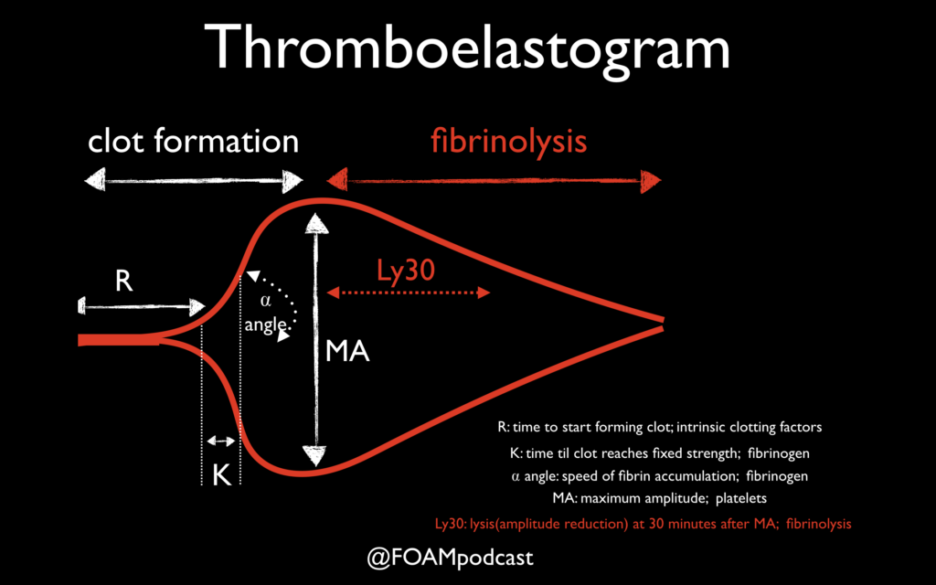
One of the primary advantages to TEG/ROTEM is the ability to target transfusion related therapies to the patient’s overall coagulation profile. Below are some common patterns that emerge and the recommended therapies.

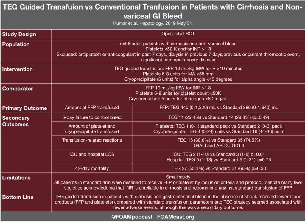
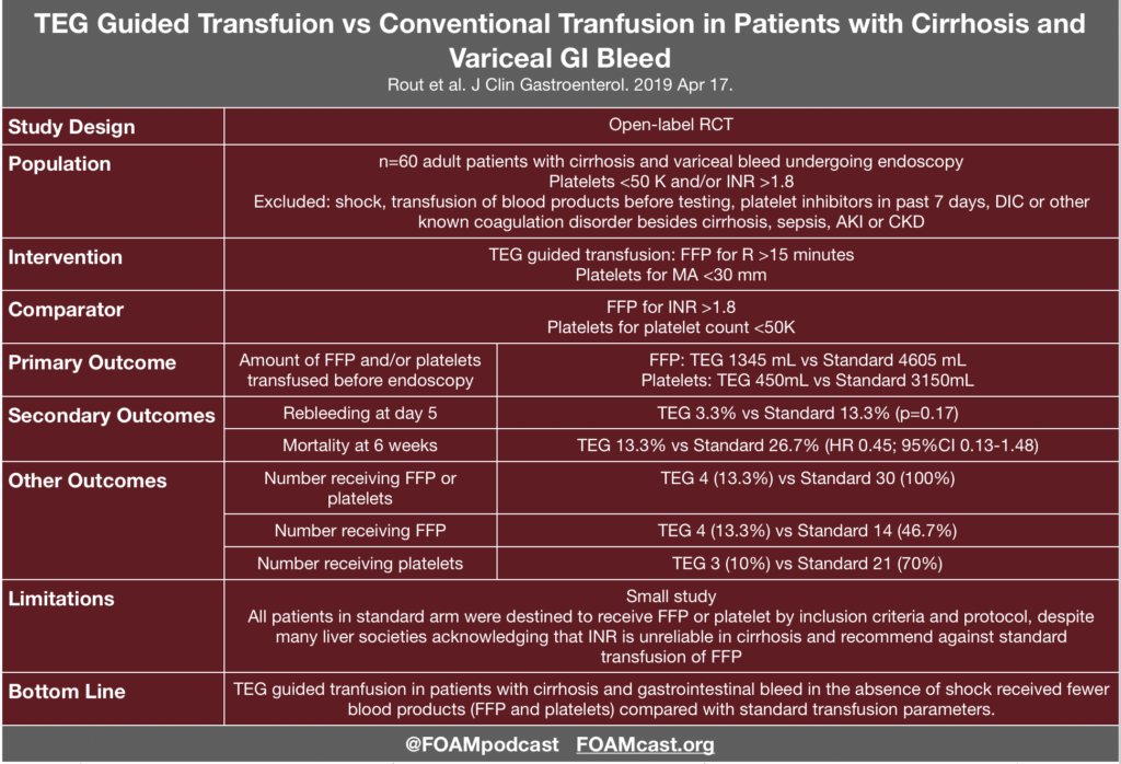
Evidence for TEG in Cirrhosis

Additional FOAM resources: PulmCrit

References:

  1. Wikkelsø A, Wetterslev J, Møller AM, Afshari A. Thromboelastography (TEG) or thromboelastometry (ROTEM) to monitor haemostatic treatment versus usual care in adults or children with bleeding. Cochrane Database Syst Rev. 2016;(8):CD007871.
  2. Kumar M, Ahmad J, Maiwall R, et al. Thromboelastography-Guided Blood Component Use in Patients With Cirrhosis With Nonvariceal Bleeding: A Randomized Controlled Trial. Hepatology. 2019; In Press
  3. Rout G, Shalimar, Gunjan D, et al. Thromboelastography-guided Blood Product Transfusion in Cirrhosis Patients With Variceal Bleeding: A Randomized Controlled Trial. J Clin Gastroenterol. 2019; In Press.
  4. Goodman MD, Makley AT, Hanseman DJ, Pritts TA, Robinson BR. All the bang without the bucks: Defining essential point-of-care testing for traumatic coagulopathy. J Trauma Acute Care Surg. 2015;79(1):117-24.

You are called to the bedside for a postoperative patient who is hypotensive, febrile, and has acute onset of respiratory distress following the initiation of a blood transfusion. Which of the following is the most appropriate initial action in the management of this patient?

A. Apply oxygen

B. Call the blood bank

C. Order steroids

D. Stop the transfusion

[accordion]
[toggle title=”Answer” state=”closed”]

Transfusion-related acute lung injury is a blood transfusion complication characterized by a rapid onset of non-cardiogenic pulmonary edema. The pathogenesis is thought to be a two-part mechanism involving neutrophil sequestration with priming in the lung microvasculature followed by neutrophil activation by a factor in the blood product. Pre-transfusion risk factors include current smoking, chronic alcohol use, liver transplantation surgery, positive fluid balance, shock, and higher ventilated peak airway pressures. Though there is an association of transfusion-related acute lung injury with all blood products, high-plasma-volume products (plasma, apheresis platelet concentrations, and whole blood) have the greatest risk. Clinical presentation may occur immediately after the initiation of the blood transfusion although it can be delayed up to six hours. The patient may rapidly develop acute respiratory distress syndrome, with symptoms that may include hypoxemia, fever, hypotension, cyanosis, pulmonary infiltrates on chest imaging, and if intubated, pink frothy secretions when suctioned. Treatment involves immediate discontinuation of the transfusion followed by supportive care of the acute respiratory distress syndrome. This includes oxygen supplementation and hemodynamic support. Apply oxygen (A), call the blood bank (B), and order steroids (C) are treatments for transfusion-related acute lung injury but are not the most appropriate in the initial management. Discontinuing the transfusion, which is the cause of the acute lung injury, is the immediate need upon identifying the condition.

[/toggle]
[/accordion]
Top Literature of 2019 – Mid Year Review

iTunes or Listen Here

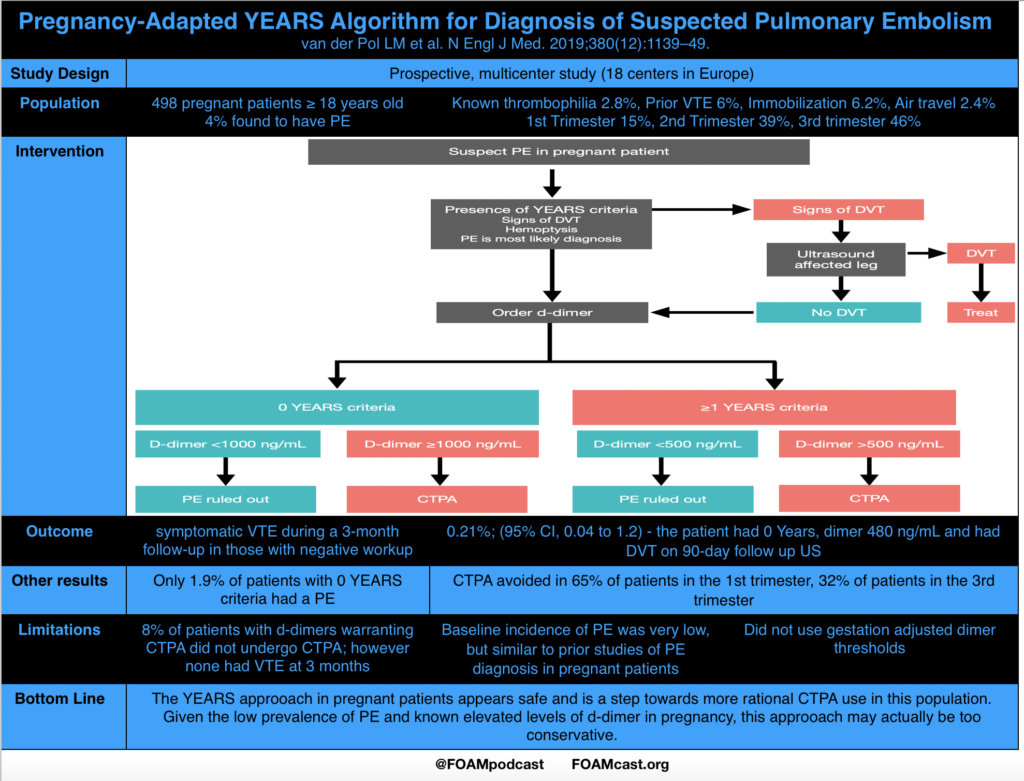
Risk Stratification and D-dimer in Pregnant Patients With Suspected Pulmonary Embolism (PE)

Van der pol LM, Tromeur C, Bistervels IM, et al. Pregnancy-Adapted YEARS Algorithm for Diagnosis of Suspected Pulmonary Embolism. N Engl J Med. 2019;380(12):1139-1149.

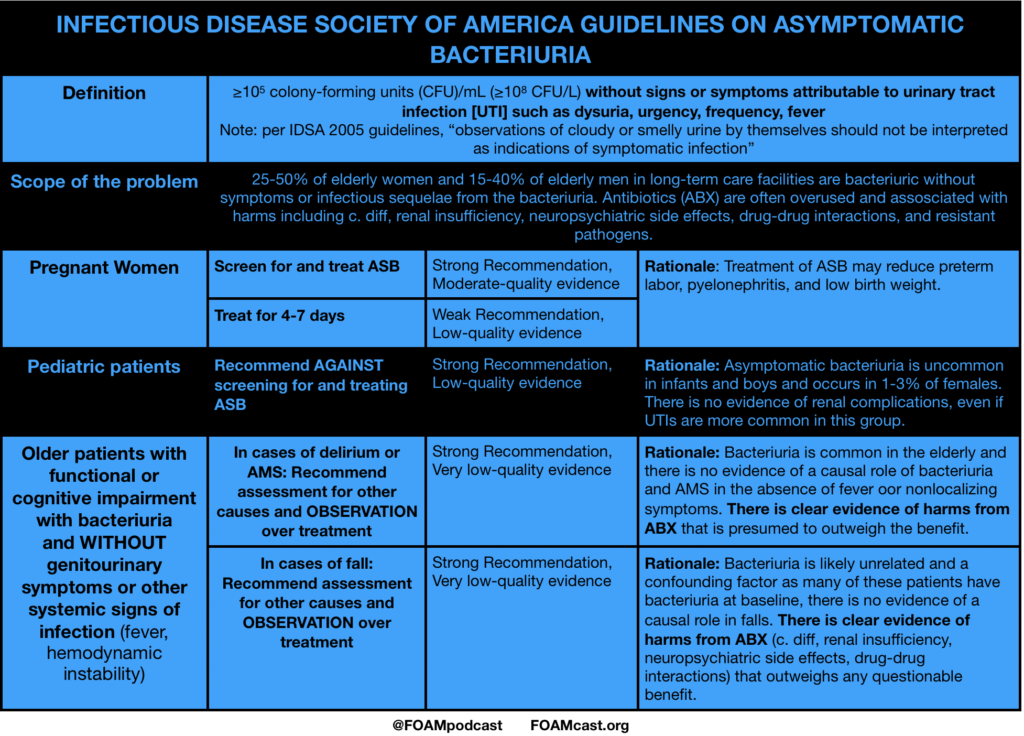
Infectious Disease Society of America (IDSA) Guidelines for Asymptomatic

Nicolle LE, Gupta K, Bradley SF, et al. Clinical Practice Guideline for the Management of Asymptomatic Bacteriuria: 2019 Update by the Infectious Diseases Society of America. Clin Infect Dis. 2019.

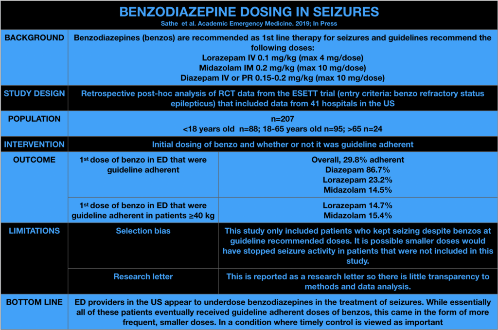
Benzodiazepine dosing for seizures

Sathe AG, Tillman C, Coles LD, et al. Underdosing of benzodiazepines in patients with status epilepticus enrolled in Established Status Epilepticus Treatment Trial. Acad Emerg Med. 2019 Jun 4.

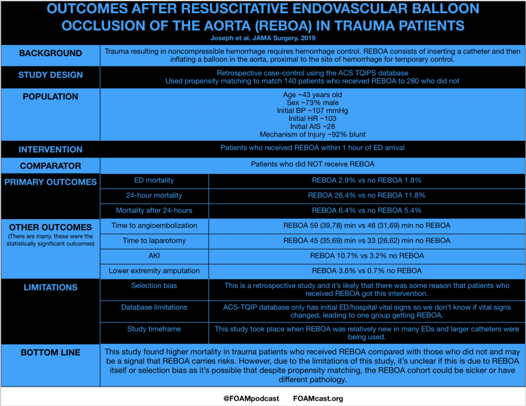
  • Outcomes after Resuscitative Endovascular Balloon Occlusion of the Aorta (REBOA) in Trauma Patients

Joseph B et al. Nationwide analysis of resuscitative endovascular balloon occlusion of the aorta in civilian trauma. JAMA Surg 2019. Mar 20.

Additional References:

  1. Brophy GM, Bell R, Claasen J, et al. Guidelines for the evaluation and management of status epilepticus. Neurocrit Care. 2012;17(1):3-13.
  2. Glauser T, Shinnar S, Gloss D, et al. American Epilepsy Society Guideline Evidence-Based Guideline: Treatment of convulsive status epilepticus in children and adults: report of the guideline committee of the American Epilepsy Society. Epilepsy Curr. 2016;16(1):48-61.

#dasSMACC – Endocarditis, Statistics, and Sickle Cell

ITUNES OR LISTEN HERE

We are at #dasSMACC in Berlin thanks to the Rosh Review.  All talks will be released for free on the SMACC podcast over the next year, but meanwhile, we wanted to share some pearls from the conference.

Dr. Brian Burns (@HawkmoonEMS) on trauma

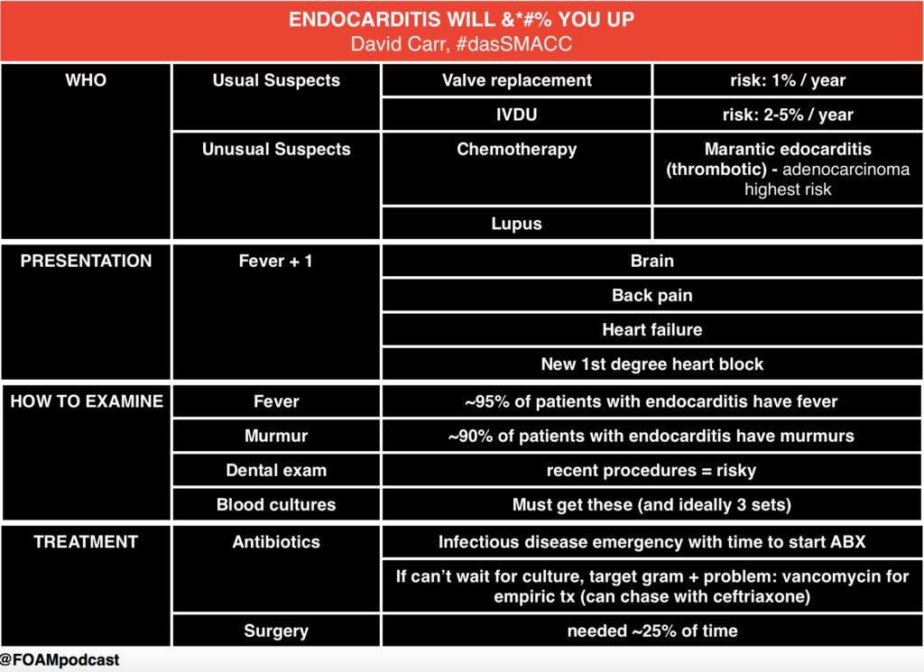
Dr. David Carr (@davidcarr333)

Endocarditis

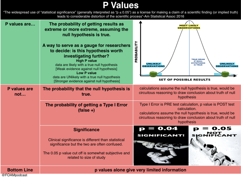
Dr. Paul Young (@DogICUma)

p values

Dr Jess Mason (@JessMasonMD)

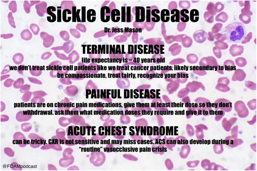
sickle cell

Episode 65 – Contrast-Induced Nephropathy and Genitourinary Trauma

iTunes or Listen Here

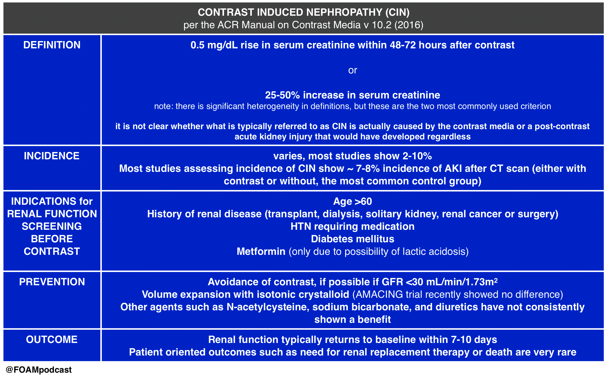
We cover Free Open Access Medical Education (FOAM) on Contrast-Induced Nephropathy (CIN).  A large retrospective study by Hinson and colleagues in Annals of Emergency Medicine, reignited enthusiasm in the FOAM world about the questionable entity (and clinical significance) of CIN. The data on CIN is somewhat difficult to parse out as the data consists entirely of retrospective cohort and case-control studies. The highest risk of CIN seems to come from large volume contrast procedures such as percutaneous coronary angiography. Studies looking at the risk of CIN after contrast-enhanced CT scan have been less conclusive.

The American College of Radiology (ACR) Manual on Contrast Induced Nephropathy

CIN

This Emergency Medicine Literature of Note post covers the AMACING  trial, which looks at intravenous fluid administration (0.9% NaCl) versus usual care in patients “at risk” for CIN undergoing contrast-enhanced CT scan.   The study found a difference of -0.1% (95% CI -2.25 to 2.06), which was below the non-inferiority margin. Prior literature shows similar results, with no clear-cut efficacy from prevention strategies for CIN.

This post by Dr. Joel Topf (nephrologist @kidneyboy) on the Precious Bodily Fluids blog discusses a nephrologist’s take on CIN.

EM topics post on fluids in CIN.

Core Content

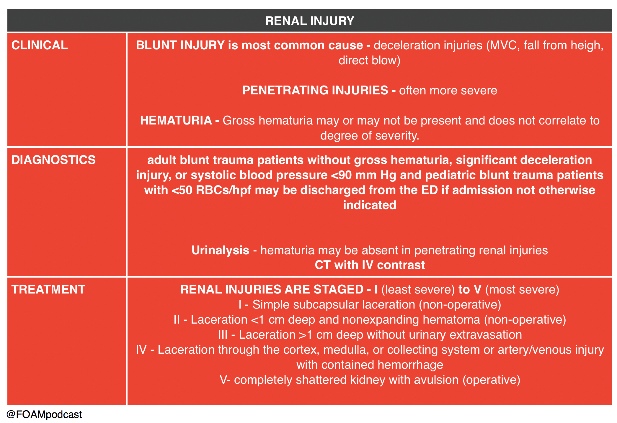
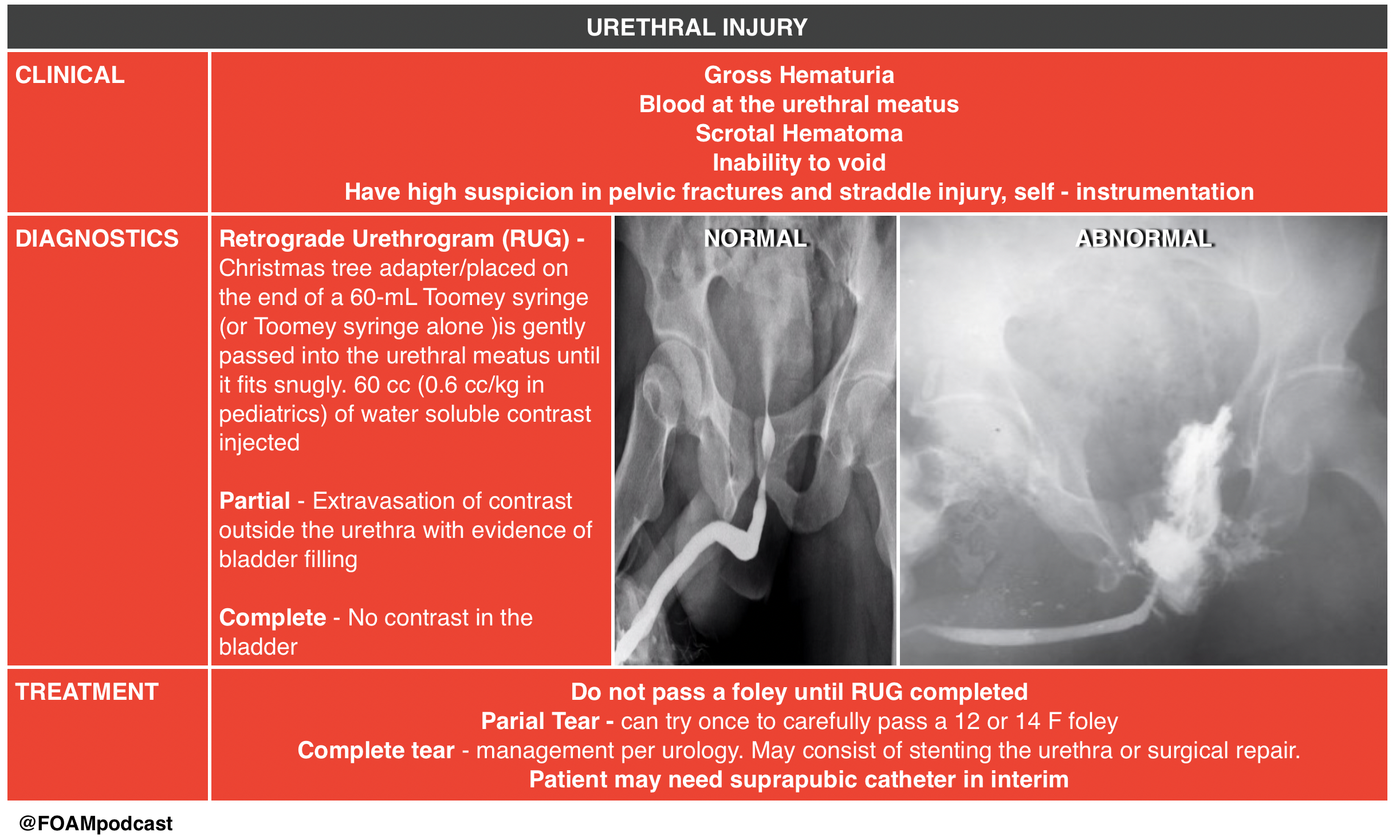
We then delve into core content on genitourinary trauma using Rosen’s Chapter 47 and Tintinalli’s Chapter265 as a guide.

Screen Shot 2017-02-28 at 1.43.40 PM Screen Shot 2017-02-28 at 1.44.33 PM

 Rosh Review Emergency Board Review Questions

An 18-year-old man involved in a motor vehicle collision is transferred from a rural facility after being diagnosed with a pelvic fracture. After your initial assessment and stabilization, a secondary survey is performed. On examination, he is noted to have blood at his urethral meatus as well as a scrotal hematoma. Which of the following is the most appropriate next step?

A. Retrograde cystogram

B. Retrograde urethrogram

C. Suprapubic catheter placement

D. Transurethral urinary catheter placement

[accordion]
[toggle title=”Answer” state=”closed”]

B. The patient should undergo a retrograde urethrogram to rule out an underlying urethral injury. In patients with a pelvic fracture and signs of urethral injury, a retrograde urethrogram should be performed in the supine position prior to urethral instrumentation. If a partial disruption is identified, one attempt to place a 12- or 14-French Foley or coude catheter can be made. If unsuccessful or a complete tear is diagnosed, then a suprapubic catheter will need to be placed. Pelvic fractures with displacement of the pubic symphysis can cause laceration or avulsion of the prostatic urethra. The three classic findings of urethral injury include blood at the urethral meatus, a high-riding prostate and a scrotal or perineal hematoma. Failure to recognize a urethral injury can lead to urethral stricture formation and urinary incontinence.

Urethral manipulation can convert a partial urethral tear to a complete tear, therefore transurethral urinary catheter placement (D) should be avoided until urethral integrity is known. Suprapubic catheter placement (C) may ultimately be necessary if complete urethral injury is diagnosed, however in this case a retrograde urethrogram should be performed prior to proceeding with suprapubic placement. The patient may also have a bladder injury and may require a retrograde cystogram (A), however urethral integrity must be evaluated by a retrograde urethrogram prior to performing a cystogram

[/accordion]

A 29-year-old man presents to the ED after a MVC. A pelvic fracture is identified on radiography. His vital signs are stable. The decision is made to place a Foley catheter, but blood is noted at the urethral meatus. Which of the following is an appropriate next step?

A. Consult a urologist

B. Obtain a CT scan to evaluate for urethral injury

C. Perform a retrograde urethrogram

D. Place a condom catheter

[accordion]
[toggle title=”Answer” state=”closed”]

C. Perform a retrograde urethrogram. In general, a Foley catheter should not be placed in the setting of suspected urethral injury. In such cases, it is recommended that further testing be performed to evaluate for urethral injury. A retrograde urethrogram should be performed. If there is no contrast extravasation, then a Foley catheter can safely be inserted.  A urologist should be consulted (A) if a urethral injury is confirmed by the retrograde urethrogram. A CT scan (B) is a poor study to identify urethral injuries. A condom catheter (D) does not allow for accurate urine output measurements and may delay identification of a urethral injury.

[/accordion]

References:

  1. Hinson JS, Ehmann MR, Fine DM, et al. Risk of Acute Kidney Injury After Intravenous Contrast Media Administration. Ann Emerg Med. 2017
  2. ACR Manual on Contrast Media.  v10.2. 2016. p33-40
  3. “Genitourinary System.”  Rosen’s Emergency Medicine, 8th ed. Chapter 47, 479-499.e1
  4. “Genitourinary Trauma.” Tintinalli’s Emergency Medicine: A Comprehensive Review.  8th ed.  Chapter 265

Episode 51 – Ocular Trauma

(ITUNES OR LISTEN HERE)

The Free Open Access Medical Education (FOAM)

We cover this short video from EMRAP on lateral canthotomies.  This is one of those rare procedures that is vision saving; hence, it is worthy of frequent review.

Lateral Canthotomy and Cantholysis

Indication:  Suspect orbital compartment syndrome -afferent pupillary defect (APD), “tense eye” with taught lids, and high intraocular pressure (IOP) ≥ 40 mmHg. The APD is really a critical feature, as it is your indicator that there is optic nerve compromise from the intraorbital pressure

Clinical presentation:  History of orbital trauma, often in the setting of orbital fractures with decreased visual acuity, proptosis, chemosis

Procedure: Call ophthalmology.

  • Analgesia – inject lidocaine with epinephrine into the area of the lateral canthus
  • Devascularize – use straight kelly clamps to crush the lateral canthus for 1-2 minutes
  • Incise – Use scissors to cut the lateral canthus 1-2 cm. Then,  find the inferior crus of the lateral canthus ligament (looks kind of like a wishbone from a turkey but you rarely are going to be able to see the tendon so you’re “strumming” it with the closed tips of your scissors inferomedially toward the nose) – cut the inferior crus of the ligament to release pressure (this is the key step)

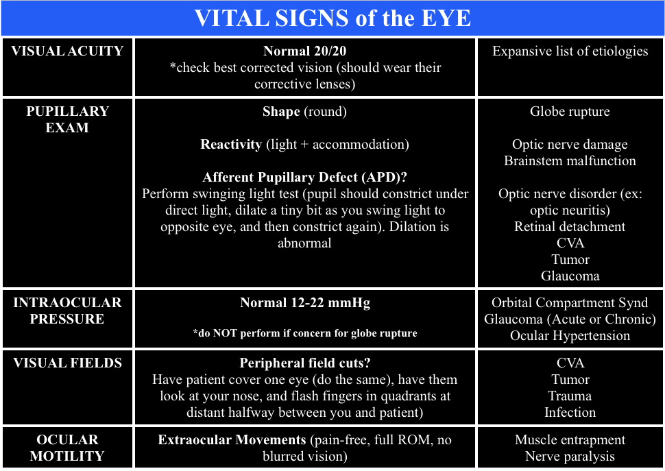
When calling ophthalmology, ensure you have the “Vital Signs of the Eye”

  • Vital Signs of the Eye - @FOAMpodcast
    Vital Signs of the Eye – @FOAMpodcast

Core Content

We delve into core content on eye trauma using Rosen’s (8th edition) Chapter 71 and Chapter 241 in Tintinalli (8th edition)

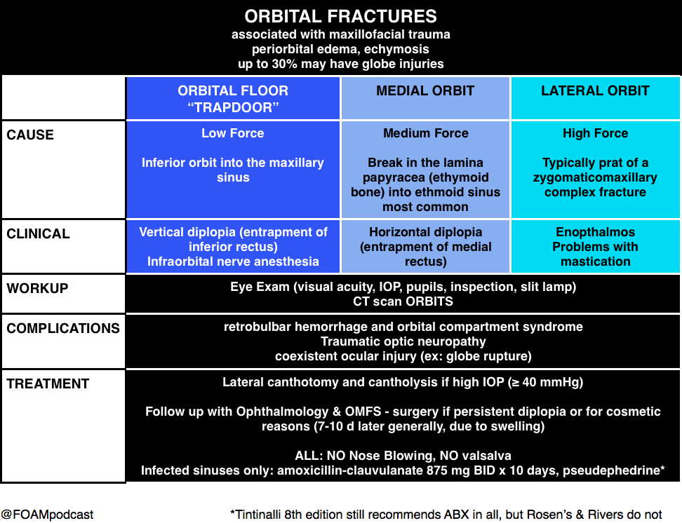
Orbital Fractures

Orbital Fractures

Note: entrapment is a clinical diagnosis.  A CT cannot comment on function, only structure.  Extraocular movements are a critical part of the fracture exam.

Pearl: young people are prone to “greenstick” fractures where the floor can actually pinch the muscle and trigger the oculocardiac reflex, which can be deadly.  They often have a “white eye” without too much impressive on exam but refuse to look in a certain direction due to nausea.

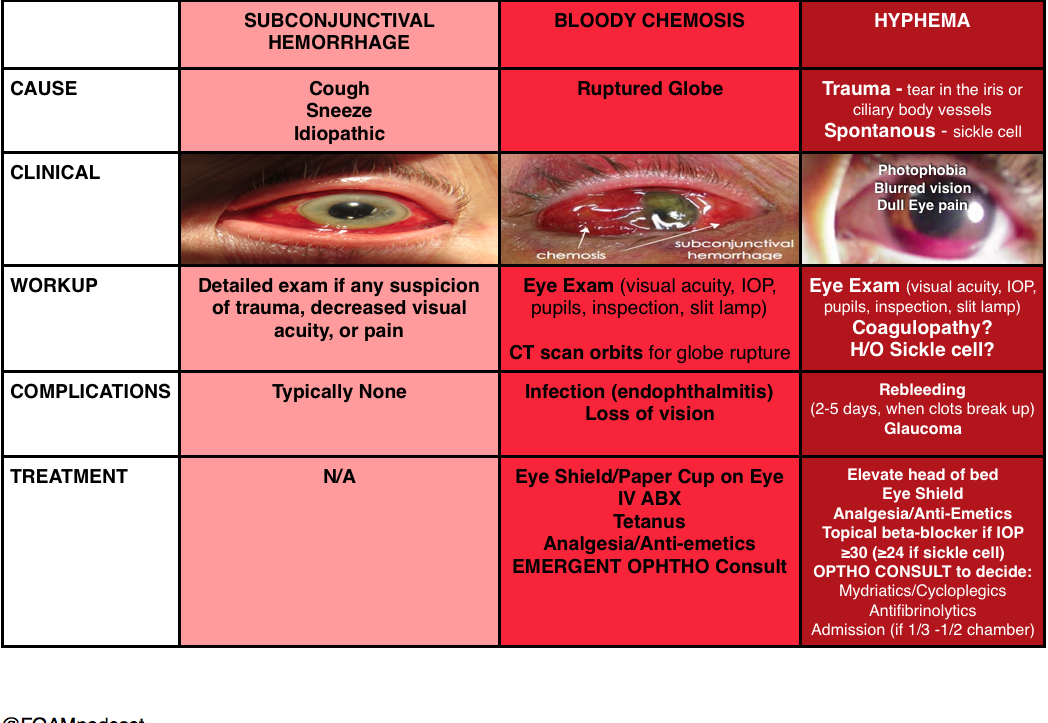
Hyphema

(and things confused with hyphema)

Hyphema

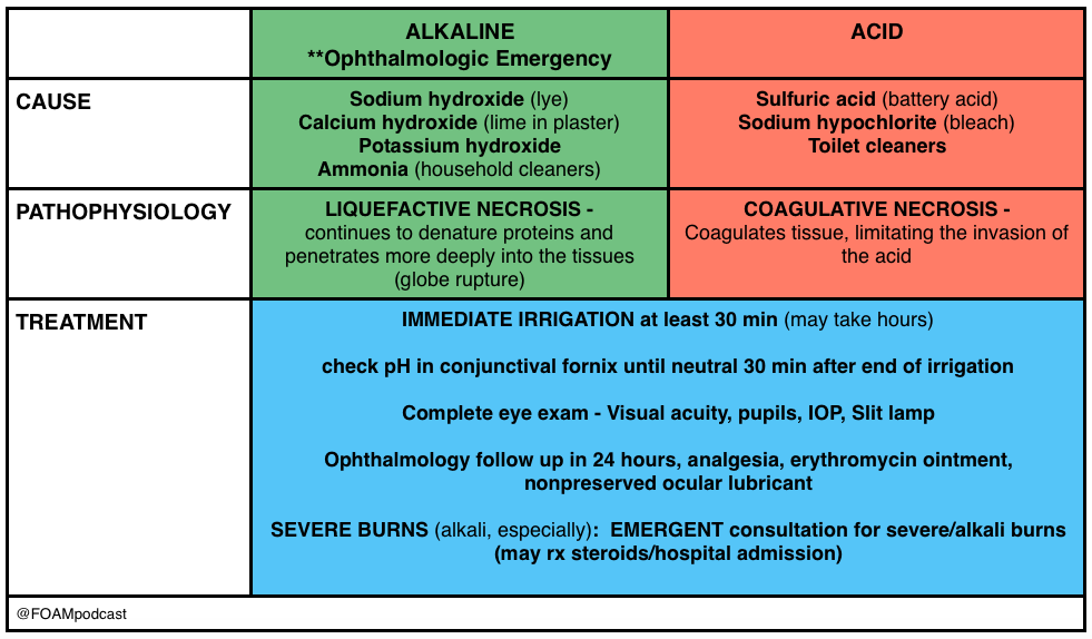
Burns

Ocular Burns

Pearl: An important thing to keep in mind is that a “white” eye after such an exposure is actually an ominous sign, as it implies ischemia of the limbal blood vessels, which portends a pretty bad prognosis.  People usually think that the more red an eye, the worse when, in truth, it’s often the other way around.

Generously Donated Rosh Review Questions

A 43-year-old construction worker presents with right eye pain. He states he was using a nail gun when he felt something hit his eye. Visual examination reveals a small nail penetrating the globe. What management should be pursued?

A. CT scan of the orbit and ophthalmology consultation

B. Measure intraocular pressure and consult ophthalmology

C. Perform lateral canthotomy and consult ophthalmology

D. Remove the foreign body, start topical antibiotics and send to ophthalmology for follow up

[accordion]
[toggle title=”Answer” state=”closed”]

This patient presents with a globe injury and should have a protective shield placed, intravenous antibiotics started, CT scan of the orbit performed and ophthalmology consulted emergently. Foreign body penetration of the globe is often associated with hammering, drilling, mechanical grinding or sanding. Any patient who presents with a foreign body sensation after one of these activities should increase suspicion for a penetrating injury of the globe. CT scan, MRI and ultrasound can all be used for diagnosis but MRI should be avoided if the suspected foreign body is metal containing. Many of these patients will require operative management and should be kept NPO. A protective shield should be placed to protect the eye but patching should be avoided as it may increase pressure on the eye. Emergent ophthalmologic consultation should be obtained

[/toggle]
[/accordion]

A 23-year-old man presents after a fight. His eye is seen below. Physical examination reveals intact extraocular movements, normal fluorescein staining, normal intraocular pressure, and normal visual acuity.

content_image-_medial_lower_lid_laceration

What management is indicated?

A. Delayed closure

B.  Laceration repair by the Emergency Physician

C. Ophthalmology consultation for repair

D. Tissue adhesive for repair

[accordion]
[toggle title=”Answer” state=”closed”]

C. Ophthalmology consultation for repair. This patient presents with a complex eyelid laceration possibly involving the canalicular system and should have a consultation with either ophthalmology or plastic surgery regarding repair. It is important to search for a penetrating globe injury in any patient with an eyelid laceration because of the proximity of structures. Simple horizontal and partial thickness lid lacerations can be repaired primarily by an Emergency Physician. However, more complicated lacerations should be considered for specialist repair because of the high likelihood of cosmetic or functional complications, or both. In general, lacerations through the orbital septum, lacerations with tissue loss, lacerations involving the lid margins, lacerations involving the levator or canthal tendons and those involving the canalicular system should be repaired by a skilled ophthalmologist or plastic surgeon. Injury to the canalicular system should be suspected in any laceration involving the medial lower eyelid. Tissue adhesive (D) is contraindicated this close to the eye and should not be used in an injury that requires precision alignment of tissue. Delayed closure (A) will likely lead to worse cosmetic outcomes. Primary repair by the Emergency Physician (B) is not recommended if the canalicular system may be involved.

[/toggle]
[/accordion]
 

Thanks to our peer reviewer, Michael Westafer, MD – Ophthalmologist and Glaucoma Fellow at Mayo Clinic

References

  1. “Eye Emergencies.”  Chapter 241.  Tintinalli’s Emergency Medicine: A Comprehensive Review. 8th edition.  
  2. Sharma R and Brunette D.  “Ophthalmology.”  Chapter 71.  Rosen’s Emergency Medicine.  8th edition, 909-930.

Episode 37 – Lacerations

(ITUNES OR LISTEN HERE)

The Free Open Access Medical Education (FOAM)

We cover a trick of the trade from Dr. Brian Lin, posted on the Academic Life in Emergency Medicine (ALiEM site) on hemostasis in finger tip avulsions. Dr. Lin also has his own excellent FOAM site on all things laceration – LacerationRepair.com.

We also cover FOAM on dogma of wound care from Dr. Ken Milne’s The Skeptic’s Guide to Emergency Medicine, Episode #63

Core Content – Wounds and Laceration Care

Tintinalli (7e) Chapter 44, “Wound Preparation.” Rosen’s  (8e) Chapter 59, “Wound Management Principles.”

Laceration Care:

  • Use gloves, they don’t have to be sterile [1].
  • Anesthetize (lidocaine with epinephrine is just fine).
  • Irrigate copiously. It’s estimated that one needs ~60 mL/centimeter of wound or at least 200 mL.
    • You can irrigate with water or saline. Potable tap water is fine [2,3]
  • For a cornucopia of laceration techniques visit LacerationRepair.com
  • No clear “golden period” for laceration repair [4-6]. Rosen’s and Tintinalli recommend using clinical judgment as a guide.

Risks for Infection:

  • Diabetes
  • Length of laceration (>5 cm)
  • Location of the wound
  • Degree of contamination [6]

Age of wound when approximated (i.e. “golden period”) has not been found to be an independent risk factor). Rosen’s sites use of epinephrine as a risk but only cites a paper by Barker et al from 1982 in which tetracaine/epinephrine/cocaine was applied to wounds inflicted by researchers that were inoculated by s. aureus.

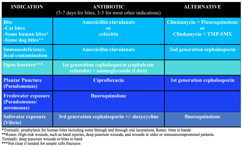
Prophylactic antibiotics:

Screen Shot 2015-11-15 at 5.34.59 PM

Generously Donated Rosh Review Questions 

Question 1.  An 18-year-old woman presents with a laceration to her face from a dog bite that occurred 24 hours ago. The patient owns the dog. Examination reveals a 4 cm laceration to the left cheek with no signs of infection. [polldaddy poll=9180209]

Question 2A 30-year-old man presents with a 2 cm linear laceration through his right eyebrow that he sustained after hitting his head on the kitchen cabinet. You determine that the wound will require repair with sutures. [polldaddy poll=9180210]

Answers

  1. Mammal bites to any part of the body should be copiously irrigated and explored followed by an assessment for primary closure. In this patient, primary closure is recommended as the laceration is on the face. Canine bites often involve laceration as well as crush injury to tissue depending on the size of dog. The presence of a crush injury may make primary wound repair difficult. Additionally, devascularization of the tissue may make primary closure contraindicated as the risk of infection increases. Classically, it was taught that lacerations sustained from dog bites should be irrigated, given antibiotics and not primarily repaired because of these risks. However, more recent literature has shown that the risk of infection was no different for primary closure versus healing by secondary intention. Additionally, if the laceration is to a cosmetic area like the face, primary repair should be attempted. As with any laceration, tetanus status should be updated. Copious irrigation and wound exploration is central to good wound care. Exploration should pay particular attention to the presence of foreign bodies especially teeth, which may break off during the bite. Antibiotics (A & C) are not routinely needed for dog bites despite classic teaching. Antibiotics should be reserved for patients with signs of infection, multiple comorbidities or large wounds with gross contamination. If antibiotics are given, they should primarily cover Staphylococcus and Streptococcus species, as these are the predominant organisms in the canine oral cavity. Eikenella and Pasturella are less commonly responsible for infections. Irrigation and antibiotics alone (A) would be indicated for dog bites that are grossly infected or have large defects that cannot be primarily closed. Wound closure and antibiotics without irrigation (D) is also contraindicated as copious irrigation is central to proper wound management.
  2. A pair of clean, non-sterile gloves can be worn by the physician (and any assistants) during laceration repair. The use of sterile gloves has not been proven to be associated with lower infection rates and is not required. Wounds must be prepped prior to closure. This generally involves cleaning and draping the wound, providing local or regional anesthesia, copious irrigation and exploring the wound to evaluate the integrity underlying structures and identify any foreign bodies. The skin surrounding a wound should be cleansed with either 10% povidone-iodine (C) or chlorhexidine gluconate solution. In general, these commercially available antiseptics should not be used for wound irrigation, as they can be toxic to the tissues. Irrigation should then follow with copious amounts of tap water or saline (at least 250 mL). This is best achieved with a large volume syringe attached to an 18-gauge needle or another commercially available irrigation device that achieves adequate pressure for irrigation. Alternatively, patients can irrigate at the sink if the laceration is in area that allows for this. Shaving of hair been shown to increase the risk of infection and should generally be avoided. It is best to apply a small amount of petroleum- or water-based lubricant to the hair to keep it out of the wound. Alternatively, hair can be clipped with scissors when necessary. Eyebrows (B) in particular should not be shaved as they provide anatomic landmarks that aid in wound approximation and removal results in poor short- and long-term cosmetic effect. In general, non-complex facial wounds are closed with nonabsorbable suture material, such as nylon or polypropylene. Most commonly this will be done with 6-0 suture, as it provides the best cosmetic effect. The use of 3-0 (D) and 4-0 suture is reserved for repair of fascia or wounds that are under high stress, such as those that overly major joints or involve the scalp.

References:

  1. Perelman VS, Francis GJ, Rutledge T, et al. Sterile versus nonsterile gloves for repair of uncomplicated lacerations in the emergency department: a randomized controlled trial. Annals of emergency medicine. 43(3):362-70. 2004
  2. Fernandez R, Griffiths R. Water for wound cleansing. The Cochrane database of systematic reviews. 2:CD003861. 2012.
  3. Weiss EA, Oldham G, Lin M, Foster T, Quinn JV. Water is a safe and effective alternative to sterile normal saline for wound irrigation prior to suturing: a prospective, double-blind, randomised, controlled clinical trial. BMJ open. 3(1):. 2013.
  4. American College of Emergency Physicians: Clinical policy for the initial approach to patients presenting with penetrating extremity trauma. Annals of emergency medicine. 33(5):612-36. 1999. [pubmed] **A past policy, no current clinical policy
  5. Zehtabchi S, Tan A, Yadav K, Badawy A, Lucchesi M. The impact of wound age on the infection rate of simple lacerations repaired in the emergency department. Injury. 43(11):1793-8. 2012.
  6. Quinn JV, Polevoi SK, Kohn MA. Traumatic lacerations: what are the risks for infection and has the ‘golden period’ of laceration care disappeared? Emergency medicine journal : EMJ. 31(2):96-100. 2014.

Episode 36 – Rib and Sternal Fractures

(ITUNES OR Listen Here)

The Free Open Access Medical Education (FOAM)

This week we cover a joint piece between the Ultrasound Podcast and SonoIn5 on diagnosis of rib and sternal fractures with ultrasound.

Technique:  Linear probe, in line with the long axis of the bone (vertical for sternum, horizontal-ish for ribs).

Diagnosis: Cortical disruption (step off). Excellent sensitivity for sternal fractures [1-3]

Sternal Fracture
Sternal Fracture
  • Caution with sternal fractures as the sternomanubrial joint can mimic fracture, but looks more “bumpy” (see below)

Core Content – Rib and Sternal Fractures

Tintinalli (7e) Chapters 258, 259; Rosen (8e) Chapter 45 

Rib Fractures

Diagnosis:

  • Chest x-ray initial test of choice – may miss 50% of fractures, unclear if this is clinically significant [6]
  • Ultrasound has found to have excellent sensitivity [7]
  • Rib films are NOT recommended [4-6].

Complications: Traumatic rib fractures may be associated with other traumatic injuries such as pneumothorax, hemothorax, or in the case of lower rib fractures, intra-abdominal injury. However, rib fractures themselves have been associated with mortality, most often as sequelae of pulmonary embarrassment including pneumonia, intubation, and death. Mortality in elderly patients with rib fractures is significantly higher than the younger counterparts at 22% and 10% respectively [8,9].

  • Mortality is between 3-13%
  • Risk stratification (see this post): Battle and colleagues developed a prognostic scoring system, not externally validated and unclear if it would change practice, that highlights common sense predictors of poorer outcomes:
    • Age (>65)
    • Higher number of rib fractures
    • Chronic lung disease
    • Hypoxia (<90%)
    • Pre-injury anticoagulant use [11]

Treatment

  • Analgesia:
    • Often includes NSAIDS (ibuprofen), acetaminophen, and narcotics +/- gabapentin (ibuprofen and gabapentin depending on renal function)
    • Epidural analgesia – highly recommended in the EAST guidelines [14].
    • Paracostal analgesia (ex: ON-Q pump) – not sufficient evidence for EAST recommendation (2005) [14]
  • Pulmonary Hygiene (formerly pulmonary toilet):  involved incentive spirometry, coughing, mobilization (up, out of bed), and possibly chest physical therapy
  • ORIF, “rib fixation” or “rib plating,” is increasingly common in the US and studies have found improvements in ICU LOS and ventilator days [15]

Disposition

  • Many rib fracture patients will need to be admitted to the hospital for pain control, observation, and pulmonary hygiene.
    • Some rib fracture patients may benefit from care at trauma centers.  Lee et al  wrote that 3+ rib fractures exists as an indication for transfer to a level 1 trauma center and many places ascribe to this, it depends on the hospital and physicians.
    • While patients in the ED may look good, patients may benefit from high intensity floors (ie stepdown units) and many patients get observed in ICUs, again, depending on local practice patterns. Some protocols risk stratify patients (i.e. to the ICU vs floor) by incentive spirometry.
  • Patients with adequate pain control who are low risk (younger, <3 rib fractures, good effort on incentive spirometry) may be discharged from the ED with analgesia and education on importance of pulmonary hygiene

Sternal Fractures – more common with ubiquity of airbags and seatbelts.

Diagnosis:  Classically the “gold standard” has been lateral x-ray. However, CT technology has improved since those studies. Ample literature suggests that ultrasound has excellent sensitivity [1-3].

Complications: Historically, sternal fractures were associated with injuries of the great vessels, high mortality, and blunt cardiac injury (BCI) [16-18].  The most recent iteration of the EAST guidelines states, “the presence of a sternal fracture alone does not predict the presence of BCI and thus should not prompt monitoring in the setting of normal ECG result and troponin I level” (Level 2) [18].

Treatment: Analgesia. Most patients with isolated sternal fractures (no pneumothorax, hemothorax, BCI, or hemodynamic instability) that have adequate pain control can be discharged from the ED [1-2].

Blunt Cardiac Injury

A broad category including a range of injuries from clinically silent dysrhythmias to cardiac wall rupture or vasospasm. BCI often results from high impact injury and should be considered in patients with significant thoracic trauma including rib fractures, sternal fracture, pneumothorax, hemothorax, and pulmonary contusion.

Diagnosis: There is no gold standard test.  One can rule out BCI with a normal ECG and a single normal troponin I [18].

Management: If an ECG or troponin is abnormal, admit to telemetry for monitoring and echo.

Generously Donated Rosh Review Questions 

Question 1.  A 23-year-old man presents with chest pain after a motor vehicle collision. The patient’s chest struck the steering wheel. He has no other complaints or injuries. Chest X-ray is unremarkable. ECG shows sinus tachycardia with anterior ST depressions. A troponin is sent and is positive at 3.50 mg/dl. [polldaddy poll=9134639]

Question 2A 20-year-old man presents with left rib pain after falling while playing soccer and striking his chest. Vital signs are normal. On physical examination, the patient has tenderness to palpation over the 4th rib in the midaxillary line. [polldaddy poll=9134640]

Question 3.  A 32-year-old woman was the restrained driver involved in a head-on motor vehicle collision (MVC) 2 days prior to presentation. She is complaining of chest pain and bruising to her chest. Her blood pressure is 118/78 mm Hg, pulse is 88 beats/minute, respirations are 18 breaths/minute and oxygen saturation is 96% on room air. You note bony tenderness and ecchymosis to her sternum. You order a chest X-ray and diagnose a non-displaced sternal fracture. [polldaddy poll=9134643]

Answers

  1. This patient presents with a myocardial contusion and should have an echocardiogram performed to look for any cardiac damage. Myocardial contusion describes a nebulous condition. It can occur through several mechanisms including a direct blow to the chest and compressive force over a prolonged period of time. Histologically, the disorder has similar findings to those seen after acute myocardial infarction. The majority of contusions heal spontaneously but small pericardial effusions may develop. Delayed rupture after resorption of hematoma is feared but rare complication. Patients with myocardial contusion will present after trauma with external signs of trauma and typically have other concomitant thoracic lesions (pulmonary contusion, pneumothorax, hemothorax). Patients will typically have tachycardia (up to 70%). ECG may show dysrrhythmia or ST changes but may also be normal. Although it is not effective to admit all patients for workup for myocardial contusion and the disease has a very low rate of cardiac complications, in the presence of ECG changes and elevated biomarkers, observation and echocardiography are a reasonable approach. Echocardiogram can be used to diagnose pericardial effusion, thrombi formation and valvular disruption.Cardiac catheterization (A) is not necessary after a myocardial contusion as coronary artery obstruction is not part of the pathophsyiology. The patient should not be discharged home (B)without an echocardiogram. Pericardiocentesis (D) is only necessary in the presence of a large pericardial effusion or one causing cardiac tamponade.
  2. This patient presents with signs and symptoms consistent with a rib facture. A chest X-ray should be performed to rule out any other pathology including pneumothorax and pulmonary contusion. Rib fractures are a common injury after thoracic trauma and the incidence increases with increasing age. They may be associated with a number of potential complications including pulmonary contusions, hemothorax, penumothorax and post-traumatic pneumonia. Fractures are most common at the posterior angle, which represents the weakest area. The ribs most commonly fractured are the 4th – 9th ribs. The 9th – 11th ribs are mobile, which reduces the risk of fracture. However, fractures of these ribs are more likely to be associated with intraabdominal injuries. Rib fractures should be suspected based on history and clinical evaluation. Patients will present with chest pain and tenderness over the area. Imaging should be obtained to rule out the more serious associated complications of pneumothorax, hemothorax and pulmonary contusion. Chest X-ray is the appropriate modality for this but often will not demonstrate the presence of a single rib fracture when it is in fact present. This is particularly true of non-displaced fractures. Rib belts (B) are discouraged as they may decrease the depth of respiration and lead to atelectasis and pneumonia. CT scan of the chest (D) is not routinely required for management of a simple rib fracture. Analgesia and discharge home (A) is likley to occur once more serious pathology is ruled out with a chest X-ray. Patients with rib fractures should also receive an incentive spirometer to help reduce the complication of pneumonia.
  3. Isolated, non-displaced sternal fractures are associated with low overall mortality rates. Fractures and dislocations of the sternum are caused primarily by anterior blunt chest wall trauma during a head-on MVC. Isolated fractures of the sternum most commonly occur when the chest wall is thrust against a diagonal seatbelt strap during rapid deceleration in a frontal impact MVC. They are more common in older individuals and women. Most fractures are transverse and non-displaced and can be diagnosed on a lateral chest radiograph. Although a fracture of the sternum can be seen following major thoracic trauma, its presence alone does not indicate severe underlying thoracic injury. However, if other significant underlying thoracic injuries are suspected, a CT-scan of the thorax should be performed

References:

  1. You JS, Chung YE, Kim D, Park S, Chung SP. Role of sonography in the emergency room to diagnose sternal fractures. Journal of clinical ultrasound : JCU. 38(3):135-7. 2010. [pubmed]
  2. Engin G, Yekeler E, Güloğlu R, Acunaş B, Acunaş G. US versus conventional radiography in the diagnosis of sternal fractures. Acta radiologica (Stockholm, Sweden : 1987). 41(3):296-9. 2000. [pubmed]
  3. Jin W, Yang DM, Kim HC, Ryu KN. Diagnostic values of sonography for assessment of sternal fractures compared with conventional radiography and bone scans. J Ultrasound Med. 2006 Oct. 25(10):1263-8; quiz 1269-70.
  4. ”Pulmonary Trauma” Tintinalli’s Emergency Medicine: A Comprehensive Study Guide.  7th ed. Ch 258.
  5.  “Thoracic Trauma” Rosen’s Emergency Medicine. 8th ed. Chapter 45.
  6. Henry TS, Kirsch J. ACR Appropriateness Criteria® rib fractures. Journal of thoracic imaging. 29(6):364-6. 2014. [pubmed]
  7. Chan SS. Emergency bedside ultrasound for the diagnosis of rib fractures. The American journal of emergency medicine. 27(5):617-20. 2009. [pubmed]
  8.  Ziegler DW, Agarwal NN. The morbidity and mortality of rib fractures. J. Trauma. 1994;37(6):975–9.
  9. Bulger EM, Arneson M a, Mock CN, Jurkovich GJ. Rib fractures in the elderly. J. Trauma. 2000;48(6):1040–6
  10. Flagel BT, Luchette F a, Reed RL, et al. Half-a-dozen ribs: the breakpoint for mortality. Surgery. 2005;138(4):717–23; discussion 723–5.
  11. Battle CE, Hutchings H, Evans P. Risk factors that predict mortality in patients with blunt chest wall trauma: a systematic review and meta-analysis. Injury. 2012;43(1):8–17.
  12. Livingston DH, Shogan B, John P, Lavery RF. CT diagnosis of Rib fractures and the prediction of acute respiratory failure. The Journal of trauma. 64(4):905-11. 2008. [pubmed]
  13. Battle CE, Hutchings H, Lovett S.  Predicting outcomes after blunt chest wall trauma: development and external validation of a new prognostic model Critical Care 2014, 18:R98
  14. Pain Management in Blunt Thoracic Trauma (BTT)J Trauma. 59(5):1256-1267, November 2005.
  15. Doben AR, Eriksson EA, Denlinger CE. Surgical rib fixation for flail chest deformity improves liberation from mechanical ventilation. Journal of critical care. 29(1):139-43. 2014. [pubmed]
  16. Screening for Blunt Cardiac Injury. J Trauma. 73(5):S301-S306, November 2012
  17. Karangelis D, Koufakis T, Spiliopoulos K, Tsilimingas N, Bouliaris K, Desimonas N. Management of isolated sternal fractures using a practical algorithm. J Emerg Trauma Shock. 7(3):170-. 2014. [article]
  18. Dua A, McMaster J, Desai PJ et al. The Association between Blunt Cardiac Injury and Isolated Sternal Fracture. Cardiology Research and Practice. 2014:1-3. 2014. [article]

Episode 27 – Burns

(ITUNES OR Listen Here)

The Free Open Access Medical Education (FOAM)

Burns are incredibly noticeable but the accompanying inhalational injury gets less attention. The amazing Maryland Critical Care Project features a neat review, N-acetylcysteine for Inhalational Burn Injury

  • Occurs in ~20% of all burn patients
  • Mortality is 30%
  • Major mechanisms of injury:
    • Temperature – often causing burns down to the level of the vocal cords, but not below.
    • Toxins – products of combustion such as cyanide, carbon monoxide, and hydrogen sulfide are asphyxiants and impair oxygen utilization
    • Irritants – inhaled particulates irritate airways
  • Airway edema peaks at 24-48 hours
  • Diagnosis made on bronchoscopy

Nebulized heparin, N-acetylcysteine, and albuterol protocol

May be beneficial, but protocols based on small studies. Rosen gives a head nod to this regimen, yet the evidence is questionable [1-3].

Nebulized heparin can be used at 10,000 international units every 4 hours, followed by nebulized NAC & albuterol 2 hours later for a total of 7 days.

  • Nebulized heparin may help prevent formation of airways casts.
  • NAC may have a mucolytic effect and help scavenge free radicals.
  • Albuterol added to prevent bronchospasm
  • Alternating nebulized heparin and NAC may:
    • Improve P/F ratio (not a patient centered outcome)
    • Decrease ventilator days
    • Reduce the development of acute lung injury

The Bread and Butter

We cover burns including nomenclature, fluid resuscitation, burn center referral criteria, and more. We do this based on Rosen’s and Tintinalli. But, don’t just take our word for it. Go enrich your fundamental understanding yourself.

Burn Depth

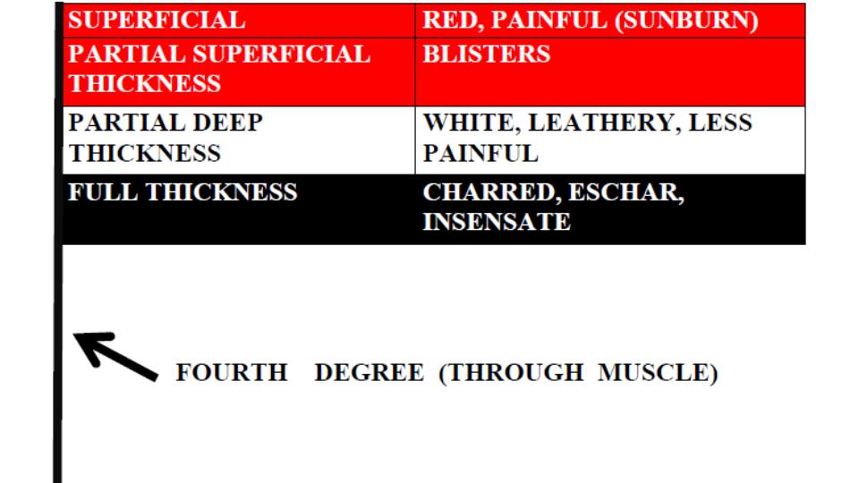
Classically, burns were described in degrees but are now described by the degree of thickness (except, apparently, fourth degree burns). At FOAMcast, we like to remember these distinctions by thinking about the Egyptian flag, with the flag pole representing fourth degree burn or burns essentially down to the bone.

Burn Degree
Burn Degree

Fluid Resuscitation

The Parkland Formula is probably one of the most well known but both this formula and the modified Brooke formula have led to fluid “creep” or over-resuscitation, which may have lasting consequences. Major guidelines and Rosenalli approve another method, the “Rule of Tens.”

Rule of Tens

  • Estimate burn size (TBSA) to the nearest 10%.
  • Multiply %TBSA x 10 = Initial fluid rate in mL/hr (for adult patients weighing 40 kg to 80 kg).
  • For every 10 kg above 80 kg add 100 mL/hr to the rate
  • Limitation: Overestimates fluids 100 [Alvarado]

Consider Referral to Burn Center

Bad Burn

  • Partial thickness burns > 10% TBSA
  • Full thickness burns
  • Electrical burns or chemical burns
  • Inhalation injury

Bad Place – Burns in the sensitive places such as face or genitals or important areas such as hands or major joints .

Bad Protoplasm – Preexisting medical disorders that could complicate management or prolong recovery (such as immunocompromised HIV patients).

Generously Donated Rosh Review Questions

Question 1. [polldaddy poll=8816037]

Question 2. A 52-year-old woman is brought to the emergency department with burns from a house fire. Physical exam reveals superficial burns over her entire left arm and partial-thickness and full-thickness burns covering her entire right arm, her anterior right leg and anterior trunk. [polldaddy poll=8824315]

Answers

1. C. Burn classification is based on burn depth. Second-degree burns are classified into superficial and deep partial-thickness burns. Deep partial-thickness burns extend into thereticular dermis. Skin color is usually a mixture of red and blanched white, and capillary refill is slow. Blisters are thick-walled and commonly ruptured and the skin may appear leathery white. Two-point discrimination may be diminished, but pressure and pinprick applied to the burned skin can be felt. Whereas superficial partial-thickness burns usually re-epithelialize 7-10 days after injury; so the risk of hypertrophic scarring is very small. For deep partial-thickness burns, tissue may undergo spontaneous epithelialization from the few viable epithelial appendages at this deepest layer of dermis and heal within 3-6 weeks. Because these burns have less capacity for re-epithelializing, a greater potential for hypertrophic scar formation exists. In deep partial-thickness burns, treatment with topical antimicrobial dressings is necessary to prevent infection as the burn wound heals. Contraction across joints, with resulting limitation in range of motion, is a common sequela. Splash scalds often cause second-degree burns.

2. B. The extent of burn size in this patient is 36% total body surface area. A thorough and accurate estimation of burn size is essential to guide therapy and to determine when to transfer a patient to a burn center. The extent of burns is expressed as the total percentage of body surface area. Superficial burns are not included in the burn assessment. For adult assessment, the most expeditious method to estimate total percentage of body surface area is the Rule of Nines. This method only takes into account partial-thickness and full-thickness burns. Each leg represents 18% total percentage of body surface area; each arm represents 9% total percentage of body surface area; the anterior and posterior trunk each represent 18% total percentage of body surface area; and the head represents 9% total percentage of body surface area. This patient has partial-thickness and full-thickness burns covering her entire right arm, her anterior right leg and anterior trunk, which calculates to 36%. The superficial burn on her left arm is not included in the calculation. According to the Rule of Nines, the percentage in this patient can be calculated as follows: entire right arm = 9%; anterior right leg = 9%; anterior trunk 18%. 9+9+18= 36%.

References:

1. Mlcak RP, Suman OE, Herndon DN. Respiratory management of inhalation injury. Burns. 2007;33:(1)2-13. [pubmed]

2. Kashefi NS, Nathan JI, Dissanaike S. Does a Nebulized Heparin/N-acetylcysteine Protocol Improve Outcomes in Adult Smoke Inhalation? Plast Reconstr Surg Glob Open. 2014;2:(6)e165. [pubmed]

3. Elsharnouby NM, Eid HE, Abou Elezz NF, Aboelatta YA. Heparin/N-acetylcysteine: an adjuvant in the management of burn inhalation injury: a study of different doses. J Crit Care. 2014;29:(1)182.e1-4. [pubmed]

Episode 26 – The Spinal Cord

(ITUNES OR Listen Here)

The Free Open Access Medical Education (FOAM)

In January 2015, ACEP recommended against the use of long backboards by EMS, “Backboards should not be used as a therapeutic intervention or as a precautionary measure either inside or outside the hospital or for inter-facility transfers.”

We review the use of longboards and cervical collars for spinal immobilization using posts by Thomas D of ScanCrit (Curse of the Cervical Collar, Cervical Collar RIP,  Cervical Collars Slashed From Guidelines), a post by Dr. Minh Le Cong from PHARM, and this Medest118 post.

The bottom line:

  • The benefits of devices to aid in spinal immobilization such as cervical collars and long backboards are controversial.  Guidelines and protocols are continuing to recommend judicious use of these devices.  Examples include:
    • Clearing collars in obtunded blunt trauma patients with negative high quality CT [EAST]
    • Selective application of cervical collars [ILCOR]
    • No backboards and selective pre-hospital immobilizaiton [ACEP]

The Bread and Butter

We differentiate between spinal shock and neurogenic shock, cover the incomplete cord syndromes (anterior cord, central cord, Brown-Sequard Syndrome), and fly through some of the cover using Tintinalli (7e) Chapter 255; Rosen’s (8e) Chapter 43, 106  But, don’t just take our word for it.  Go enrich your fundamental understanding yourself.

Spinal shock – Reduced reflexes following think of this as a stunning of the spinal cord.

Neurogenic shock –  This is loss of sympathetic innervation from injury to the cervical or thoracic spine, typically from a cervical or upper thoracic spinal cord injury, resulting in bradycardia and hypotension.

  • Warm, peripherally vasodilated , and hypotensive  from loss of sympathetic arterial tone with a relative bradycardia from unopposed parasympathetic (vagal) tone
  • Typically presents within 30 minutes, can last 6 weeks
  • Diagnose only after excluding other sources of shock
  • Treatment: crystalloid, vasopressors

Incomplete Cord Syndromes

 Better prognosis than complete cord syndromes. Means there is some sensory or motor preserved distal to lesion (i.e. rectal tone or perineal sensation)

Anterior Cord Syndrome 

  • Complete loss of motor, pain, and temperature below but retain posterior columns (position and vibration)
  • Flexion injury or decreased perfusion (aortic surgery or injury)
  • Paralysis and hypalgesia below the level of injury with preservation of posterior column (position and vibration)

Central Cord Syndrome

  • Sensory and motor deficit, often associated with hyperextension injuries (think whiplash)
  • Affects arms>legs
  • Think MUDE (pronounced muddy): Motor, Upper, Distal, Extension (injury)

Brown-Sequard Syndrome

  • Classically associated with a stab wound
  • Loss of motor function and position and vibration on ipsilateral side with contralateral loss of pain and temperature (fibers cross)

 Reflex Review

C4 Spontaneous breathing: “3-4-5 keep the diaphragm alive”
C5 Shoulder shrug
C6 Flexion at elbow:  think flexing your elbow up to drink before…
C7 Extension at elbow: …extending it to set a drink down.
C8-T1 Flexion of fingers
T1-T12 Intercostal and abdominal muscles
L1-L2 Flexion at hip
L3 Adduction at hip
L4 Abduction at hip
L5 Dorsiflexion of foot
S1-S2 Plantar flexion of foot
S2-S4 Rectal sphincter tone: “2-3-4 keeps your junk off the floor”

Generously Donated Rosh Review Questions 

Question 1. A patient arrives to the ED 15 minutes after being involved in a MVC. He is conscious, and there is no obvious trauma. He is immobilized on a long spine board with a cervical collar in place. His BP is 60/40 mm Hg and HR is 60 bpm. His skin is warm.

[polldaddy poll=8775757]

Question 2. [polldaddy poll=8776250]

Answers

1.  A. Loss of deep tendon reflexes is expected. Neurogenic shock occurs after an injury to the spinal cord. Sympathetic outflow is disrupted resulting in unopposed vagal tone. The major clinical signs are hypotension and bradycardia. Patients are generally hypotensive with warm, dry skin because the loss of sympathetic tone impairs the ability to redirect blood flow from the periphery to the core circulation. The most commonly affected area is the cervical region, followed by the thoracolumbar junction, the thoracic region, and the lumbar region. The anatomic level of the injury to the spinal cord impacts the likelihood and severity of neurogenic shock. Injuries above the T1 level have the capability of disrupting the spinal cord tracts that control the entire sympathetic system leading to the loss of deep tendon reflexes.Neurogenic shock must be differentiated from “spinal” shock, which refers to neuropraxia (B) associated with incomplete spinal cord injuries. This state is transient (C) and resolves in 1 to 3 weeks. Alpha-1 vasopressors (e.g., phenylephrine), in addition to dopamine, norepinephrine, and epinephrine (D), should be used to maintain blood pressure and ensure organ perfusion.

2. C.  In the anterior spinal cord syndrome, just the posterior columns are preserved and so patients lose all pain and temperature sensation as well as motor function. Most cases of anterior cord syndrome follow aortic surgery, but it has also been reported in the setting of hypotension, infection, vasospasm, or anterior spinal artery ischemia or infarct. In trauma, typically hyperflexion of the cervical spine causes the injury to the spinal cord.

Loss of all motor and sensory function (B) occurs with a complete transection of the spinal cord. Most commonly this occurs after a significant trauma. Isolated motor function loss (A) is not a classic syndrome and would result from a small area of injury on the cord just involving the corticospinal tract. Upper greater than lower motor weakness occurs (D) with a central cord syndrome. Sensory involvement is variable although burning dysesthesias in the upper extremities may occur. Most commonly the syndrome occurs after a fall or motor vehicle accident.