Emergency Medicine Updates – Hot Literature

iTunes or Listen Here

We co-hosted (with John Vassiliadis) the #smacc EM Updates half-day conference. We had amazing speakers. Salim Rezaie spoke on TXA for Everything, Ken Milne spoke on hot papers from 2018, and we learned about when ultrasound may be helpful in pediatric lumbar punctures. In addition, Jeremy spoke on what is usual care in sepsis and Lauren spoke on pulmonary embolism: the next generation. In this short podcast we highlight some of our other talks.

Aidan Baron (@Aidan_Baron) on Prehospital Updates in Cardiac Arrest

This talk focused on focusing on things that are most likely to make a difference in OHCA (bystander CPR and defibrillation) rather than on fun interventions like intubation and adrenaline (epinephrine). Aidan suggests that the future debates and questions in OHCA will be largely philosophical – what outcomes do we care about: neuro intact survival or ROSC or survival?

  1. Jabre P, Penaloza A, Pinero D, et al. Effect of bag-mask ventilation vs endotracheal intubation during cardiopulmonary resuscitation on neurological outcome after out-of-hospital cardiorespiratory arrest a randomized clinical trial. JAMA -2018;319(8):779–87.
  2. Benger JR, Kirby K, Black S, et al. Effect of a Strategy of a Supraglottic Airway Device vs Tracheal Intubation During Out-of-Hospital Cardiac Arrest on Functional Outcome: The AIRWAYS-2 Randomized Clinical Trial. JAMA. 2018;320(8):779-791.
  3. Wang HE, Schmicker RH, Daya MR, et al. Effect of a Strategy of Initial Laryngeal Tube Insertion vs Endotracheal Intubation on 72-Hour Survival in Adults With Out-of-Hospital Cardiac Arrest: A Randomized Clinical Trial. JAMA. 2018;320(8):769-778.

Low-Risk Chest Pain and the HEART Score by Barbra Backus

Modified Heart Score (redefining the T or troponin based on newer assays) results in a NPV of 99.8% and classifies 48% of patients as low-risk.

Clinically Relevant Adverse Cardiac Events (CRACE) is way less common than major adverse cardiac events (MACE). HEART score of ≤3 ? CRACE is 0.05%

Hot Literature in 2019

Lemkes JS, Janssens GN, van der Hoeven NW, et al. Coronary Angiography after Cardiac Arrest without ST-Segment Elevation. N Engl J Med. 2019;NEJMoa1816897

Pluymaekers NAHA, Dudink EAMP, Luermans JGLM, et al. Early or Delayed Cardioversion in Recent-Onset Atrial Fibrillation. N Engl J Med .2019;NEJMoa1900353.

Special thanks to the Rosh Review for supporting our trip to SMACC!

Supraventricular Tachycardia Treatment

iTunes or Listen Here

We have previously podcasted on tachyarrythmias (Episode 34 Tachyarrhythmias), but in this episode, we focus specifically on the treatment of supraventricular tachycardia  (SVT),  specifically paroxysmal SVT.

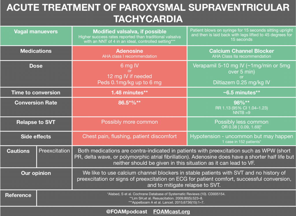
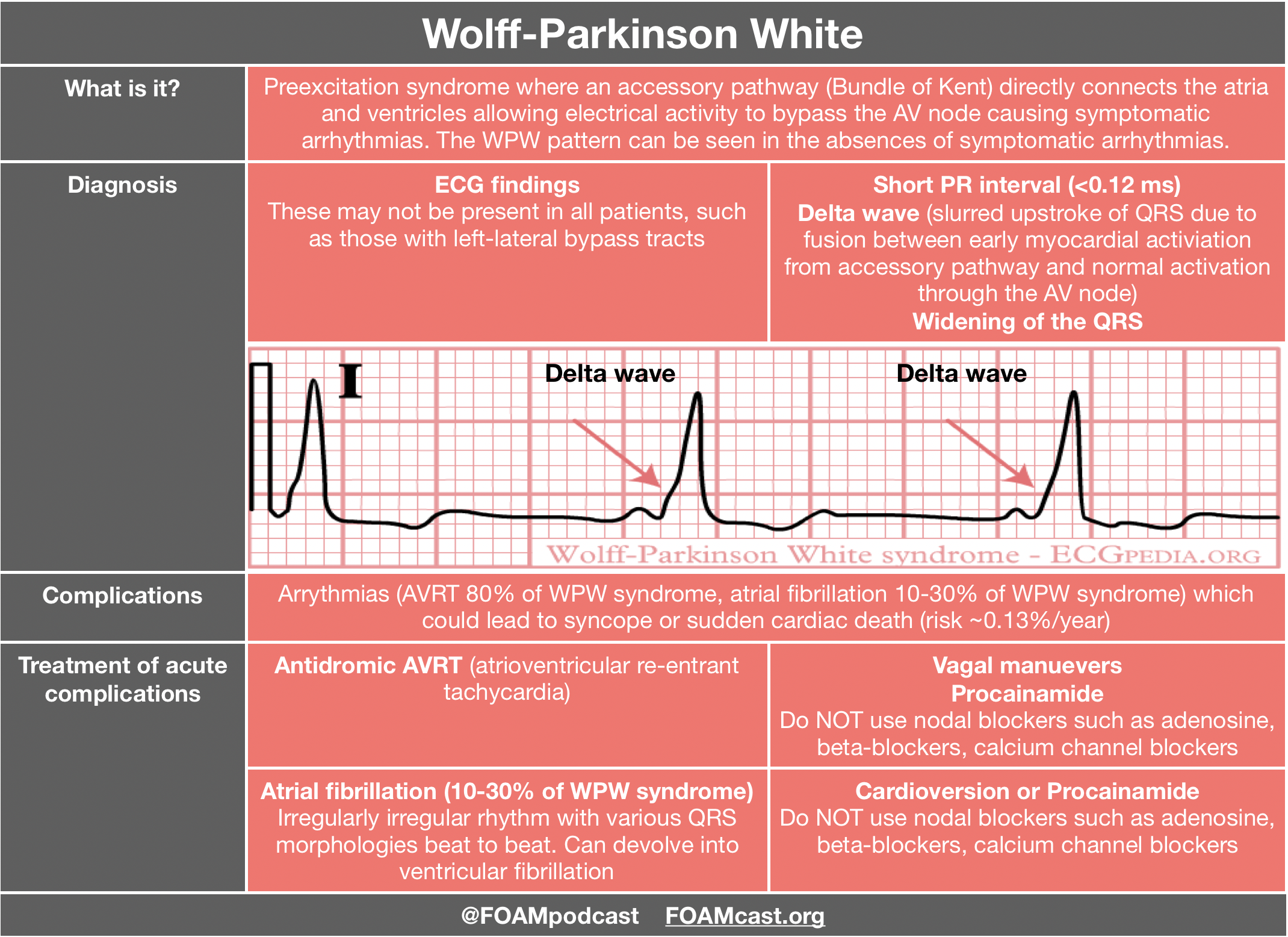
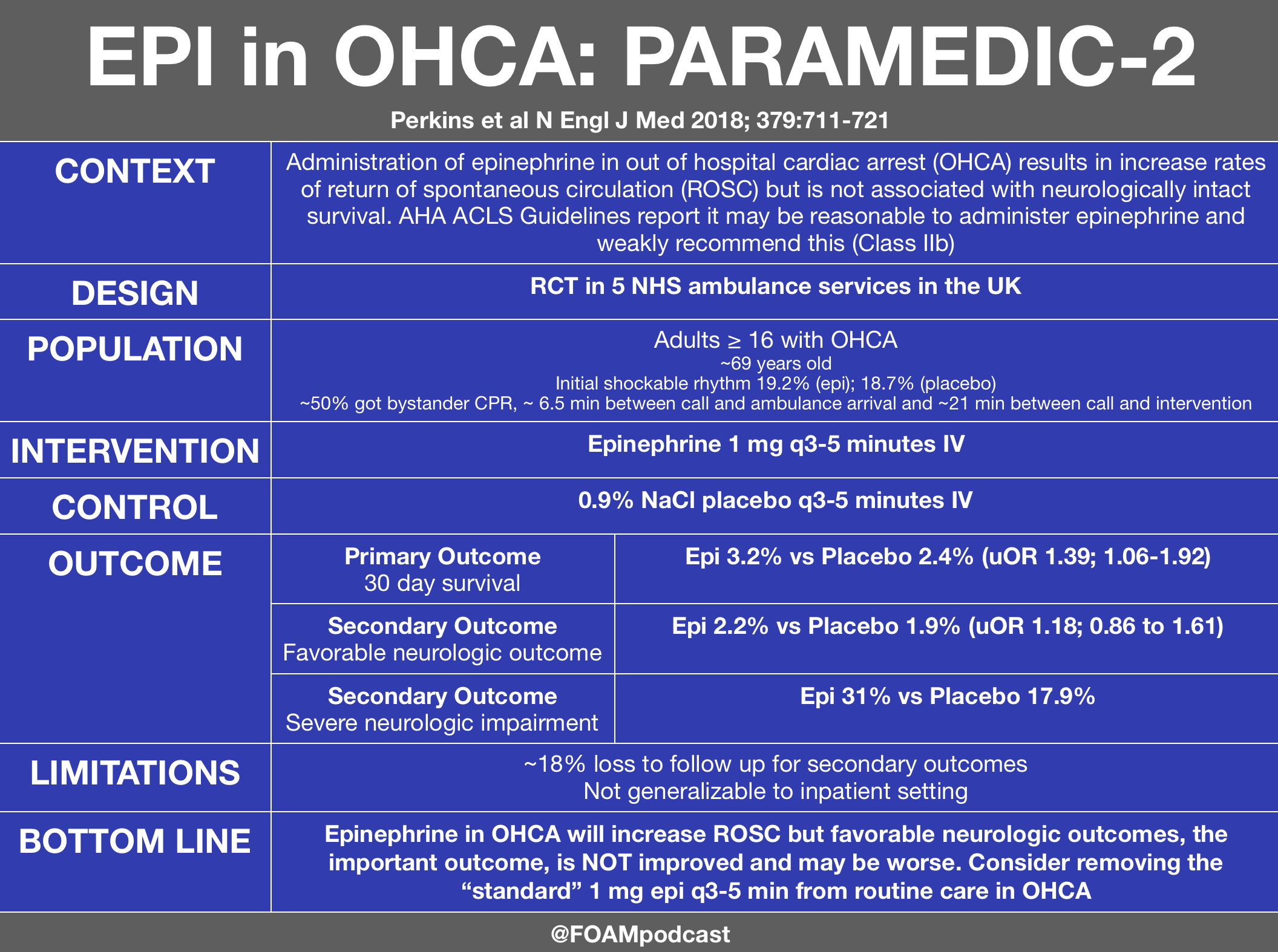
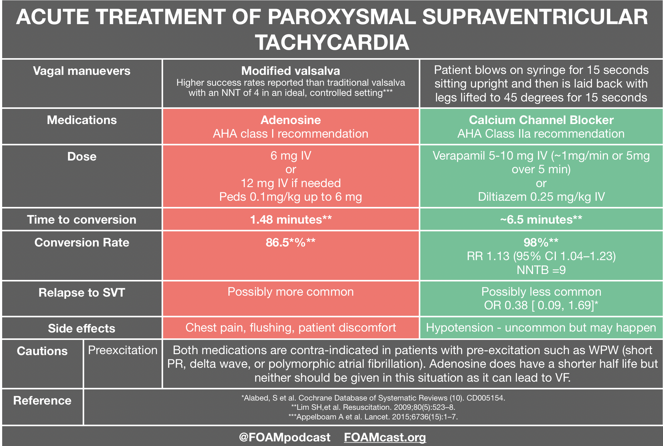
Adenosine and calcium channel blockers are both commonly used in the treatment of SVT; however, practice often varies by region. Advantages to adenosine are the short half-life but this comes with a trade-off of patients experiencing terrifying feelings as they have a sinus pause. Calcium channel blockers have the advantage of not causing those side effects and may prevent recurrence, but patients may infrequently experience hypotension [3,4]. Adenosine and calcium channel blockers are both contra-indicated in pre-excitation syndromes as they may precipitate ventricular fibrillation. A case series is often cited as a reason to not give calcium channel blockers in SVT; however, in these cases 4 of the 5 patients were treated for atrial fibrillation with rapid ventricular response NOT SVT and all patients had signs of preexcitation on their ECG in which use of nodal blockers are discouraged [5].  Both adenosine and calcium channel blockers are recommended by the AHA [1]. In fact, the AHA recommendation for calcium channel blockers in SVT is higher than that for epinephrine in out of hospital cardiac arrest (which is a Class IIb recommendation).

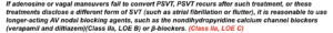
Personally, we like calcium channel blockers for the treatment of stable SVT in patients who do not have signs of pre-excitation.


A 2-month-old male infant presents with rapid heart rate from the pediatrician’s office. The baby’s blood pressure is normal for age and he appears interactive with mom. ECG confirms your suspicion of supraventricular tachycardia. What is the mechanism of action of the medication of choice?

A. Blocks accessory pathway re-entry

B. Decreases conduction through the accessory pathway

C. Decreases conduction through the atrioventricular node and blocks atrioventricular nodal re-entry

D. Increases conduction through the atrioventricular node and blocks atrioventricular nodal re-entry

[accordion]
[toggle title=”Answer” state=”closed”]

C. Adenosine is the pharmacologic treatment of choice for supraventricular tachycardia (SVT). It is a purine nucleoside that decreases the rate of conduction through the atrioventricular (AV) node and blocks AV nodal re-entry. It works directly on adenosine receptors at the AV node. By decreasing the rate of conduction, adenosine effectively slows the anterograde entry of SVT. By blocking AV nodal re-entry, it effectively stops retrograde conduction through the AV node. Thus, adenosine works well for SVT with and without accessory pathways. Adenosine is a very short acting medication and is metabolized quickly, so it should be given as close to the heart as possible. It should be administered through a large gauge peripheral IV, with a flush of 5 to 10 mL of normal saline, traditionally using a 3-way stopcock. SVT is the most common dysrhythmia in childhood. Often, children with SVT are relatively stable. Heart rates for infants are generally over 220 beats per minute and over 180 beats per minute in young children under 2 years old. In children who have normal mentation and stable blood pressures, IV access should be obtained and vagal maneuvers may be attempted. If a child is unstable, synchronized cardioversion at 0.5-1 J/kg should be performed as soon as possible. Cardiology should be consulted for further management, including accessory pathway ablation in many cases.  

Adenosine does not block accessory pathway re-entry (A). Adenosine also does not slow forward conduction of the accessory pathway (B). It only works at the AV node and blocks AV re-entry. Adenosine is effective only at the adenosine receptors on the AV node. Adenosine does not increase conduction through the atrioventricular node (D) but rather decreases conduction at the AV node.

/toggle]
[/accordion]

References:

  1. Page RL, Joglar JA, Caldwell MA, et al. 2015 ACC/AHA/HRS Guideline for the Management of Adult Patients With Supraventricular Tachycardia: A Report of the American College of Cardiology/American Heart Association Task Force on Clinical Practice Guidelines and the Heart Rhythm Society. J Am Coll Cardiol. 2016;67(13):e27-e115.
  2. Appelboam A, Reuben A, Mann C, Gagg J, Ewings P, Barton A, et al. Postural modification to the standard Valsalva manoeuvre for emergency treatment of supraventricular tachycardias ( REVERT ): a randomised controlled trial. Lancet. 2015;6736(15):1–7.
  3. Alabed, S. orcid.org/0000-0002-9960-7587, Sabouni, A., Providencia, R. et al. (3 more authors) (2017) Adenosine versus intravenous calcium channel antagonists for supraventricular tachycardia. Cochrane Database of Systematic Reviews (10). CD005154. https://doi.org/10.1002/14651858.CD005154.pub4
  4. Lim SH, Anantharaman V, Teo WS, Chan YH. Slow infusion of calcium channel blockers compared with intravenous adenosine in the emergency treatment of supraventricular tachycardia. Resuscitation. 2009;80(5):523–8.
  5. Mcgovern B, Garan H, Ruskin JN. Precipitation of cardiac arrest by verapamil in patients with Wolff-Parkinson-White syndrome. Ann Intern Med. 1986;104(6):791-4.

Fluoroquinolones and Aortic Dissection

iTunes or Listen Here

The US Food and Drug Administration (FDA) recently released another warning for fluoroquinolones, this time regarding aortic dissection and aneurysm.

Much of the evidence behind this warning comes from these studies, released in 2015 and 2018 [1-3].

We also review the ACEP Clinical Policy on thoracic aortic dissections [4].

Check out Figure 1 cases on aortic dissection (free educational app for healthcare providers)

A 75-year-old man presents to the emergency department with acute onset of chest pain that radiates to his upper back. Which of the following features would help support a diagnosis of an aortic dissection?

A. A difference of 24 mm Hg in the systolic blood pressures between the upper extremities

B. An increase in heart rate of 120 beats per minute with standing

C. Auscultation of a carotid bruit

D. Chest pain that worsens with inspiration and improves with expiration

[accordion]
[toggle title=”Answer” state=”closed”]

A. An aortic dissection is relatively rare, but it can be life-threatening, so early diagnosis and intervention are critical. They occur more commonly in men aged 60 to 80 years old. Dissections occur secondary to tears in the aortic intima. They can develop due to trauma or degeneration of the aortic media. This deficit in the aorta causes blood to pass into the aortic media, which separates the intima from the surrounding media or adventia. A false lumen then results. Death can occur if the the dissection continues into the aortic valve causing severe aortic regurgitation or if the dissection ruptures into the pericardium causing cardiac tamponade. Other factors that increase mortality include obstruction of the abdominal aortic branch vessels causing end-organ failure or obstruction of the coronary artery causing myocardial infarction. Aortic dissections may be classified as acute or chronic. The acute phase is defined as the first two weeks, and during this time period, the risk of complications or aortic rupture is much higher compared to the chronic phase. Patients will commonly complain of symptoms in the early morning hours as this may be the time of day when the blood pressure is elevated. Chest or back pain is the most common presenting symptom. Patients with a prior history of diabetes or an aortic aneurysm may present with heart failure, syncope, or stroke symptoms. The pain associated with an aortic dissection usually begins abruptly and is severe and sharp in nature with radiation to the back. Weak pulses may occur secondary to impaired blood flow to the peripheral vessels. Often, patients may maintain a variation of over 20 mm Hg in systolic blood pressure between the arms. If the dissection involves the aortic valve, a diastolic murmur associated with aortic regurgitation may be appreciated at the right sternal border. Hypertension is more common in patients with descending aortic dissection compared to hypotension, which is a more classic presentation of ascending aortic dissectionsAn increase in heart rate of 120 beats per minute with standing (B) would likely be associated with postural orthostatic tachycardia syndrome (POTS). Tachycardia may occur as a compensatory mechanism to the hypotension associated with a dissection but it would not be impacted by position changes. Postural orthostatic tachycardia syndrome tends to be more common in teenage girls and will present with anxiety, palpitations, or dizziness. The auscultation of a carotid bruit (C) would classically correlate with carotid artery disease and occurs secondary to turbulent blood flow. It is not commonly associated with an aortic dissection, however, it can correlate with an increased risk of stroke. Chest pain that worsens with inspiration and improves with expiration (D) often correlates with a pulmonary embolism. This type of pain is referred to as pleuritic. While this symptom is not sensitive or specific for an aortic dissection, any patient with chest pain should be thoroughly evaluated for potential cardiac pathology. [/toggle]
[/accordion]

  1. Lee CC, Lee MT, Chen YS, et al. Risk of Aortic Dissection and Aortic Aneurysm in Patients Taking Oral Fluoroquinolone. JAMA Intern Med. 2015;175(11):1839-47.
  2. Lee CC, Lee MG, Hsieh R, et al. Oral Fluoroquinolone and the Risk of Aortic Dissection. J Am Coll Cardiol. 2018;72(12):1369-1378.
  3. Pasternak B, Inghammar M, Svanström H. Fluoroquinolone use and risk of aortic aneurysm and dissection: nationwide cohort study. BMJ. 2018;360:k678.
  4. Clinical Policy: Critical Issues in the Evaluation and Management of Adult Patients With Suspected Acute Nontraumatic Thoracic Aortic Dissection. Ann Emerg Med. 2015; 65(1)

ACEP Non-ST Elevation Acute Coronary Syndrome Guidelines

iTunes or Listen Here

We cover the clinical policy from the American College of Emergency Physicians – Clinical Policy: Critical Issues in the Evaluation and Management of Emergency Department Patients With Suspected Non–ST-Elevation Acute Coronary Syndromes

Check out our sponsor for this show, Figure 1, a free open access application for providers using images and cases.


Rosh Review Emergency Board Review Questions

An elderly man presents with 4 episodes of angina in the past 24 hours. His medical history includes diabetes and asthma. Based on initial testing, you diagnose non-ST-elevation myocardial infarction. You are waiting for the cardiac team to admit him to the critical care unit. In the interim, which of the following is the most appropriate medication to begin?

A. Atelplase

B. Clopidogrel

C. Digoxin

D. Metoprolol

[accordion]
[toggle title=”Answer” state=”closed”]

B. Non-ST-elevation myocardial infarction (NSTEMI) treatment begins with a basic anti-ischemic regimen consisting of oxygen, morphine, nitrates, and possibly beta-blockers and ACE-inhibitors. Antiplatelet medications are then considered. Choices include aspirin, clopidogrel, and prasugrel. NSTEMI treatment is rounded out with anticoagulants such as enoxaparin, bivalirudin, and fondaparinux. Based on risk stratification, definitive treatment may include medications-alone, angiography, percutaneous cardiac intervention or coronary artery bypass surgery. Clopidogrel acts by irreversibly inhibiting a platelet receptor that is needed for activation, thereby inhibiting platelet function. Thrombolytics (fibrinolytics), such as alteplase (A), reteplase and tenecteplase, are contraindicated in the treatment of NSTEMI, as they have shown worse outcomes with their use. Digoxin (C), a cardiac glycoside, is used in treating certain dysrhythmias and heart failure, not myocardial infarction. Beta-blockers such as metoprolol (D) carry a relative contraindication in patients with severe COPD, asthma, atrioventricular block, hypotension or bradycardia.

[/toggle]
[/accordion]

Medicated Assisted Therapy

iTunes or Listen Here

We discussed Medicated Assisted Treatment for opioid use disorder (OUD), focusing on buprenorphine initiated MAT from the ED. In the United States, providers must obtain a DEA X waiver in order to prescribe buprenorphine for OUD For $199, physicians can take a fully online training course, advanced practitioners require additional training. 

Protocol specifics vary by institution. Below is an example of a protocol. The clinical opiate withdrawal scale (COWS) can be found on MDCalcYale’s protocol, along with extensive resources for MAT from the ED can be found here.  Washington ACEP also has online resources .

Check out our sponsor for this show, Figure 1, a free open access application for providers using images and cases.

References:

  1. Berg ML, Idrees U, Ding R, Nesbit SA, Liang HK, and McCarthy ML. Evaluation of the use of buprenorphine for opioid withdrawal in an emergency department. Drug Alcohol Depend. 2007;86(2-3):239-44. doi:10.1016/j.drugalcdep.2006.06.014.
  2. Mattick RP, Breen C, Kimber J, Davoli M. Buprenorphine maintenance versus placebo or methadone maintenance for opioid dependence. Cochrane Database Syst Rev. 2014;(2):CD00220
  3. Pierce M, Bird SM, Hickman M, Marsden J, Dunn G, Jones A, and Millar T. Impact of treatment for opioid dependence on fatal drug-related poisoning: a national cohort study in England. Addiction. 2016;111(2):298-308. doi:10.1111/add.13193.
  4. Clark RE, Samnaliev M, Baxter JD, and Leung GY. The evidence doesn’t justify steps by state Medicaid programs to restrict opioid addiction treatment with buprenorphine. Health Aff (Millwood). 2011;30(8):1425-33. doi:10.1377/hlthaff.2010.0532
  5. Martin SA, Chiodo LM, Bosse JD, Wilson A. The Next Stage of buprenorphine Care for Opioid Use disorder. Annal of Internal Medicine

Magnesium

iTunes or Listen Here

The Free Open Access Medical Education (FOAM) world is often obsessed with magnesium. There are two situations that get unequivocal, high recommendations to give intravenous (IV) magnesium – eclampsia and polymorphic ventricular tachycardia. In this show we address the use of magnesium for various applications.

Magnesium for Acute Atrial Fibrillation with Rapid Ventricular Response (RVR)

  1. Bouida et al. LOw dose MAGnesium sulfate versus HIgh dose in the early management of rapid atrial fibrillation: randomised controlled double blind study. Acad Emerg Med. 2018 Jul 19.
  2. Ho KM, Sheridan DJ, Paterson T. Use of intravenous magnesium to treat acute onset atrial fibrillation: a meta-analysis. Heart. 2007;93(11):1433-40.
  3. Davey MJ, Teubner D. A randomized controlled trial of magnesium sulfate, in addition to usual care, for rate control in atrial fibrillation. Ann Emerg Med. 2005;45(4):347-53.

Magnesium for Migraine

  1. Corbo J, Esses D, Bijur PE, Iannaccone R, Gallagher EJ. Randomized clinical trial of intravenous magnesium sulfate as an adjunctive medication for emergency department treatment of migraine headache. Ann Emerg Med. 2001;38(6):621–7.
  2. Bigal ME, Bordini CA, Tepper SJ, Speciali JG. Intravenous magnesium sulphate in the acute treatment of migraine without aura and migraine with aura. A randomized, double-blind, placebo-controlled study. Cephalalgia. 2002;22(5):345-53.
  3. 1Cete Y, Dora B, Ertan C, Ozdemir C, Oktay C. A randomized prospective placebo-controlled study of intravenous magnesium sulphate vs. metoclopramide in the management of acute migraine attacks in the Emergency Department. Cephalalgia. 2005;25(3):199–204.
  4. Shahrami A, Assarzadegan F, Hatamabadi HR, Asgarzadeh M, Sarehbandi B, Asgarzadeh S. Comparison of therapeutic effects of magnesium sulfate vs. dexamethasone/metoclopramide on alleviating acute migraine headache. J Emerg Med. 2015;48(1):69–76.
  5. Orr SL, Friedman BW, Christie S, Minen MT, Bamford C, Kelley NE, et al. Management of Adults with Acute Migraine in the Emergency Department: The American Headache Society Evidence Assessment of Parenteral Pharmacotherapies. Headache. 2016;56(6):911–40.

Magnesium for Acute Asthma Exacerbation

Griffiths B, Kew KM. Intravenous magnesium sulfate for treating children with acute asthma in the emergency department. Cochrane Database Syst Rev. 2016;4:CD011050.
Kew KM, Kirtchuk L, Michell CI. Intravenous magnesium sulfate for treating adults with acute asthma in the emergency department. Cochrane Database Syst Rev. 2014;(5):CD010909.

2018 Literature Review

iTunes or Listen Here

In this episode we highlight key articles in Emergency Medicine that came out, thus far, in 2018.

D’souza et al. Effects of prophylactic anticholinergic medications to decrease extrapyramidal side effects in patients taking acute antiemetic drugs: a systematic review and meta-analysis Emerg Med J. 2018;35(5):325-331.

Driver et al. Effect of Use of a Bougie vs Endotracheal Tube and Stylet on First-Attempt Intubation Success Among Patients With Difficult Airways Undergoing Emergency Intubation: A Randomized Clinical Trial. JAMA. 2018;319(21):2179-2189.Atkinson PR, Milne J, Diegelmann L, et al. Does Point-of-Care Ultrasonography Improve Clinical Outcomes in Emergency Department Patients With Undifferentiated Hypotension? An International Randomized Controlled Trial From the SHoC-ED Investigators. Ann Emerg Med. 2018; In press.

Perkins GD et al. A Randomized Trial of Epinephrine in Out-of-Hospital Cardiac Arrest. N Engl J Med 2018; 379:711-721

Meltzer et al. Effect of Tamsulosin on Passage of Symptomatic Ureteral Stones: A Randomized Clinical Trial. JAMA Intern Med. 2018 Aug 1;178(8):1051-1057

Kabrhel et al Multicenter Evaluation of the YEARS Criteria in Emergency Department Patients Evaluated for Pulmonary Embolism. Acad Emerg Med. 2018;25(9):987-994

Myths in imaging and pediatric emergency medicine

iTunes or Listen Here

We are in San Diego for the American College of Emergency Physicians (ACEP) Scientific Assembly and are bringing you daily pearls from the lectures.

References

  1. Chen F, Shen YH, Zhu XQ, Zheng J, Wu FJ. Comparison between CT and MRI in the assessment of pulmonary embolism: A meta-analysis. Medicine. 2017;96(52):e8935.
  2. Akhter M et al. Ruling out Pulmonary Embolism in Patients with High Pretest Probability. Western Journal of Emergency Medicine. 2018; (18)3.
  3. Hogg K, Brown G, Dunning J, et al. Diagnosis of pulmonary embolism with CT pulmonary angiography: a systematic review. Emerg Med J. 2006;23(3):172-8.
  4. Mccrindle BW, Rowley AH, Newburger JW, et al. Diagnosis, Treatment, and Long-Term Management of Kawasaki Disease: A Scientific Statement for Health Professionals From the American Heart Association. Circulation. 2017;135(17):e927-e999.
  5. Schwartz RH, Kim D, Martin M, Pichichero ME. A Reappraisal of the Minimum Duration of Antibiotic Treatment Before Approval of Return to School for Children With Streptococcal Pharyngitis. Pediatr Infect Dis J. 2015;34(12):1302-4.

Problems with CYA medicine and biomarkers

iTunes or Listen Here

We are in San Diego for the American College of Emergency Physicians (ACEP) Scientific Assembly and are bringing you daily pearls from the lectures.

We cover a lecture by Nathan Schlicher on problems with CYA medicine including admissions (which can be harmful), imaging (also can be harmful), and polypharmacy.

We also cover a lecture by Haney Mallemat on the utility of common biomarkers.

Severe pediatric head trauma, cutting edge aortic dissection diagnostics, and chest tube pearls

iTunes or Listen Here

We are in San Diego for the American College of Emergency Physicians (ACEP) Scientific Assembly and are bringing you daily pearls from the lectures.

Nazerian P, Mueller C, Dematosoeiro A, et al. Diagnostic Accuracy of the Aortic Dissection Detection Risk Score Plus D-Dimer for Acute Aortic Syndromes: The ADvISED Prospective Multicenter Study. Circulation. 2018 Jan 16;137(3):250-258

Check out this post from Highland on regional anesthesia for rib fractures