Lyme Disease (Borreliosis)

iTunes or Listen Here

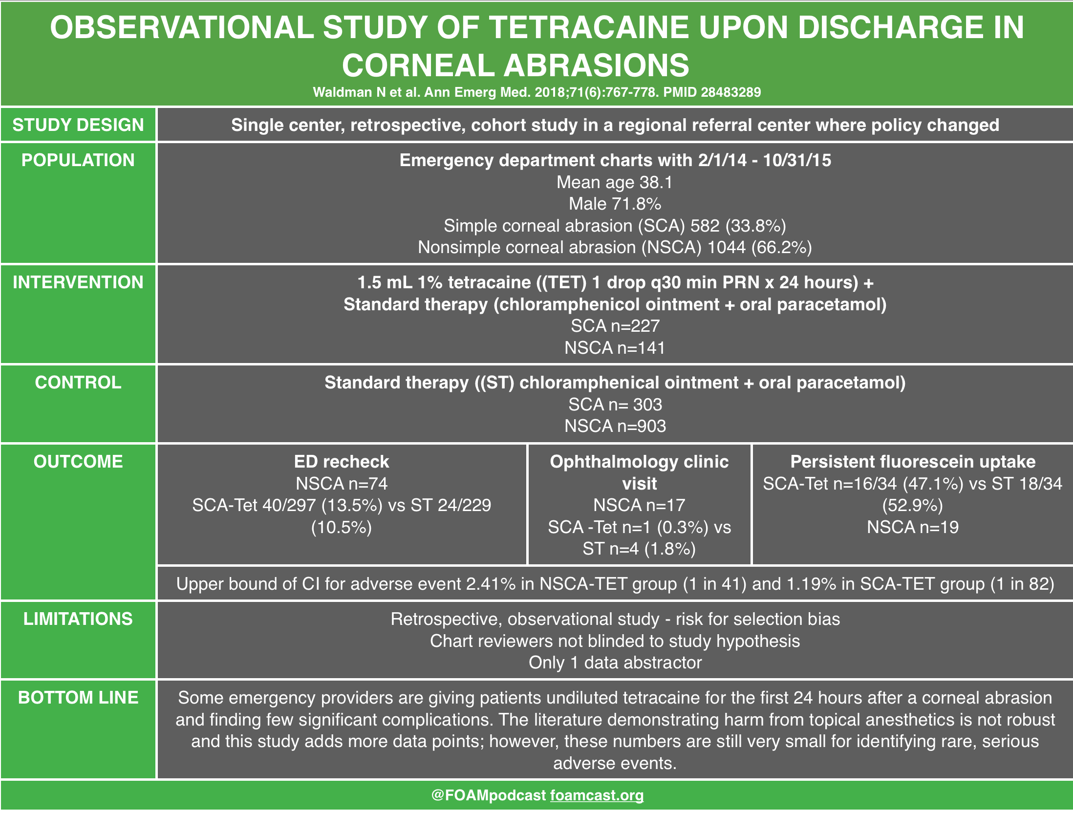
We cover a JAMA Clinical Reviews podcast on lyme disease, including some myth-busters.

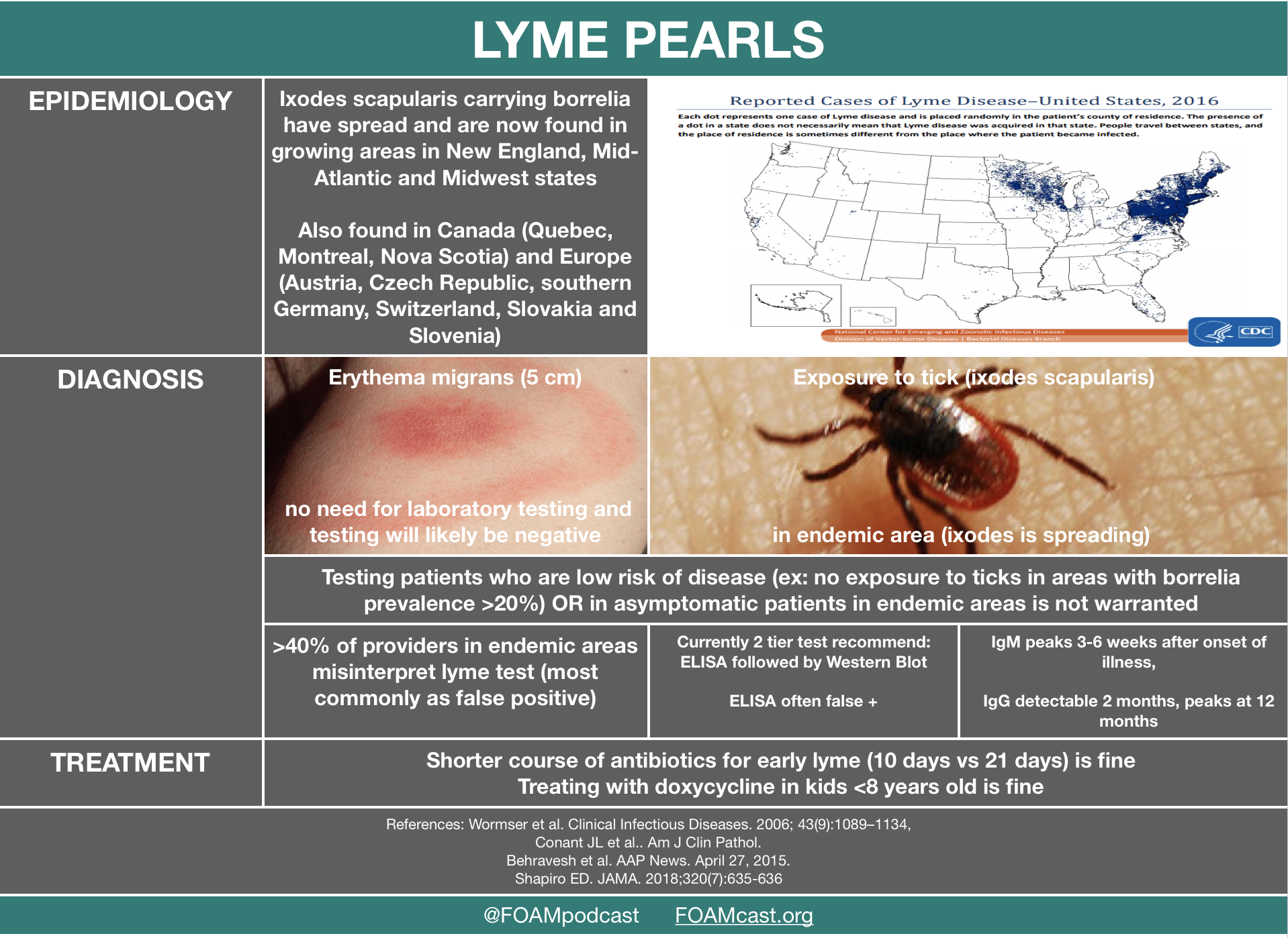
  • Doxycycline can be used, safely, in kids < 8 years old [1].
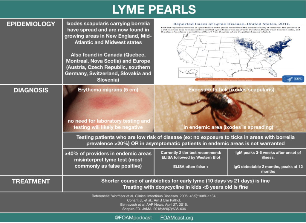
  • Testing for lyme is a mess because :
    • (1)  we test patients with an ultra low probability of disease
    • (2) we test patients who shouldn’t be tested (i.e. have erythema migrans and thus very high probability)
    • (3) the tests are a pain to interpret and many clinicians (42.4% in lyme endemic Vermont) misinterpret tests, most commonly as false positive [2].
  • Lyme disease is spreading further south and west in the US, into Canada, and it’s also increasingly found in Europe [3,4].


Rosh Review Emergency Board Review Questions

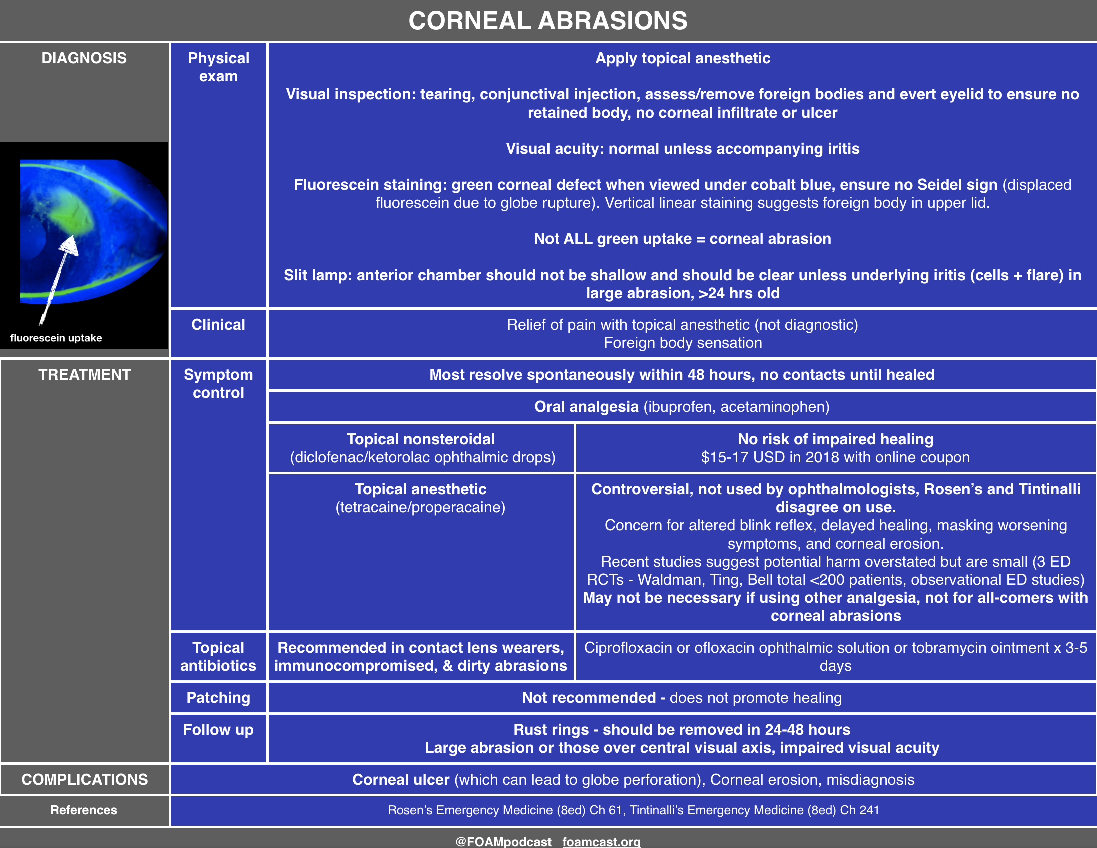
A 32-year-old man with a history of hypertension and sickle cell disease presents to the ED for intermittent fevers. He has been feeling ill for the past few weeks with intermittent headaches, night sweats, and abdominal pain. He recently returned from Maine after a trip to see the fall colors. His vital signs are only remarkable for a temperature of 100.7oF. Physical exam reveals mild scleral icterus and hepatomegaly. The patient’s Wright stain shows intraerythrocytic rings. What co-infection is common in this disease?

A. Babesia microti

B. Borrelia burgdorferi

C. Francisella tularensis

D. Rickettsia rickettsii

[accordion]
[toggle title=”Answer” state=”closed”]

A.This patient’s presentation is consistent with acute babesiosis infection. Patients are commonly co-infected with Borrelia burgdorferi (Lyme disease) or ehrlichiosis. Risk factors include functional or surgical asplenia, immunocompromised state, and advanced age. This patient likely has functional asplenia given his age and history of sickle cell disease. Diagnosis is confirmed with a Wright or Giemsa stain showing intraerythrocytic rings, similar to malaria. Babesiosis is due to Babesia parasite and transmitted via the Ixodes deer tick. Patients present with a wide array of symptoms from a vague viral syndrome-type symptoms and spiking fevers to hepatomegaly and hemolytic anemia. Patients with risk factors tend to have more severe features including severe hemolysis, jaundice, renal failure, and acute respiratory distress syndrome. Treatment is with atovaquone and clindamycin or azithromycin.

[/toggle]
[/accordion

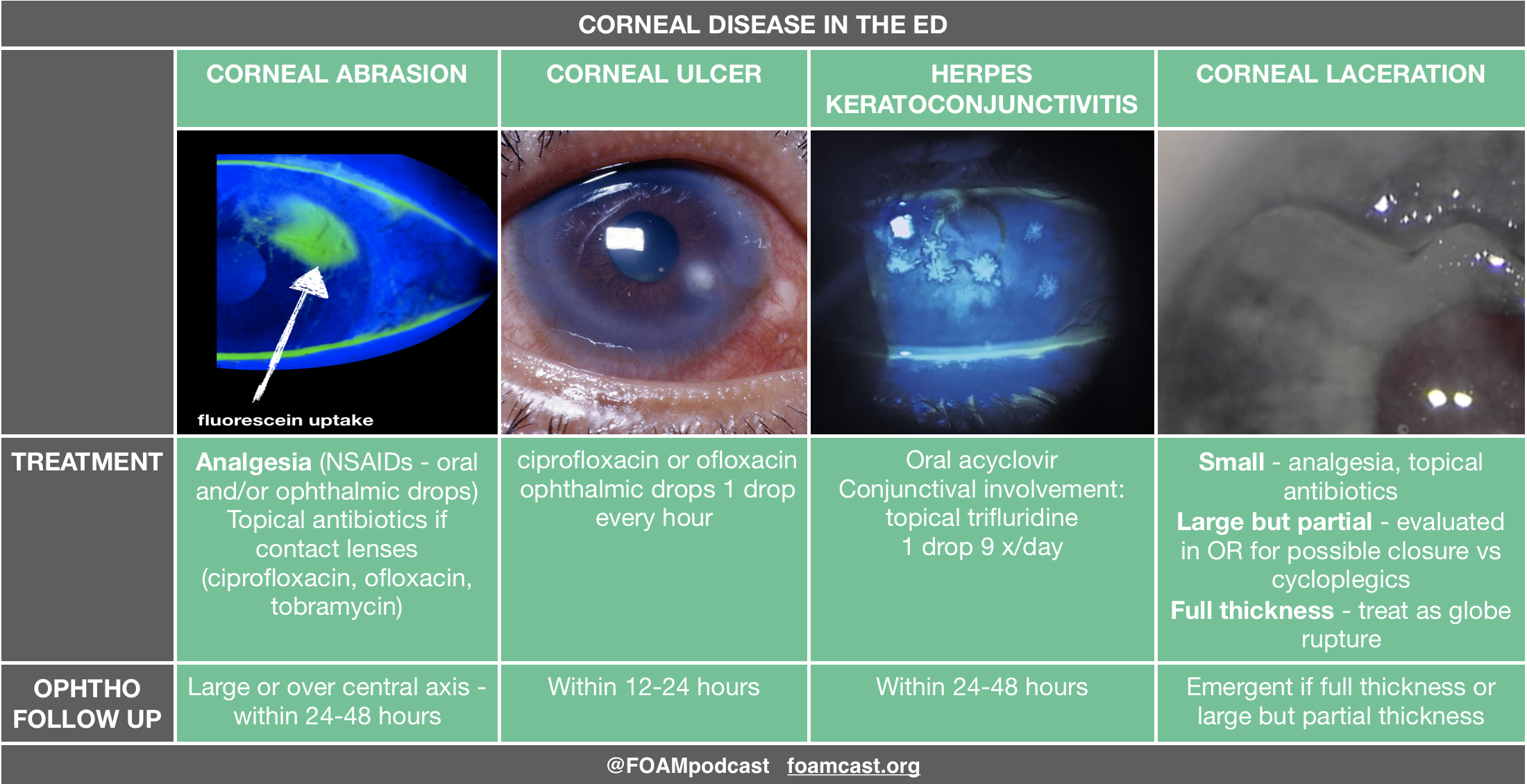
Which of the following would be the best antibiotic choice for first-line treatment of a 5-year-old who presents to your office with multiple erythema migrans lesions but no cardiac or neurological symptoms?

A. Amoxicillin PO for 14 to 21 days

B. Azithromycin PO for 14 to 21 days

C. Doxycycline PO for 10 to 21 days

D. Ceftriaxone IM for 14 days

[accordion]
[toggle title=”Answer” state=”closed”]

***This is an example of the difference between practice and board exams, which tend to lag behind current knowledge.

Correct-Answer: Amoxicillin PO for 14 to 21 days. Lyme disease is a tick-borne illness that is caused by Borrelia burgdorferi, making it a spirochetal infection. When deciding the medical therapy for Lyme disease, staging is important. Early localized disease usually presents with a “bullseye rash,” otherwise known as erythema migrans. Patients in this stage of disease may have a few constitutional symptoms, such as fever, fatigue, headache, and myalgias. Early disseminated disease is present if patients have multiple erythema migrans, cardiac or neurologic findings. Late disease usually involves persistent arthritis of a large joint or more severe neurological findings such as encephalopathy or polyneuropathy. In the case above, amoxicillin is the best choice of treatment because the patient is under 8 years of age, and, while multiple lesions are present, the patient does not have neurological or cardiac involvement and should be treated with the same therapy as if he had a single lesion. Recommended duration of therapy is between 14 and 21 days. The goal of therapy is to reduce the risk of developing late Lyme disease and to shorten the duration of symptoms. Azithromycin PO for 14 to 21 days (B) is not recommended as a first-line treatment for Lyme disease because it is less effective than amoxicillin. There has been documented resistance to macrolides by some strains of Borrelia burgdorferi. Doxycycline PO for 10 to 21 days (C) is not recommended in this case because of the patient’s age. Tetracyclines are not recommended for children under 8 years of age because they can lead to permanent staining of their teeth. Ceftriaxone IM for 14 days (D) is not recommended because oral antibiotics, such as doxycycline, are just as effective for treatment of erythema migrans and make for easier administration.

[/toggle]
[/accordion

References:

  1. Todd S et al. No Visible Dental Staining in Children Treated with Doxycycline for Suspected Rocky Mountain Spotted Fever. The Journal of Pediatrics. May 2015. Volume 166, Issue 5, Pages 1246–1251
  2. Conant JL, Powers J, Sharp G, Mead PS, Nelson CA. Lyme Disease Testing in a High-Incidence State: Clinician Knowledge and Patterns. Am J Clin Pathol. 2018;149(3):234-240.
  3. European Centre for Disease Prevention and Control Accessed 9.22.2018
  4. Infectious Disease Society of America’s  2006 Lyme Guidelines
  5.  Min Han J et al. Comparative effectiveness and toxicity of oral antibiotics for early Lyme disease associated with erythema migrans: a systematic review and network meta-analysis. ECCMID abstract . 2017.
  6. Tintinalli’s Emergency Medicine: A Comprehensive Review. Chapter 160 “Zoonotic infections.”
  7. Rosen’s Emergency Medicine. (8 ed) . Chapter 126 “Tickborne Illnesses”

Penile fracture and priapism

iTunes or Listen Here

We review the American Urological Association guidelines on penile fracture and priapism. We supplement this with core content from Tintinalli and Rosen’s.

Rosh Review Emergency Board Review Questions

A 16-year-old boy with sickle cell disease presents to the emergency department having intermittent but frequent short and painful erections. He denies current pain but reports having had four unwanted painful erections beginning this morning, each becoming progressively longer with the most recent lasting one hour. On exam he is well appearing and in no distress with a flaccid non-tender penis on genitourinary exam. Which of the following is the most appropriate next step?

A. Corporal aspiration

B. Oxygen

C. Pseudoephedrine

D. Warm penile compress

[accordion]
[toggle title=”Answer” state=”closed”]

C. Priapism is a painful erection unrelated to sexual stimulation. It is most common in sickle cell patients and is usually an ischemic, lowflow process resulting in venous stasis and sickling of cells. Irreversible tissue damage begins at six hours, so patients with a painful erection are taught to seek medical attention with erections lasting more than four hours. Optimal management is not well defined and local expert opinion may vary. Brief episodes can often be managed at home with oral analgesics, brief exercise, warm/cold compresses, or showering. Recurrent episodes of priapism such as seen in our patient are referred to as stuttering priapism which may herald a longer event. The first-line treatment for stuttering priapism is with an oral alpha-adrenergic agent such as pseudoephedrine. Patients with priapism lasting more than four hours are treated with a combination of intravenous fluids, intravenous analgesia, and emergent urology consult for corporal aspiration. Conscious sedation may be indicated depending on patient age and anxiety. Once low-flow priapism is confirmed, intracavernosal injection of an alpha-adrenergic receptor agent such as phenylephrine can help to achieve detumescence. If multiple attempts at corporal aspiration fail, a surgical shunt may be indicated.

Corporal aspiration (A) is not recommended in patients with current detumescence as in our patient. Oxygen (B) is recommended if hypoxic but has not been shown to assist in achieving detumescence. Warm penile compresses (D) are a recommended home therapy to achieve detumescence early in a painful erection but will not help between recurrent stuttering priapism.

[/toggle]
[/accordion]

A 21-year-old man presents to the Emergency Department with sudden onset penile pain that occurred while having intercourse with his wife. Physical examination reveals an angulated circumcised penis with fusiform swelling and ecchymosis. Which of the following is the next best step in management?

A. Analgesic therapy and outpatient urology follow up

B. Foley catheter placement

C. Intracavernosal phenylephrine injection

D. Urologic consultation for surgical repair

[accordion]
[toggle title=”Answer” state=”closed”]

D. A urologic consultation for surgical repair is required to optimize functional outcome in the case of penile fracture. Penile fractures occur during direct penile trauma and involves rupture of Buck’s fascia, tunica albuginea, or corpora cavernosa. Symptoms include acute pain and swelling, angulation of the penis, and loss of erection. The penis may take on a characteristic “eggplant deformity” due to swelling and ecchymosis from extravasated blood that collects within Buck’s fascia. Complications include deep dorsal vein injury and partial or complete urethral rupture. Sexual intercourse is the most common cause, but other causes include animal bites, ballistic injuries, stab wounds, and vigorous masterbation. Management involves early surgical repair of the tunica albuginea (within 24-36 hours after the injury) to improve functionality.

[/toggle]
[/accordion]

Corneal Emergencies

iTunes or Listen Here

We review one of the editors top picks from Annals of Emergency Medicine, which are free for 6 months from issue release.  The article covered in this podcast is Waldman et al, is a study of tetracaine in corneal abrasions.

Review from First10EM on topical anesthetics and corneal abrasions.

Rosh Review Emergency Board Review Questions

A 21-year-old man presents to the emergency department complaining of left eye pain. Fluorescein staining reveals a dendritic lesion over the cornea. What is the most likely diagnosis based on this finding?

A. Bacterial conjunctivitis

B. Corneal abrasion

C. Herpes keratitis

D. Retinal detachment

[accordion]
[toggle title=”Answer” state=”closed”]

A dendritic lesion of the cornea detected by fluorescein staining indicates the presence of herpetic keratitis. Herpetic keratitis is caused by herpes simplex virus and can lead to blindness because of scarring and opacification of the cornea. A typical presentation includes a painful eye, blurred vision or excessive tearing. Diagnosis can be made with fluorescein staining which gives the classic dendritic appearance. The presence of a dendritic lesion indicates active replication of the virus. Superficial epithelial herpetic keratitis can be successfully treated in two to three weeks with topical antivirals such as ganciclovir. Topical steroids must be avoided in patients with herpetic keratitis as the herpetic lesions can become much deeper and threaten the patient’s vision. Oral antivirals have not been studied comparatively with topical antivirals. Risk of transmission of ocular herpes simplex virus to another person is thought to be extremely low.

Bacterial conjunctivitis (A) can cause eye discomfort but typically presents with copious purulent discharge from one or both eyes. Fluorescein staining of the eye will be negative for any defect. Corneal abrasions (B) present with a superficial epithelial lesion that is usually linear in shape. The cornea is expected to be clear. Retinal detachment (D) is painless and does not cause a change in the appearance of the surface of the eye. Fundoscopic exam will often show rugae indicating the edge of the retina floating in the vitreous humor.

[accordion]
[toggle title=”Answer” state=”closed”]

A 20-year-old woman presents to the emergency department for left eye pain. Yesterday she scratched her eye while putting in her contact lens. She has had constant left eye pain and tearing since. She has no foreign body sensation. On exam, her visual acuity is at baseline and equal in both eyes. Her fluorescein exam findings are shown below. Which of the following is the most appropriate treatment for this patient’s condition?

A. Erythromycin ophthalmic ointment

B. Gentamicin-prednisolone ophthalmic suspension

C. Tetracaine ophthalmic solution

D. Tobramycin ophthalmic ointment

[accordion]
[toggle title=”Answer” state=”closed”]

D. This patient’s fluorescein exam is consistent with a corneal abrasion. It is caused by direct mechanical damage, leading to a partial-thickness corneal injury. Athletes, contact lens wearers, welders, and glass workers may present more often with these injuries. Diagnosis is confirmed when fluorescein dye highlights the usually linear or punctate abrasions. Multiple vertical corneal abrasions may indicate a retained foreign body beneath the eyelid. Corneal abrasion fluorescein exam findings are not to be confused with open globe injuries (streaking of the dye from the site of injury), corneal ulcers (circular patches of dye uptake with ragged, “heaped up” edges), and herpes simplex keratitis (dendritic pattern uptake). Patients commonly present with eye pain and tearing with a usually known mechanism of injury. The mainstay of treatment includes pain control, infection prophylaxis, and preventative care to avoid abrasions in the future. Pain control includes cycloplegics like homatropine and cyclopentolate and nonsteroidal anti-inflammatory medications. Commonly prescribed antibiotic ointments include erythromycin, ciprofloxacin, tobramycin, and gentamicin. Notably, erythromycin does not cover pseudomonal infections, which are more likely to occur in contact lens wearers.  Erythromycin (A) does not have appropriate pseudomonal coverage for this patient who wears contact lenses and has a corneal abrasion. Ophthalmic medications containing steroids, like gentamicin-prednisolone (B), should not be prescribed in corneal abrasions or ulcers as they may decrease healing rates. While in the emergency department, tetracaine (C) may be used to more easily evaluate the patient’s eye. However, ocular anesthetics should not be prescribed as they decrease healing rates and block the basic corneal reflex to avoid further injury.

[/toggle]
[/accordion]

References:

  1. Waldman N, Winrow B, Densie I, et al. An Observational Study to Determine Whether Routinely Sending Patients Home With a 24-Hour Supply of Topical Tetracaine From the Emergency Department for Simple Corneal Abrasion Pain Is Potentially Safe. Ann Emerg Med. 2018;71(6):767-778.
  2. Ball IM, Seabrook J, Desai N, Allen L, Anderson S. Dilute proparacaine for the management of acute corneal injuries in the emergency department. CJEM. 2010;12:(5)389-96.
  3. Ting JY, Barns KJ, Holmes JL. Management of Ocular Trauma in Emergency (MOTE) Trial: A pilot randomized double-blinded trial comparing topical amethocaine with saline in the outpatient management of corneal trauma. J Emerg Trauma Shock. 2009;2:(1)10-4.
  4. Swaminathan A, Otterness K, Milne K, Rezaie S. The safety of topical anesthetics in the treatment of corneal abrasions: A review. J Emerg Med 2015
  5. Calder LA, Balasubramanian S, Fergusson D. Topical nonsteroidal anti-inflammatory drugs for corneal abrasions: meta-analysis of randomized trials. Acad Emerg Med. 2005;12(5):467-73.
  6. Galuma K, Lee J. “Ophthalmology.” Rosen’s Emergency Medicine: Concepts and Clinical Practice, Chapter 61, 790-819.e3

Clostridium difficile (c diff)

iTunes or Listen Here

The Infectious Disease Society of America (IDSA)/SHEA released new  clostridium difficile (c diff) guidelines in 2017.


Rosh Review Emergency Board Review Questions

An 85-year-old woman who was recently treated with ciprofloxacin for a urinary tract infection presents to the emergency department with diarrhea and lightheadedness. Her vital signs are heart rate 95 bpm, blood pressure 84/50 mm Hg, and temperature 38.9°C. Laboratory studies are notable for a white blood cell count of 19,000 and creatinine of 2.8 mg/dL. On abdominal X-ray, the transverse width of the patient’s colon is 7 cm. Chest X-ray and urinalysis are unremarkable. In addition to fluid resuscitation, what is the most appropriate treatment for this patient?

A. Intravenous and rectal vancomycin

B. Intravenous vancomycin and oral ciprofloxacin

C. Oral metronidazole and surgical consultation

D. Oral vancomycin and surgical consultation

[accordion]
[toggle title=”Item 1 Title” state=”closed”]

D. This patient presents with severe, life-threatening Clostridium difficile colitis. This is evidenced by a history of severe diarrhea preceded by recent antibiotic use. When the normal microbial flora of the colon is disrupted by exposure to antibiotics, Clostridium difficile can opportunistically dominate. It produces several toxins, which cause inflammation of the colon (colitis). Clinically, these patients present with watery diarrhea and crampy abdominal pain. Fever is often present, particularly in severe cases. This is a severe case of colitis due to the white blood cell count greater than 15,000, impaired renal function and presence of severe sepsis. This patient should receive empiric treatment with oral vancomycin. Given the dilated colon on abdominal X-ray, this patient likely has toxic megacolon and requires early surgical consultation to evaluate for surgical colectomy (if condition is unresponsive to medical management). Clostridium difficile is a species of gram-positive spore-forming bacteria that can exist in the human colon. A novel macrocyclic antibiotic called fidaxomicin was recently approved for the treatment of Clostridium difficile colitis. However, there is a lack of evidence to support its use in life-threatening illness.

The use of oral metronidazole (C) is appropriate for mild infections, but its use in severe disease is strongly discouraged. Intravenous vancomycin (A and B) is not a recommended treatment for Clostridium difficile colitis, as bactericidal concentrations are not achieved in the colon. Moreover, ciprofloxacin is a cause of Clostridium difficile colitis not a treatment. Rectal vancomycin can be considered as an adjunct to oral vancomycin when ileus is present.

[/toggle]
[/accordion]

Pulmonary Embolism Risk Stratification and ACEP Clinical Policy

iTunes or Listen Here

We cover the American College of Emergency Physicians clinical policy on Acute Venous Thromboembolic Disease (i.e. PE) [1]

Risk Stratification in Pulmonary Embolism

 In the United States, the workup rate for PE is astonishingly high, with 1-2% of all ED patients receiving a CTPA for PE and a low yield (~5-10% of CTPAs are positive for PE). Thus, major medical societies such as ACEP and the American College of Physicians recommend the use of risk stratification tools [1,2]. For some reason, approximately 25% of patients who are low risk AND PERC negative still receive d-dimer or imaging for PE, despite the risk of PE being exceptionally low. To be clear, use of clinical decision tools are not mandatory and do not necessarily perform better than clinical gestalt [9]. These tools may serve as a “reality check” for some, reminding them that patients may be at lower risk of PE than they think

Rosh Review Emergency Board Review Questions

References:

  1. Acute Venous Thromboembolic Disease. ACEP Clinical Policy. 2018
  2. Raja AS et al. Evaluation of patients with suspected acute pulmonary embolism: Best practice advice from the Clinical Guidelines Committee of the American College of Physicians. Ann Intern Med. 2015;163(9):701–11.
  3. Bariteau A, Stewart LK, Emmett TW, Kline JA. Systematic Review and Meta-analysis of Outcomes of Patients With Subsegmental Pulmonary Embolism With and Without Anticoagulation Treatment. Acad Emerg Med. 2018 (in Press)
  4. Kline JA, Mitchell AM, Kabrhel C, Richman PB, Courtney DM. Clinical criteria to prevent unnecessary diagnostic testing in emergency department patients with suspected pulmonary embolism. J Thromb Haemost JTH [Internet]. 2004;2(8):1247–55.
  5. Buchanan I, Teeples T, Carlson M, Steenblik J, Bledsoe J, Madsen T. Pulmonary Embolism Testing Among Emergency Department Patients Who Are Pulmonary Embolism Rule-out Criteria Negative. Acad Emerg Med. 2017;24(11):1369–76.
  6. Courtney DM, Miller C, Smithline H, Klekowski N, Hogg M, Kline JA. Prospective multicenter assessment of interobserver agreement for radiologist interpretation of multidetector computerized tomographic angiography for pulmonary embolism. J Thromb Haemost. 2010;8(3):533–9.
  7. Wells PS, et al. Derivation of a simple clinical model to categorize patients probability of pulmonary embolism: increasing the models utility with the SimpliRED D-dimer. Thromb Haemost. 2000;83:416-20. [PMID: 10744147]
  8. Gibson NS et al; Christopher study investigators. Further validation and simplification of the Wells clinical decision rule in pulmonary embolism. Thromb Haemost. 2008;99:229-34. [PMID: 18217159]
  9. Penaloza A, Verschuren F, Meyer G, Quentin-Georget S, Soulie C, Thys F, et al. Comparison of the Unstructured Clinician Gestalt, the Wells Score, and the Revised Geneva Score to Estimate Pretest Probability for Suspected Pulmonary Embolism. Ann Emerg Med [Internet]. 2013 Aug;62(2):117–124.e2. Available from: http://linkinghub.elsevier.com/retrieve/pii/S0196064412017180

Emergency Care of Lactating Patients

iTunes or Listen Here

Emergency providers receive training to care for pregnant patients but little training to care for breastfeeding patients. Many myths persist, particularly urging patients to “pump and dump” (pump and discard milk instead of feeding the baby) after medications or illness that are, in fact, completely safe to continue breastfeeding through . We review popular medications used in the emergency department (ED) as well as other pearls regarding the ED care of lactating patients (references for drugs come from LactMed, other references are listed at bottom of page).

Medications During Breastfeeding

We often obsess over medications that are safe in pregnancy but may not have much of an idea of what medications are ok in patients who are breastfeeding. Rare medications are unsafe in breastfeeding (chemotherapy, radioactive isotopes) but most that we prescribe in the emergency department are fine (including anesthesia/procedural sedation meds – see this Anesthesia graphic). Please review medications prior to prescribing to ensure that they are safe for the infant and will have no deleterious effects on lactation. For example, pseudoephedrine, a seemingly benign medication, can dry up a patient’s milk supply.

LactMed is a searchable database provided by the National Institutes of Health that summarizes the safety of medications in breastfeeding and the effects of medications on lactation. There is also a free LactMed app. Providers and patients can also call InfantRisk( 806) 352-2519  regarding specific medications (Mon-Fri 9am-6pm CST)

**Note: Some sources cite different safety profiles so we have tried to synthesize this information but depending on the patient and the source, the risk profile may be a bit different.

Medical illnesses specific to the lactating patient

General support for the lactating patient

Thanks to the physicians and lactation experts of Dr. Milk (@DoctorDrMilk) for providing peer review.

Rosh Review Emergency Board Review Questions

A 36-year-old woman presents to the office for a painful right breast for the past two days. She is one month postpartum. The patient reports swelling and redness of the right breast in addition to pain. She is febrile to 101.2°F. On physical examination, the lower lateral quadrant of the right breast is erythematous, firm, warm, and markedly tender to palpation. Enlarged right axillary lymph nodes are noted on exam. In addition to warm compresses, which of the following would be the most appropriate course of treatment?

A. Cease breastfeeding from the affected breast

B. Incision and drainage

C. Initiation of dicloxacillin 500 mg four times daily

D. Initiation of trimethoprim-sulfamethoxazole 800 mg-160 mg two times daily

[accordion]
[toggle title=”Answer” state=”closed”]

C. Initiation of dicloxacillin 500 mg four times daily is the treatment of choice for postpartum women with lactational mastitis who do not have a penicillin allergy. Mastitis is an infection of the breast commonly seen in breastfeeding women from milk duct blockage, prolonged breast engorgement, and nipple trauma. The most common bacterial pathogen implicated in mastitis is Staphylococcus aureus. Patients most commonly complain of a swollen, painful, red breast. Diagnosis is a clinical one, but a breast milk culture can be performed in severe infections or infections refractory to initial treatment. For worsening symptoms or infection that is not responsive to usual treatment, an ultrasound of the breast may be indicated to rule out a breast abscess. Treatment includes analgesics, warm compresses, and continued emptying of the breast. Antibiotic choice includes dicloxacillin, cephalexin, or clindamycin (if penicillin-allergic). For individuals with increased risk of methicillin-resistant S. aureus (MRSA), clindamycin or trimethoprim-sulfamethoxazole can be used. Sulfa-containing drugs should not be used in mothers who are nursing newborns due to risk of kernicterus.

[/toggle]
[/accordion]

References:

  1. Dobiesz VA, Robinson DW. “Drug Therapy in Pregnancy.” Rosen’s Emergency Medicine: Concepts and Clinical Practice. 9th ed. Chapter 180; p 2277-2295.e3
  2. Breastfeeding and Maternal Medication Recommendations for Drugs in the Eleventh WHO Model List of Essential Drugs
  3. Manual on Contrast Media. American College of Radiology. 2017
  4. Briggs GG, Freeman RK, Yaffe SJ. Drugs in Pregnancy and Lactation: a reference guide to fetal and neonatal risk. 8th ed. Philadelphia: Lippincott Williams & Wilkins; 2008
  5. Practice Advisory on Codeine and Tramadol for Breastfeeding Women. American College of Obstetrics and Gynecology. April 27, 2017.
  6. Hang BS. “Breast Disorders.” Tintialli’s Emergency Medicine. 8th ed. Chapter 104.
  7. Haastrup MB, Pottegård A, Damkier P. Alcohol and breastfeeding. Basic Clin Pharmacol Toxicol. 2014;114(2):168-73.

2017 Literature Review

iTunes or Listen Here

We review select articles from 2017 that are important or that got people talking.

Emergency Medicine LIterature of Note on this study

Review from The SGEM

References:

  1. Sadeghirad B et al. Corticosteroids for treatment of sore throat: systematic review and meta-analysis of randomised trials. BMJ. 2017;358:j3887.
  2. Barniol et al.Levocetirizine and Prednisone Are Not Superior to Levocetirizine Alone for the Treatment of Acute Urticaria: A Randomized Double-Blind Clinical Trial. Ann Emerg Med. 2018;71(1):125-131.e1
  3. Wilson SS, Kwiatkowski GM, Millis SR, Purakal JD, Mahajan AP, Levy PD. Use of nitroglycerin by bolus prevents intensive care unit admission in patients with acute hypertensive heart failure. Am J Emerg Med. 2017;35(1):126-131.
  4. Hu et al  Variability in Interpretation of Cardiac Standstill Among Physician Sonographers. Ann Emerg Med. 2017 Sep 1. S0196-0644(17)31376-8
  5. Clattenburg et al Point-of-care ultrasound use in patients with cardiac arrest is associated prolonged cardiopulmonary resuscitation pauses: A prospective cohort study. Resuscitation. 2018;122:65-68.
  6. Hinson JS et al. Risk of Acute Kidney Injury After Intravenous Contrast Media Administration. Ann Emerg Med. 2017
  7. Healey CD et al. Asymptomatic cervical spine fractures: Current guidelines can fail older patients. J Trauma Acute Care Surg. 2017;83(1):119-125.
  8. Crowell et al. Accuracy of Computed Tomography Imaging Criteria in the Diagnosis of Adult Open Globe Injuries by Neuroradiology and Ophthalmology. Acad Emerg Med. 2017;24(9):1072-1079.
  9. Talan et al. Subgroup Analysis of Antibiotic Treatment for Skin Abscesses. Ann Emerg Med. 2018;71(1):21-30.

Episode 78 – Influenza

iTunes or Listen Here

We tackle some free open access medical education from the CDC on the flu vaccine, including:

  • Recommendations that individuals with any severity of egg allergy (including anaphylaxis) receive the flu vaccine. The only modification that needs to occur is patients with a history of true anaphylaxis to eggs should be given the flu shot in a setting where they can be monitored by a healthcare professional if needed (i.e. a doctor’s office) [1]
  • It is very unlikely that the flu vaccine causes Guillan-Barre Syndrome (GBS). If there is any increase as a result of the flu shot, it is 1-2 in 1,000,000 [1]

Oseltamivir for Influenza

We review the CDC recommendations as well as evidence from a 2014 Cochrane Review [1,3]. The data from the most recent Cochrane review includes studies from Roche Pharmaceuticals (makers of oseltamivir) that were initially unpublished and only released with international pressure, and seemingly do not support the CDC recommendations.

Rosh Review Emergency Board Review Questions

A 74-year-old woman presents with complaints of fever, productive cough with bloody sputum, shortness of breath, and headache. These symptoms developed and worsened drastically over the past 3 days. She recently recovered from an influenza infection 1 week ago. Her medical history otherwise includes only well-controlled hypertension. Vital signs on presentation are as follows: T 39°F, HR 106, BP 110/75, RR 30, oxygen sat 95% RA. A chest radiograph is obtained and a subsequent CT scan of the chest demonstrates multiple cavitary lung lesions. Which of the following organisms is most likely responsible for this patient’s presentation?

A. Clostridum perfringens

B. Escherichia coli

C. Mycobacterium tuberculosis

D. Staphylococcus aureus

[accordion]
[toggle title=”Answer” state=”closed”]

  1. This patient’s presentation of pneumonia with multiple cavitary lesions on imaging is consistent with a post-viral secondary necrotizing pneumonia. The most common organism in necrotizing pneumonia, particularly after a viral upper respiratory infection, is S. aureus. Necrotizing pneumonia is known to be caused by a specific S. aureus strain that produces Panton-Valentine Leukocidin (PVL). Often, this infection and the ensuing pneumonia that develops, is preceded by an influenza infection. Typically this S. aureus strain is also methicillin resistant. A CT of the chest with contrast is useful in diagnosis, and empiric therapy should be initiated promptly (vancomycin or linezolid, piperacillin/tazobactam). Surgical intervention may be necessary if complications develop – such as septic shock, gross hemoptysis and empyema. The following should be considered in the differential diagnosis of pulmonary cavitation: necrotizing pneumonia, lung abscess, septic pulmonary embolism, fungal/mycobacterial infection, vasculitis, primary/metastatic tumor, rheumatoid nodules, congenital cysts. Defining characteristics of necrotizing pneumonia include: preceding influenza infection, rapid onset and progressive symptom worsening, decreased WBC count, airway hemorrhages, respiratory failure, necrotic destruction of lung parenchyma, high mortality rate. A preceding viral infection brings a large number of immune cells to the lung tissue, such that when secondary bacterial infection strikes, there is a catastrophic activation and destruction of immune mediators that damage lung tissue and lead to necrotizing pneumonia.

Clostridial gas gangrene is a highly lethal necrotizing soft tissue infection of skeletal muscle caused by toxin- and gas-producing Clostridium species. Clostridium perfringens (A), previously known as Clostridium welchii, is the most common cause of clostridial gas gangrene (80-90% of cases). Escherichia coli (B) is one of the most frequent causes of many common bacterial infections, including cholecystitis, bacteremia, cholangitis, urinary tract infection (UTI), and traveler’s diarrhea, and other clinical infections such as neonatal meningitis and pneumonia. Mycobacterium tuberculosis (C) causes cavitary lung lesions in the upper lobes and clinically manifests as hemoptysis, weight loss and night sweats. It does not have any clinical correlation with influenza.

[/toggle]
[/accordion]

References

  1. “Misconceptions about Seasonal Flu and Flu Vaccines”. CDC. Available at: https://www.cdc.gov/flu/about/qa/misconceptions.htm
  2. “Influenza Antiviral Medications: Summary for Clinicians.” CDC. Available at https://www.cdc.gov/flu/professionals/antivirals/summary-clinicians.htm
  3. Jefferson T, Jones MA, Doshi P, et al. Neuraminidase inhibitors for preventing and treating influenza in healthy adults and children. Cochrane Database Syst Rev. 2014;(4):CD008965.

Episode 77 – Alcohol Withdrawal

iTunes or Listen Here

Over at the Pulmcrit blog, Dr. Josh Farkas has proposed the use of phenobarbital monotherapy for the treatment of ethanol withdrawal. He argues that phenobarbital has the following advantages:

  • Superior neurochemistry
  • Reliable – some patients will be resistant to benzos but this does not happen as much with phenobarbital
  • Predictable pharmacokinetics

Core Content

We cover alcohol withdrawal using Rosen’s Emergency Medicine (9th ed) Chapter 142 , Tintinalli’s Emergency Medicine (8th ed) Chapter 292, and Goldfrank’s Toxicologic Emergencies (10th ed) Chapter 81  as guides.


Rosh Review Emergency Board Review Questions

A 49-year-old man presents to the Emergency Department complaining of sweating and tremors. The patient drinks a bottle of liquor per day and stopped suddenly because of a pending court case. His last alcoholic drink was 3 days ago. On physical examination, his blood pressure is 168/105 mm Hg, pulse rate is 106/minute, respirations are 22/minute, and temperature is 99.3°F. The patient appears agitated and restless with a visible tremor of bilateral hands. The triage team ordered folic acid, thiamine, and a multivitamin. Which of the following is the most appropriate disposition?

A. Admit the patient and start diazepam

B.Admit the patient and start disulfiram

C.Discharge the patient with a prescription for diazepam

D.Discharge the patient with a prescription for disulfiram

[accordion]
[toggle title=”Answer” state=”closed”] A. Admit the patient and start diazepam is the correct disposition because this patient is suffering from alcohol withdrawal, which potentially can be fatal. Withdrawal symptoms occur when a patient has alcohol use disorder and has developed a tolerance to alcohol, where an increased amount of alcohol is needed to achieve the desired effect. When tolerance has developed, cessation leads to withdrawal. Early symptoms of alcohol withdrawal include anxiety, irritability, headache, tremor, tachycardia, hypertension, hyperthermia, and hyperactive reflexes. Seizures (usually grand mal) can develop between 12-24 hours after withdrawal starts. After 24-72 hours, life-threatening delirium tremens may occur, which manifests with signs of altered mental status, hallucinations and marked autonomic instability. Treatment of alcohol withdrawal involves giving a benzodiazepine (e.g. diazepam) until symptoms lessen and then tapering the dosage over days to weeks. Thiamine, folic acid, and vitamin B12 are also administered and any electrolyte abnormalities are corrected (typically low potassium and magnesium). Following withdrawal, the patient should be referred to support groups. Long term medication used to deter use of alcohol include naltrexone, disulfiram, and acamprosate.

Admit the patient and start disulfiram (A) is incorrect because the patient needs a benzodiazepine medication to prevent delirium tremens and potentially fatal consequences. Disulfiram is a medication used in some patients for long-term adherence to alcohol abstinence. Ingestion of alcohol while taking disulfiram causes copious vomiting and potentially more severe reactions. Discharge the patient with a prescription for diazepam (C) or disulfiram (D) is incorrect because alcohol withdrawal is potentially lethal and this patient should be admitted.

[/toggle]
[/accordion]

References:

  1. “Alcohol Related Diseases.” Rosen’s Emergency Medicine. 9th ed. Chapter 142, 1838-1851.e1
  2. “Substance Use Disorders.” Tintinalli’s Emergency Medicine: A Comprehensive Review. 9th ed. Chapter 281.
  3. “Ethanol Withdrawal.” Goldfrank’s Toxicologic Emergencies. 10th ed. Chapter 81.
  4. Hendey GW, Dery RA, Barnes RL, Snowden B, Mentler P. A prospective, randomized, trial of phenobarbital versus benzodiazepines for acute alcohol withdrawal. Am J Emerg Med. 2011;29(4):382-385.
  5. Young GP, Rores C, Murphy C, Dailey RH. Intravenous phenobarbital for alcohol withdrawal and convulsions. Ann Emerg Med. 1987;16(8):847-850.