Episode 76 – Pneumoperitoneum, Gastritis, & PUD

iTunes or Listen Here

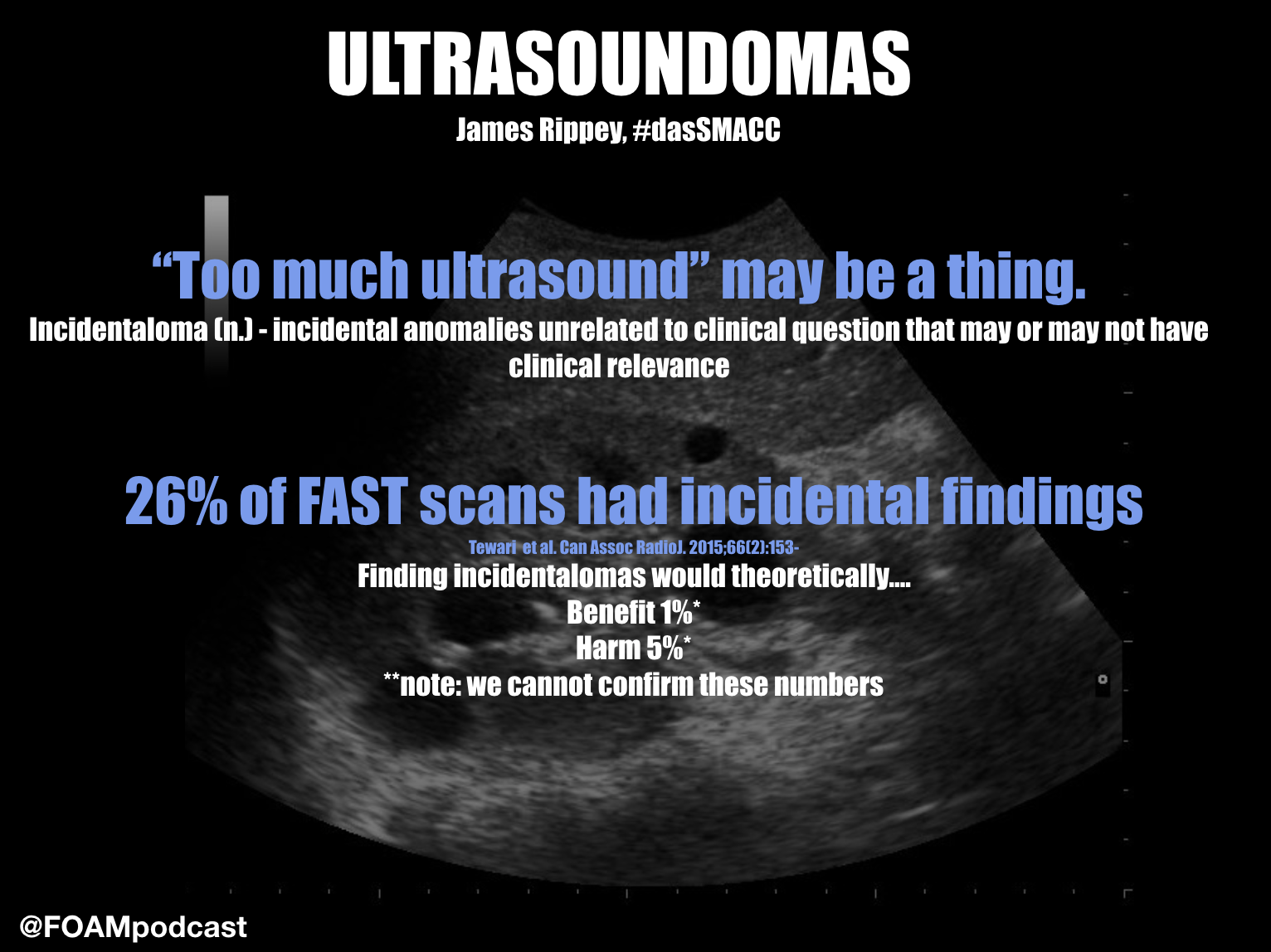
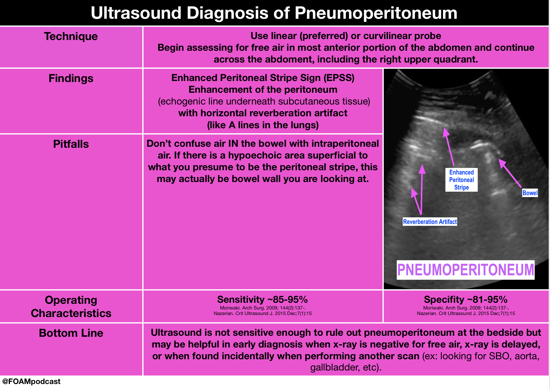
5MinSono has a great 5 minute videocast on the ultrasound diagnosis of pneumoperitoneum.

Core Content

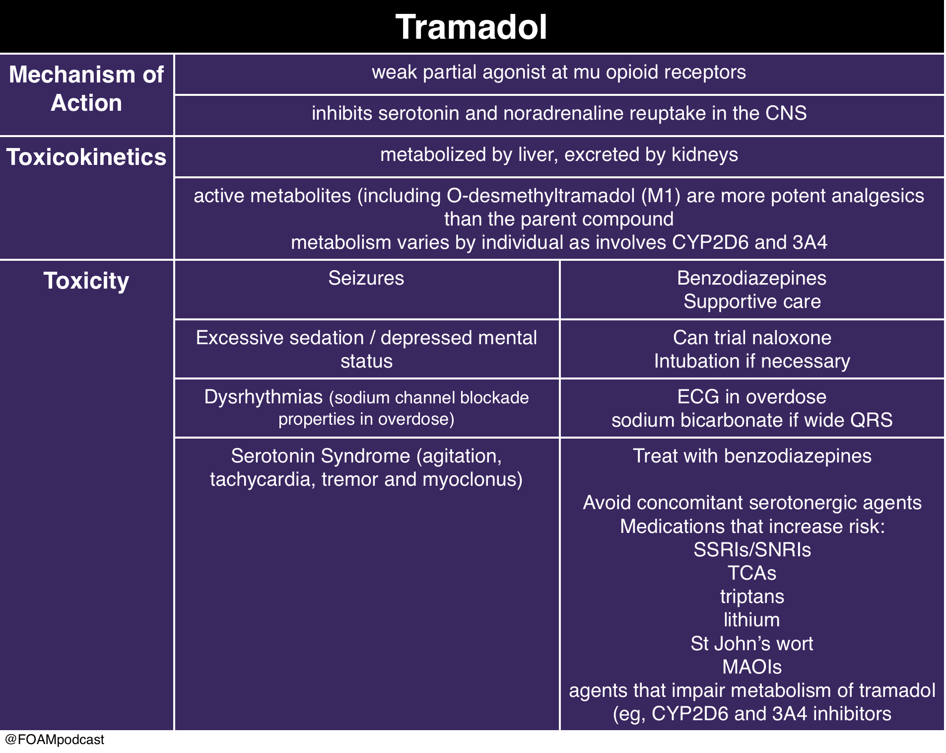
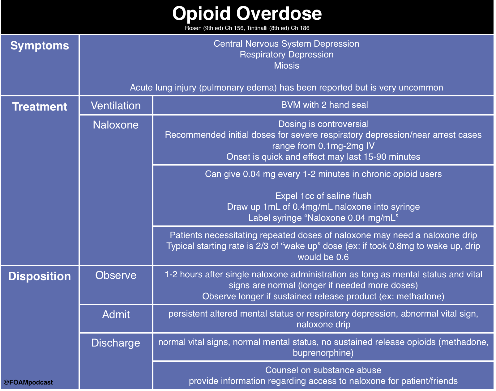
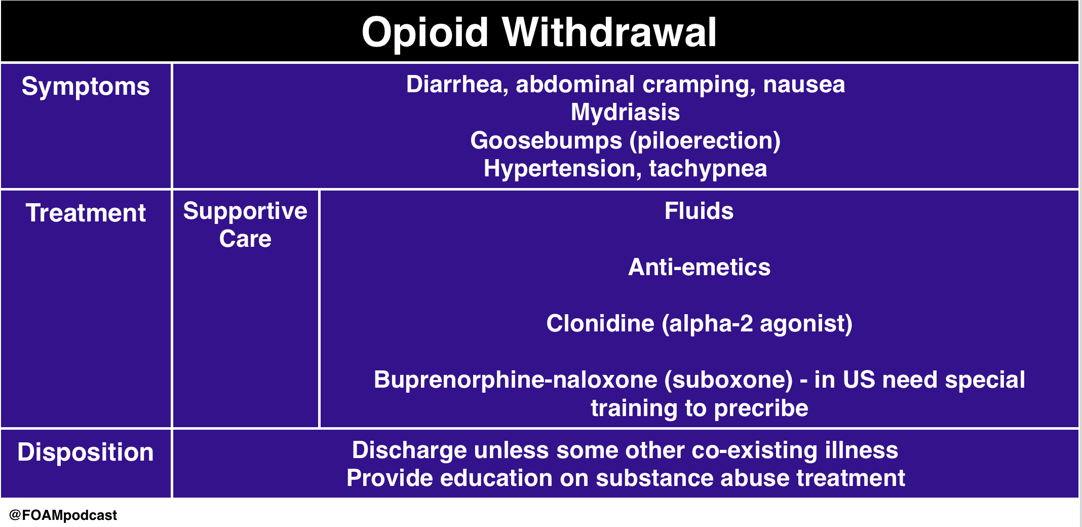
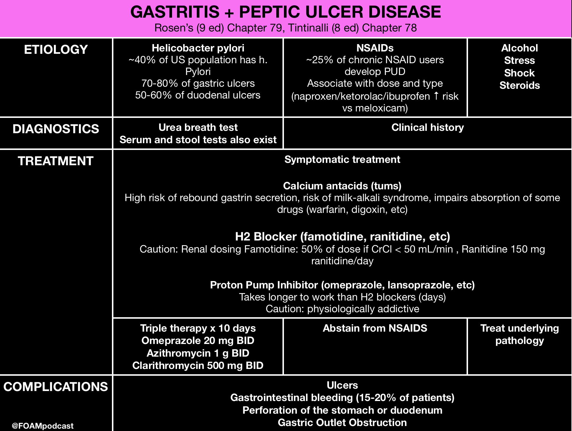
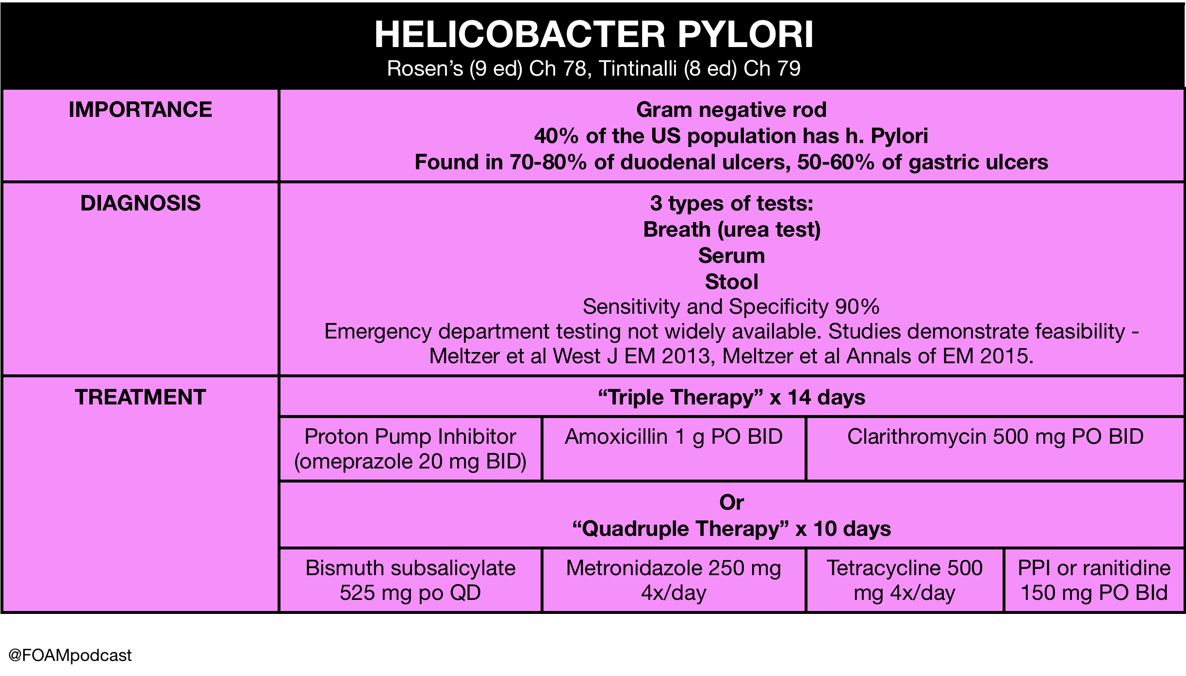
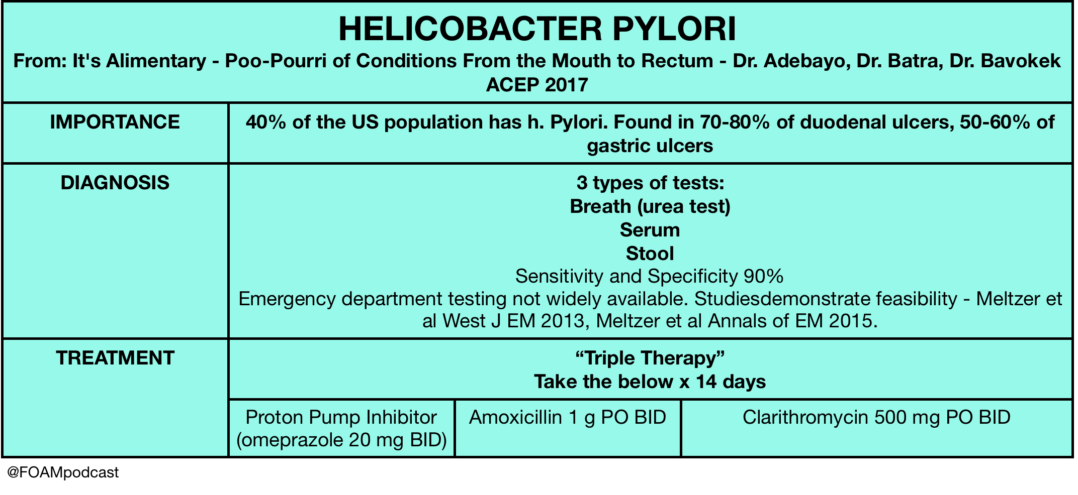
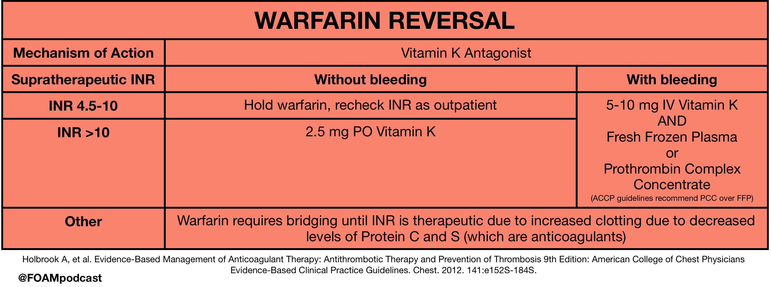
We cover gastropathies and peptic ulcer disease (PUD) using Rosen’s Emergency Medicine (9th ed) Chapter 79 and Tintinalli’s Emergency Medicine (8th ed) Chapter 78  as guides.

Rosh Review Emergency Board Review Questions

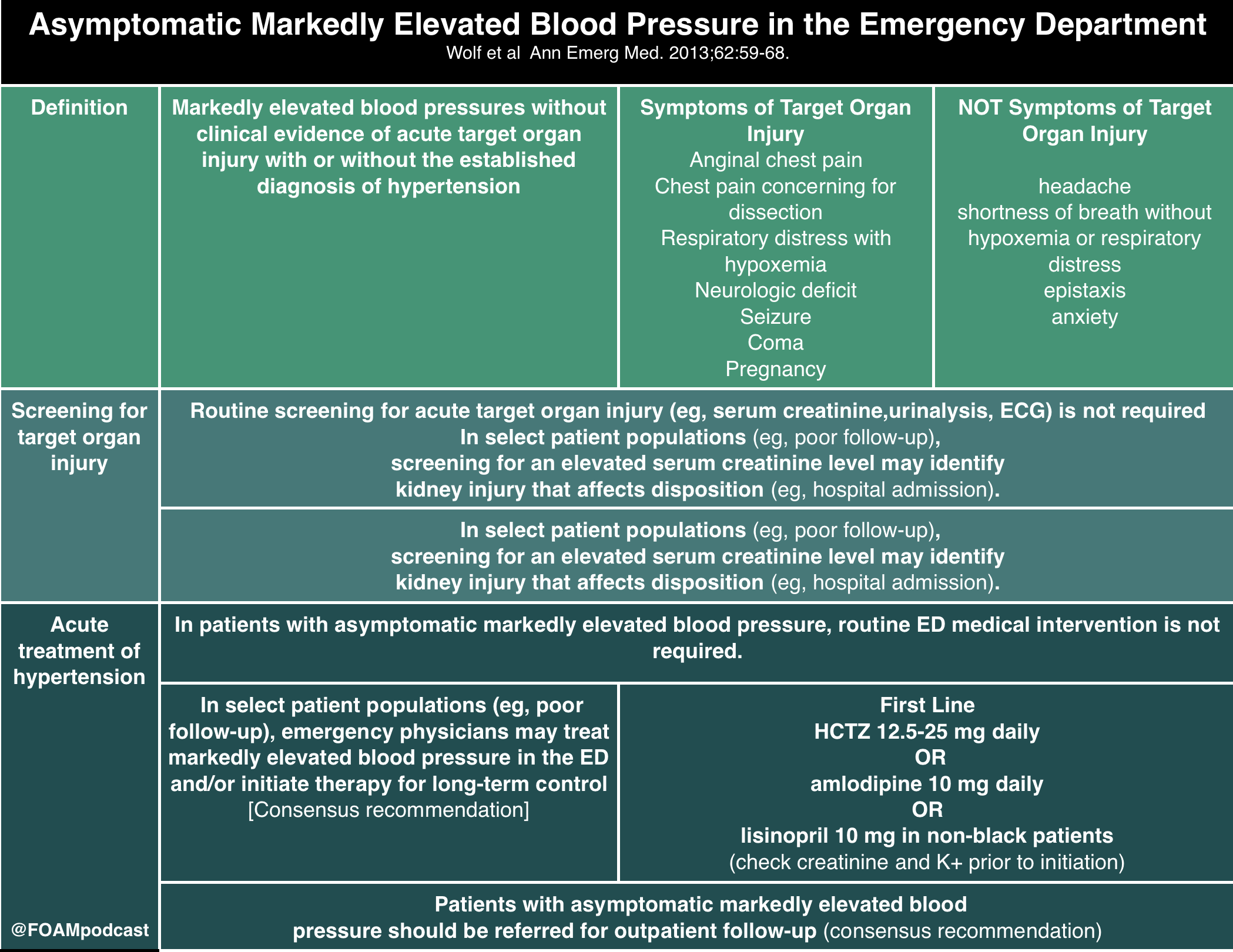
A 67-year old man with chronic osteoarthritis complains of gnawing and burning in the epigastric area that is occasionally accompanied by nausea and vomiting. His current BMI is 26 and he is physically active. What is the most probable cause for these symptoms?

A. Cholelithiasis

B. Gastric carcinoma

C. Nonsteroidal anti-inflammatory drug induced gastritis

D. Peptic ulcer disease

[accordion]
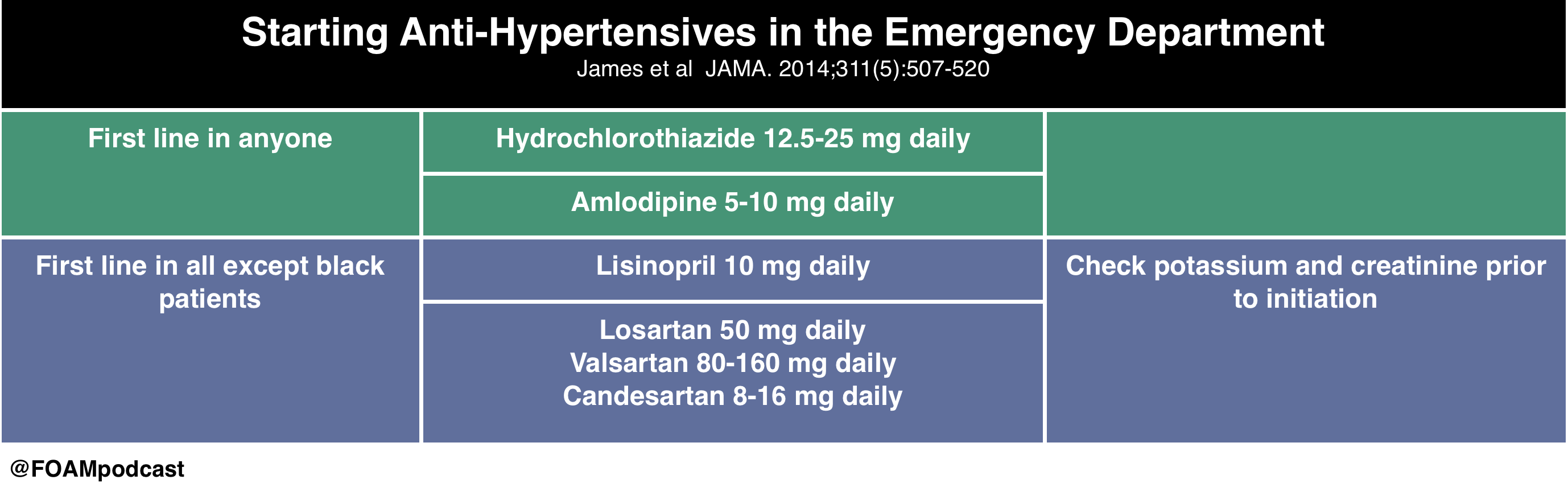
[toggle title=”Answer” state=”closed”] C. 
Nonsteroidal anti-inflammatory drug (NSAID) therapy is the first line treatment in osteoarthritis, however chronic NSAID use can often destroy the gastric mucosa leading to hemorrhage, erosions and ulcers. NSAIDs such as naproxen and ibuprofen are the most common agents associated with acute erosive gastritis. A long-term prospective study found that patients with arthritis who were older than 65 years and regularly took low-dose aspirin were at increased risk for dyspepsia severe enough to necessitate the discontinuation of NSAIDs. This suggests that better management of NSAID use should be discussed with older patients in order to reduce NSAID-associated upper GI events. COX -2 inhibitors, such as celecoxib, are an alternate therapy to NSAIDs. Other common agents that cause gastritis include alcohol and Helicobacter pylori. The mainstay treatment of erosive gastritis is to refrain from the offending agent. The gold standard for diagnoses of gastritis is an upper GI endoscopy.

Symptomatic cholelithiasis (A) is often seen in populations who have risk factors for gallstones, which include persons with diabetes mellitus, persons who are obese, women, rapid weight cyclers, and patients on hormone therapy or taking oral contraceptives. This patient does not portray the colicy pain associated with gallstones. Patients with gastric cancer (B) often present with weight loss, dysphagia, postprandial fullness and loss of appetite. Gastric cancer is multifactorial involving both inherited predisposition and environmental factors. Environmental factors implicated in he development of gastric cancer include diet, Helicobacter pylori, previous gastric surgery, pernicious anemia, chronic atrophic gastritis and radiation exposure. Smoking and smoked meats also have a high correlation with gastric cancer. Peptic ulcer disease (D) is a complication of chronic gastritis and can present in a similar manner as gastritis. Peptic ulcers include both gastric and duodenal ulcers. Peptic ulcers present with gnawing or burning sensation that occur after meals. Common risk factors include H. pylori infection and ingestion of NSAIDs. An upper GI endoscopy must be performed to visualize the ulcers. Biopsy is indicated if ulcers are seen on endoscopy in order to rule out Helicobacter pylori. Active ulcers associated with NSAID use are treated with an appropriate course of proton pump inhibitor (PPI) therapy and the cessation of NSAIDs. For patients with a known history of ulcer and in whom NSAID use is unavoidable, the lowest possible dose and duration of NSAID and co-therapy with a PPI is recommended.

[/toggle]
[/accordion]

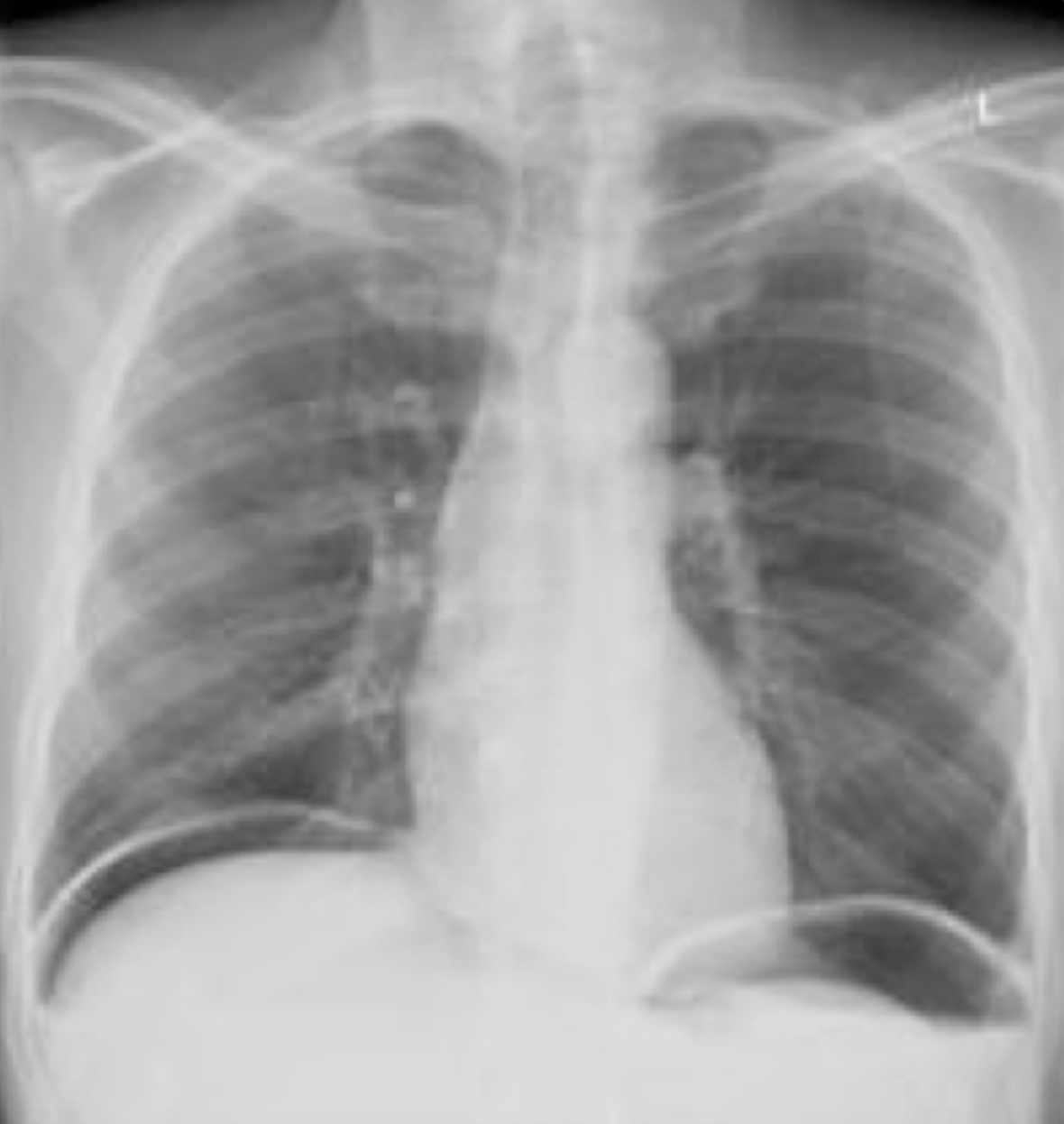
A 55-year-old man presents with severe abdominal pain and tenderness on examination that began acutely approximately 12 hours prior to arrival. His X-ray is shown below. What is the most appropriate next step?

A. Computed tomography scan of the abdomen and pelvis

B. Nasogastric tube insertion

C. Observation and serial abdominal exams

D. Surgical consultation

[accordion]
[toggle title=”Answer” state=”closed”]

D. The X-ray demonstrates free air under the diaphragm representing a perforated viscus within the intraabdominal cavity. The presence of free air is an indication for an emergent surgical consultation for repair. The emergency provider should administer broad-spectrum antibiotics covering aerobic and anaerobic organisms along with intravenous fluid resuscitation.  

A CT scan of the abdomen and pelvis (A) is indicated if the X-ray does not reveal evidence of free air and the patient has ongoing pain and tenderness requiring a diagnosis. Some perforations will not show on plain films, and as time progresses, the area of perforation may wall off and not show on X-ray. A nasogastric tube (B) is not indicated in the management of a patient with a perforated viscus. Observation and serial abdominal exams (C) are not sufficient for a patient with a perforation.  

A patient presents with hematemesis. What test is most likely to determine the etiology of the bleeding?

A. CT scan of the abdomen and pelvis

B. Nasogastric tube lavage

C. Right upper quadrant ultrasound

D. Upper endoscopy

[/toggle]
[/accordion]

[accordion]
[toggle title=”Answer” state=”closed”]

Upper endoscopy is the modality that is most likely to identify the culprit lesion in a patient with upper gastrointestinal bleeding (UGIB). UGIB is a common presentation caused by a variety of pathologies including gastritis, esophageal varices, peptic ulcer disease, Mallory-Weiss tears, arteriovenous malformations and Boerhaave’s syndrome. Of these causes, peptic ulcer disease is the most common. Regardless of the etiology, endoscopy represents the best modality for diagnosis. It allows direct visualization of the esophagus, stomach and first two sections of the duodenum. Additionally, it allows for interventions to be performed if active bleeding or stigmata of recent bleeding are found.

CT scan of the abdomen and pelvis (A) is limited in its ability to give a diagnosis. Nasogastric tube lavage (B) may show the presence of blood in the upper GI tract but cannot differentiate between causes. Right upper quadrant ultrasound (C) may give information about the patient including the presence of cirrhosis but cannot give a specific diagnosis as the cause.

[/toggle]
[/accordion]

References

  1. Nazerian P, Tozzetti C, Vanni S. Accuracy of abdominal ultrasound for the diagnosis of pneumoperitoneum in patients with acute abdominal pain: a pilot study. Critical ultrasound journal. 7(1):15. 2015. [pubmed]

Episode 75 – Mass Casualty Incidents

iTunes or Listen Here

We cover Free Open Access Medical Education (FOAM) on mass casualty incidents, an unfortunate reality in the current United States climate (and elsewhere).  There is a must read (truly, emergency providers really should read this) in EP Monthly by Dr.Kevin Menes, How One Las Vegas ED Saved Hundreds of Lives After the Worst Mass Shooting in U.S. History”. He details his process in running the Emergency Department that evening. Some of our favorite pearls

  • Plan ahead and rehearse. This means both mass casualty drills and mental rehearsal. We do this with many things in emergency medicine (thinking about how we would handle rare and critical procedures or disease processes.
  • Prepare once you have the heads up to help mitigate system induced bottlenecks.  When a mass casualty incident is expected, call for help. This means, extra staff to transport patients and techs and nurses. This also may mean calling in all trauma surgeons, anesthesiologists, and emergency providers. Additionally, bring all stretchers and wheelchairs to the ambulance bay. Consider calling for items in bulk. For example, all vials of paralytic, all chest tube trays from central supply, or large quantities of blood products.
  • Triage – according to the textbook the most senior person should be doing this. In Vegas, Dr.Menes discusses how he was needed in the and turned triage over to a senior nurse who had been assisting him in the process to that point.

We cover pearls from other great resources include a post on the St. Emlyn’s blog, “Mass Casualty Incidents: Lessons from the AAST” and a free EBMedicine article on ballistic injuries. We recommend this article by Dr. Kellerman on the reasons for the lack of firearm research in the United States.

We cover core content on triage and ED treatment pearls using Rosen’s Emergency Medicine Chapter 192 and Tintinalli Chapter 5 as guides


Rosh Review Emergency Board Review Questions

A massive explosion occurred at a nearby automotive plant injuring hundreds of employees. You are called to help as part of the disaster team. You are assigned to work on scene triaging patients according to the Simple Triage and Rapid Treatment (START) protocol. Your first victim is found unconscious with significant head and facial trauma. The patient has no spontaneous respirations. What is the most appropriate next step?

A, Assign the patient a black tag

B. Intubate the patient with an endotracheal tube

C. Oxygenate the patient with a bag-valve mask

D. Reposition the patient’s airway

[accordion]
[toggle title=”Answer” state=”closed”]

You should reposition the patient’s airway. In mass casualty situations, emergency personnel often use the Simple Triage and Rapid Treatment (START) technique that allows for rapid assessment of patient’s respirations, perfusion and mental status (RPM). Anyone that is able to walk is asked to move away from the incident site and is assigned a green tag (walking wounded). At this point emergency personnel then quickly assesses the remaining patient’s respirations, pulse and mental status, in order to assign red (immediate), yellow (delayed) or black (deceased) tags. The first step is to assess the patient’s respirations. If they have no spontaneous respirations, you make one attempt to reposition the airway. If there is no improvement, they are assigned a black tag. If they are breathing greater then 30 breaths/minute, they are assigned a red tag. If respirations are less than 30 breaths/minute, then you assess the patient’s perfusion. If their radial pulse is absent or their capillary refill is over 2 seconds, they are given a red tag. If the have a radial pulse or capillary refill less than 2 seconds, you assess their mental status. If they are able to follow commands they are assigned a yellow tag. If they cannot follow commands, they are assigned a red tag.

[/toggle]
[/accordion]

ACEP – H. Pylori, Zika, and Tox

iTunes or Listen Here

We bring you pearl from the American College of Emergency Physicians (ACEP) 2017 Scientific Assembly.

Pearls on H. Pylori from: It’s Alimentary – Poo-Pourri of Conditions From the Mouth to Rectum – Dr. Adebayo, Dr. Batra, Dr. Bavokek

References :

  1. Rosen’s 9th ed
  2. Meltzer et al. Ann Emerg Med. 2015 Aug;66(2):131-9.
  3. Meltzer et al. West J Emerg Med. 2013 May; 14(3): 278–282.

Emerging Infections: Zika and Its Friends – Dr. Joan Noelker

High Yield Toxicology – Dr Erickson, Dr Traub, Dr. Perrone

The Cutting Edge – PEs, Ultrasound, Pus, and more.

iTunes or Listen Here

In this episode, we summarize recent literature we feel is high yield, important, or frequently discussed in emergency medicine. As always, we encourage you to read the papers and not FOAM it alone.

Antibiotics and abscesses – Daum et al

FAST in hemodynamically stable patients –  Holmes et al

Haloperidol in gastroparesis – Roldan et al and Ramirez et al

Ignoring PERC – Buchanan et al

Steroids in non-asthma/COPD lower respiratory tract infections – Hay et al
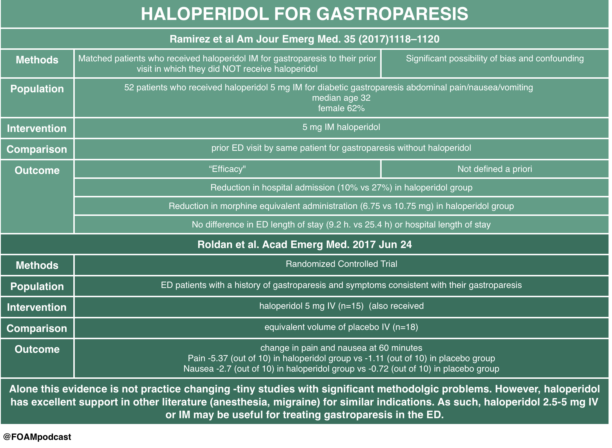
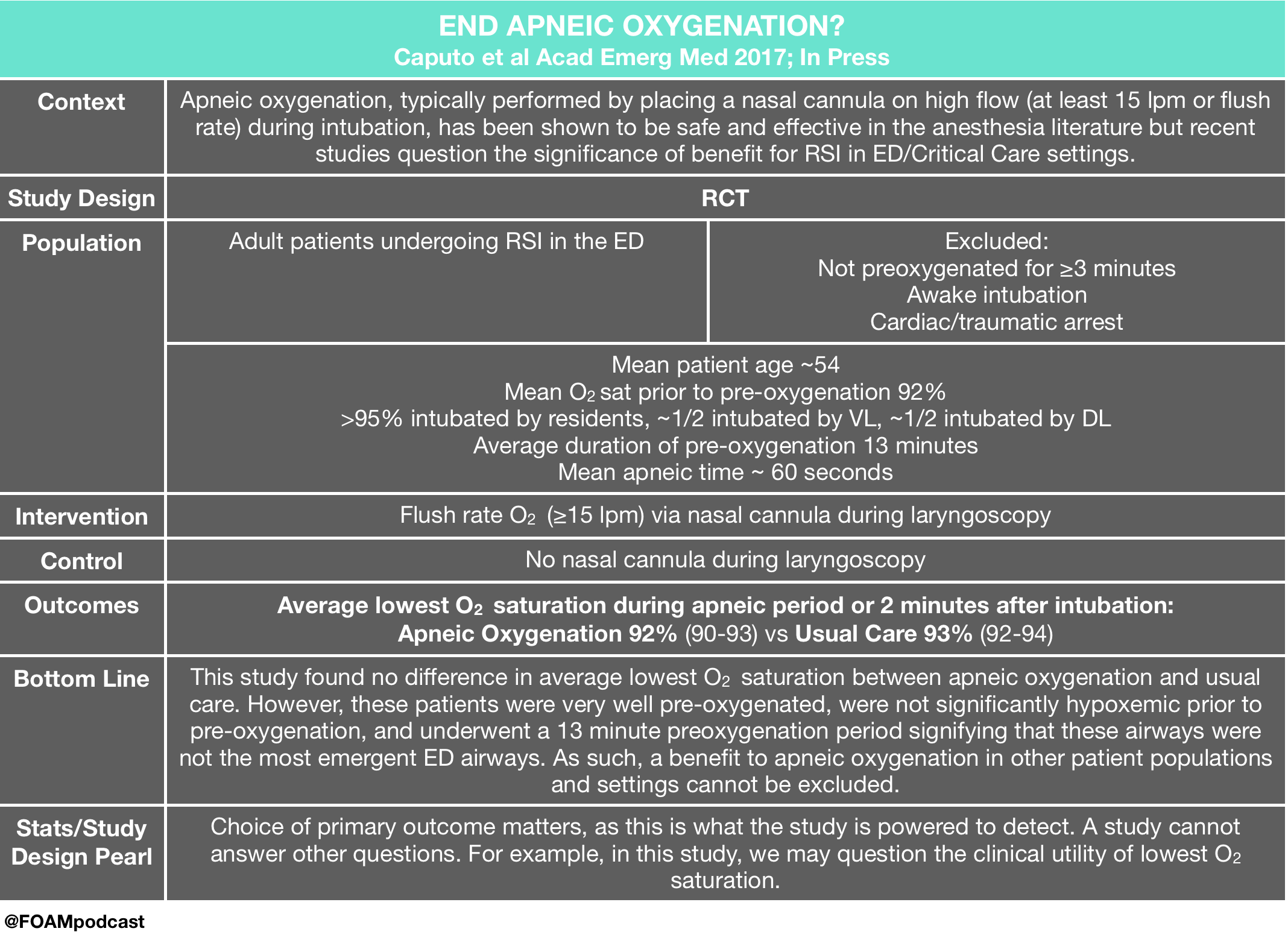
ENDAO – Caputo et al

Gender pay gap persists in academic EM (and it’s not due to hours, rank, or roles) – Madsen et al

Episode 74 – Physostigmine and Anticholinergic Toxidrome

iTunes or Listen Here

We review this blog post by Bryan Hayes, an ED pharmacist (@PharmERToxGuy), on the use of physostigmine in anticholinergic toxicity.

We review core content on anticholinergic toxicity using Rosen’s Emergency Medicine (9th ed), Tintinalli’s Emergency Medicine (8th ed) and Goldrank’s Toxicology as guides.

Rosh Review Emergency Board Review Questions

A 27-year-old man is brought to the ED by EMS after being found wandering in the street. His BP is 155/70, HR 115, T 37.5°C, RR 16, pulse ox 99% on room air, and finger stick glucose 98. On exam, the patient is confused with mumbling speech. His pupils are 7 mm and nonreactive. His face is flushed. Mucous membranes and skin are dry. Which of the following toxidromes is this patient exhibiting?

A. Anticholinergic

B. Cholinergic

C. Sedative-hypnotic

D. Sympathomimetic

[accordion]
[toggle title=”Answer” state=”closed”]

A. Anticholinergic Toxidrome refers to a constellation of physical findings that can provide important clues in a toxic ingestion. This patient exhibits an anticholinergic toxidrome. Anticholinergics are widely available as over-the-counter cold preparations and sleep aids (diphenhydramine, chlorpheniramine, doxylamine). They also are the basis of the toxicity of jimsonweed. Anticholinergics competitively inhibit acetylcholine in the central and peripheral nervous systems. This classic toxidrome is often described by the phrase “mad as a hatter (altered mental status), blind as a bat (mydriasis), red as a beet (flushed skin), hot as a hare (dry skin due to inability to sweat), dry as a bone (dry mucous membranes).” Other findings include tachycardia, delirium, seizures, and hallucinations. Treatment is mainly supportive with benzodiazepines for agitation and seizures, and cooling for temperature reduction. Gastric decontamination (whole-bowel irrigation or gastric lavage) is often recommended due to the delay in gastric emptying caused by anticholinergics. The use of physostigmine, a reversible cholinesterase inhibitor that will increase synaptic acetylcholine, is controversial and should be administered only in consultation with a toxicologist.

The cholinergic toxidrome (B) is characterized by the mnemonics SLUDGE (salivation, lacrimation, urination, defecation, GI upset, emesis) or DUMBBELS (defecation, urination, miosis, bronchospasm, bronchorrhea, emesis, lacrimation, salivation) and occurs after exposure to organophosphates or carbamates. The sedative-hypnotic toxidrome (C) is seen with benzodiazepines and barbiturates. Clinically, patients exhibit central nervous system and respiratory depression. The sympathomimetic toxidromes (D) can occur after ingestion of cocaine, amphetamines, or decongestants. It typically presents with delirium, paranoia, tachycardia, hypertension, hyperpyrexia, diaphoresis, mydriasis, seizures, and hyperactive bowel sounds. Sympathomimetic and anticholinergic toxidromes are frequently difficult to distinguish. The key difference is that sympathomimetics are associated with diaphoresis, whereas anticholinergics cause dry skin.

[/toggle]
[/accordion]

A 22-year-old man is brought into the Emergency Department by Emergency Medical Services with altered mental status. He is an agitated man and mumbling incoherent words. His pupils are 6 mm and reactive to light. His axillae are dry. His heart rate is 115 beats/minute and temperature is 101.6 oF. Which of the following is a complication of the antidote used in the treatment of this condition?

A. Asystole

B. Bladder retention

C. QT-interval prolongation

D. Respiratory depression

[accordion]
[toggle title=”Answer” state=”closed”]

Asystole. The mnemonic for the anticholinergic toxidrome is blind as a bat (mydriasis), red as a beet (vasodilation leading to flushing), hot as a hare (hyperthermia), dry as a bone (dry skin and mucosa), mad as a hatter (visual or auditory hallucinations, agitation, and mumbling or incoherent speech), bloated as a toad (ileus and urinary retention), and the heart runs alone (tachycardia). Additionally, anticholinergic toxicity can cause orthostatic hypotension (especially in the elderly), picking behavior, and can lead to seizures, coma, and even death. There are multiple anticholinergic medications, including tricyclic antidepressants, antihistamines, antiparkinson medications, antipsychotics, antispasmodics, belladonna alkaloids, skeletal muscle relaxants, and mydriatics. Antihistamine overdose is the most common cause of anticholinergic toxicity. Treatment is supportive with IV hydration. If the ingestion occurred within one hour, GI decontamination with activated charcoal is reasonable. Benzodiazepines are used for significant agitation and seizures. Good temperature control should be achieved with antipyretics and active cooling is essential. For wide complex tachyarrhythmias, treatment is with sodium bicarbonate. The antidote is physostigmine, a reversible acetylcholinesterase inhibitor. There is, however, a risk of profound bradycardia, seizures, and asystole in patients with widened QRS complexes. This risk is minimal if there is a pure anticholinergic syndrome without concomitant sodium channel blockade, but this is unfortunately not always known. Since the most common cause of anticholinergic toxicity is from diphenhydramine overdose (which causes sodium channel blockade), physostigmine should only be used with the assistance of a medical toxicologist.  

[/toggle]
[/accordion]

References:

  1. Arens AM, et al. Safety and effectiveness of physostigmine: a 10-year retrospective review. Clin Toxicol. 2017 Jul 13:1-7. [Epub ahead of print] PMID 28703024
  2. “Anticholinergics.” Rosen’s Emergency Medicine. 9th ed.  Chapter 145
  3. “Anticholinergics.” Tintinalli’s Emergency Medicine: A Comprehensive Review. 8th ed. Chapter 202.

Episode 73 – Gastroparesis & Biliary Pathology

iTunes or Listen Here

Dr. Salim Rezzaie of RebelEM wrote a blog post on HUGS – Haloperidol for Gastroparesis.  This study had significant methodologic limitations but provides some interesting insight into a practice that many providers have adopted – using haloperidol for gastroparesis. A tiny RCT by Roldan and colleagues also looked at this practice and found promising results, although the study was very small.  Haloperidol has also been used in cannabinoid hyperemesis but literature on this is limited to case reports/series. 

Core Content

We review biliary colic, cholecystitis, and cholangitis using Tintinalli (8th ed) Chapter 79 and Rosen’s Emergency Medicine (9th ed) Chapter 80 as guides.

Rosh Review Emergency Board Review Questions

A 60-year-old woman presents with two days of right upper quadrant abdominal pain constant in nature and associated with subjective fever, nausea, and vomiting. Vital signs are temperature of 38.1°C, heart rate 87, blood pressure 140/80 mm Hg, respiratory rate 14, and oxygen saturation of 99% on room air. Her abdomen is soft with right upper quadrant tenderness and a positive Murphy’s sign. Which of the following tests is most sensitive and specific in diagnosing this patient’s condition?

A. Computed tomography scan with intravenous contrast

B. Hepatobiliary iminodiacetic acid (HIDA) scan

C. MRI with gadolinium

D. Ultrasound

[accordion]
[toggle title=”Answer” state=”closed”]

This patient has suspected acute cholecystitis and requires an imaging study for confirmation. Hepatobiliary iminodiacetic acid (HIDA) scanning is considered the most sensitive and specific test for diagnosing acute cholecystitis. IDA is administered intravenously, taken up by hepatocytes, and excreted into the bile canaliculi. Failure to obtain an outline of the gallbladder within one hour proves cystic duct obstruction and, in the appropriate clinical setting, confirms the diagnosis of acute cholecystitis. Visualization of the gallbladder and common duct within one hour has a high negative predictive value. A HIDA scan is usually obtained when the ultrasound study is equivocal.

Computed tomography scan with intravenous contrast (A) can identify cholecystitis with a reported sensitivity of 92% and specificity of 99%. It is most useful in cases of emphysematous and hemorrhagic cholecystitis. MRI with gadolinium (C) provides similar diagnostic yield to CT scan. Ultrasound (D) is most useful in the ED setting because it is a quick, noninvasive test. Its sensitivity and specificity, however, are lower than a HIDA scan’s for pathology-confirmed cholecystitis. Ultrasound findings and a clinical exam consistent with acute cholecystitis are highly predictive, and many such patients will undergo cholecystectomy without further diagnostic testing.

[/toggle]
[/accordion]


References:

  1. Ramirez R, Stalcup P, Croft B, Darracq MA. Haloperidol undermining gastroparesis symptoms (HUGS) in the emergency department. Am J Emerg Med. 2017; 35(8):1118-1120. [pubmed]
  2. Roldan CJ, Chambers KA, Paniagua L, Patel S, Cardenas-Turanzas M, Chathampally Y. Randomized Controlled Double-blind Trial Comparing Haloperidol Combined With Conventional Therapy to Conventional Therapy Alone in Patients With Symptomatic Gastroparesis. Acad Emerg Med. 2017; In Press[pubmed]
  3. Trowbridge RL, Rutkowski NK, Shojania KG. Does this patient have acute cholecystitis? JAMA. 2003; 289(1):80-6. [pubmed]
  4. Jain A, Mehta N, Secko M. History, Physical Examination, Laboratory Testing, and Emergency Department Ultrasonography for the Diagnosis of Acute Cholecystitis. Acad Emerg Med.. 2017; 24(3):281-297. [pubmed]
  5. Hwang H, Marsh I, Doyle J. Does ultrasonography accurately diagnose acute cholecystitis? Improving diagnostic accuracy based on a review at a regional hospital.   J Surg. 2014 Jun; 57(3): 162–168. [pubmed]

Episode 72 – Hypertension

ITUNES OR LISTEN HERE

Picone et al published a meta-analysis in the Journal of the American College of Cardiology discussing the accuracy of cuff blood pressures.

We cover core content on hypertension using the ACEP clinical policy on hypertension, Rosen’s (9th ed) Chapter 74 “Hypertension,” and Tintinalli (8th ed),  Chapter  57 “Systemic Hypertension” Chapter 59 “Aortic Dissection and Related Aortic Syndromes” as guides. Check out this FOAM review of hypertension in the ED.


References:

  1. Zampaglione B, Pascale C, Marchisio M, Cavallo-Perin P. Hypertensive urgencies and emergencies: prevalence and clinical presentation. Hypertension. 1996;27(1):144-147.
  2. Chiang WK, Jamshahi B. Asymptomatic hypertension in the ED.Am J Emerg Med. 1998;16 (7):701-704.
  3. Masood S, Austin PC, Atzema CL. A Population-Based Analysis of Outcomes in Patients With a Primary Diagnosis of Hypertension in the Emergency Department. Ann Emerg Med. 2016;68(3):258–267.e5.
  4. Wolf SJ, Lo B, Shih RD, Smith MD, Fesmire FM. Clinical policy: Critical issues in the evaluation and management of adult patients in the emergency department with asymptomatic elevated blood pressure. Ann Emerg Med 2013;62(1):59–68.
  5. The Seventh Report of the Joint National Committee on Prevention, Detection, Evaluation, and Treatment of High Blood Pressure. National High Blood Pressure Education Program.  Bethesda (MD): National Heart, Lung, and Blood Institute (US); 2004 Aug. Report No.: 04-5230
  6. Heath I. Hypertensive Urgency — Is This a Useful Diagnosis ? JAMA Intern Med 2016;8(1):13–4.
  7. Patel KK, Young L, Howell EH, et al. Characteristics and Outcomes of Patients Presenting With Hypertensive Urgency in the Office Setting. JAMA Intern Med.  2016;176(7):981–8.
  8. Nakprasert P, Musikatavorn K, Rojanasarntikul D, Narajeenron K, Puttaphaisan P, Lumlertgul S. Effect of predischarge blood pressure on follow-up outcomes in patients with severe hypertension in the ED. Am J Emerg Med. 2016;34(5):834–9.
  9. James PA, Oparil S, Carter BL et al.  2014 Evidence-Based Guideline for the Management of High Blood Pressure in Adults:  Report From the Panel Members Appointed to the Eighth Joint National Committee (JNC 8).  JAMA. 2014;311(5):507-520.
  10. Ogedegbe G, Shah NR, Phillips C et al. Comparative Effectiveness of Angiotensin-Converting Enzyme Inhibitor-Based Treatment on Cardiovascular Outcomes in Hypertensive Blacks Versus Whites. Journal of the American College of Cardiology. 66(11):1224-1233. 2015.
  11. Flack JM, Sica DA, Bakris G et al. Management of High Blood Pressure in Blacks: An Update of the International Society on Hypertension in Blacks Consensus Statement. Hypertension. 56(5):780-800. 2010.


Episode 71 – Opioids

ITUNES OR LISTEN HERE

We cover Free Open Access Medical Education on opioids.

The Poison Review podcast reviews papers on the opioid epidemic, including a paper by Rudd et al demonstrating an increase in opioid-related deaths over the past five years as well as the following paper by Shah and colleagues.

Long term opioid use

The Dantastic Mr. Tox and Howard, a new podcast on the scene, also covers multiple topics on opioids, including common misconceptions of tramadol.


The American College of Medical Toxicology recently released a statement on fentanyl exposure, stating how absolutely unlikely it is that accidental dermal exposure would result in toxicity.  ” If bilateral palmar surfaces were covered with fentanyl patches, it would take approximately 14 minutes to receive 100 mcg of fentanyl…This extreme example illustrates that even a high dose of fentanyl prepared for transdermal administration cannot rapidly deliver a high dose.”

We cover core content on opioid overdose and withdrawal using Rosen’s Emergency Medicine (9th ed) Chapter 156 and Tintinalli’s Emergency Medicine (8th ed), Chapter 186 as a guide.

opioid withdrawal

#dasSMACC – Airway Ultrasound, Critical Care, and Dunning-Kruger

ITUNES OR LISTEN HERE

We are in Berlin for #dasSMACC and have lots of pearls to share from the speakers at this amazing conference.  Talks will be released for free on the SMACC podcast over the next year, but this podcast holds some pearls that we thought couldn’t wait.

Overall trope of the conference….

dasSMACC

for more on the Dunning-Kruger effect check out this post

Drs. Jacob Avila and Ben Smith on Airway Ultrasound

Check out this 7-minute video on ETT placement confirmation using ultrasound

Screen Shot 2017-06-29 at 8.17.49 PM

Dr. James Rippey on Point of Care Ultrasound Incidental Findings

Screen Shot 2017-06-29 at 7.43.34 PM

Dr. Jack Iwashyna on Persistent Critical Care

Screen Shot 2017-06-29 at 7.52.39 PM