Episode 70 – Nonpregnant Vaginal Bleeding

ITUNES OR LISTEN HERE

The WOMAN trial of tranexamic acid (TXA) for post-partum hemorrhage has been covered well by several free open access medical education (FOAM) sources.  We review the trial and coverage from the following excellent sites:

The Core Content

We have previously covered vaginal bleeding after the first trimester and ectopic pregnancies and first tri, ester vaginal bleeding. In this episode we cover non-pregnant vaginal bleeding using Rosen’s Emergency Medicine (8th ed), Chapter 100 , Tintialli’s Emergency Medicine (8th ed), Chapter 96 and the ACOG guidelines as guides.

Rosh Review Emergency Board Review Questions

A 32-year-old woman presents with vaginal bleeding for 2 weeks. She states she has about 1 pad of bleeding every 2-3 hours. Vital signs are stable and physical exam only reveals blood from the cervical os. The patient’s hemoglobin is 12 g/dl and her pregnancy test is negative. What treatment is indicated for this patient?

A. Admission for dilation and curettage

B. Combination oral contraceptives

C. Hysterectomy

D. Intravenous estrogen therapy

[accordion]
[toggle title=”Answer” state=”closed”] B. This patient presents with non-life threatening dysfunctional uterine bleeding (DUB), which can initially be managed with combination oral contraceptives. DUB is typically split into anovulatory (90%) and ovulatory (10%). In patients with vaginal bleeding of childbearing age, the most important first step in diagnosis is to rule out pregnancy. After this, it is important to explore other causes including medications, genital tract pathology and systemic disease. Once these are excluded, a diagnosis of DUB can be reached. Some treatments include NSAIDs that inhibit PGE1 production and can both relieve cramping and pain and also decrease bleeding. In anovulatory bleeding, combination oral contraceptive pills can aid in regulating the menstrual cycle and counteract the effects of unopposed estrogen. Typically, patients are instructed to take combination oral contraceptive pills twice a day for 5-7 days or until the bleeding stops followed by once daily dosing. Dilation and curettage (A) is typically offered to patients with heavy vaginal bleeding evidenced by hemodynamic instability. A hysterectomy (C) is rarely needed in the treatment of DUB but is indicated for patients with heavy bleeding and hemodynamic instability in which conservative management fails. Intravenous estrogen therapy (D) is effective in stopping heavy bleeding, but is not considered first line therapy.

[/toggle]
[/accordion]

A 55-year-old postmenopausal woman presents with a complaint of vaginal bleeding. Which of the following is the most appropriate next step in management?

A. Abdominal ultrasound

B. Endometrial biopsy

C. Hysterectomy

D. Watchful waiting

[accordion]
[toggle title=”Answer” state=”closed”]

Vaginal bleeding after menopause is an abnormal finding. The most common cause of vaginal bleeding after menopause is atrophy of the vaginal mucosa or endometrium, however 5-10% of postmenopausal women with vaginal bleeding have endometrial cancer. Endometrial cancer is potentially lethal, therefore any postmenopausal woman who presents with vaginal bleeding needs to be evaluated to rule out this etiology. After a careful history and physical exam, initial diagnostic testing to rule out endometrial cancer involves either endometrial biopsy or transvaginal ultrasound. Advantages of an endometrial biopsy include its high sensitivity, low cost and low incidence of complications. Women who need evaluation of the adnexa or myometrium, or who can’t tolerate endometrial biopsy should be referred for transvaginal ultrasound. If either test is inconclusive, further testing is warranted. Cervical cancer screening should also be a part of the workup for postmenopausal vaginal bleeding.

Abdominal ultrasound (A) is not recommended for women with postmenopausal vaginal bleeding. If ultrasound needs to be used, transvaginal ultrasound is the appropriate diagnostic test to order. Postmenopausal women with an endometrial thickness < 3-4 mm on transvaginal ultrasound are unlikely to have endometrial carcinoma. Hysterectomy (C) may be indicated based on the results of the diagnostic imaging, but is not an initial step in management of postmenopausal vaginal bleeding. All women who present with postmenopausal vaginal bleeding should be evaluated with either endometrial biopsy or transvaginal ultrasound, there is no role for watchful waiting (D).

[/toggle]
[/accordion]


Episode 69 – Urinary Tract Infections

ITUNES OR LISTEN HERE

We cover Free Open Access Medical Education (FOAM) from a recent Emergency Medicine Cases podcast and First10inEM blog post by Dr. Justin Morganstern regarding urinary tract infections (UTIs).   This podcast and blog tackle common issues in UTI diagnosis and treatment, including the following points:

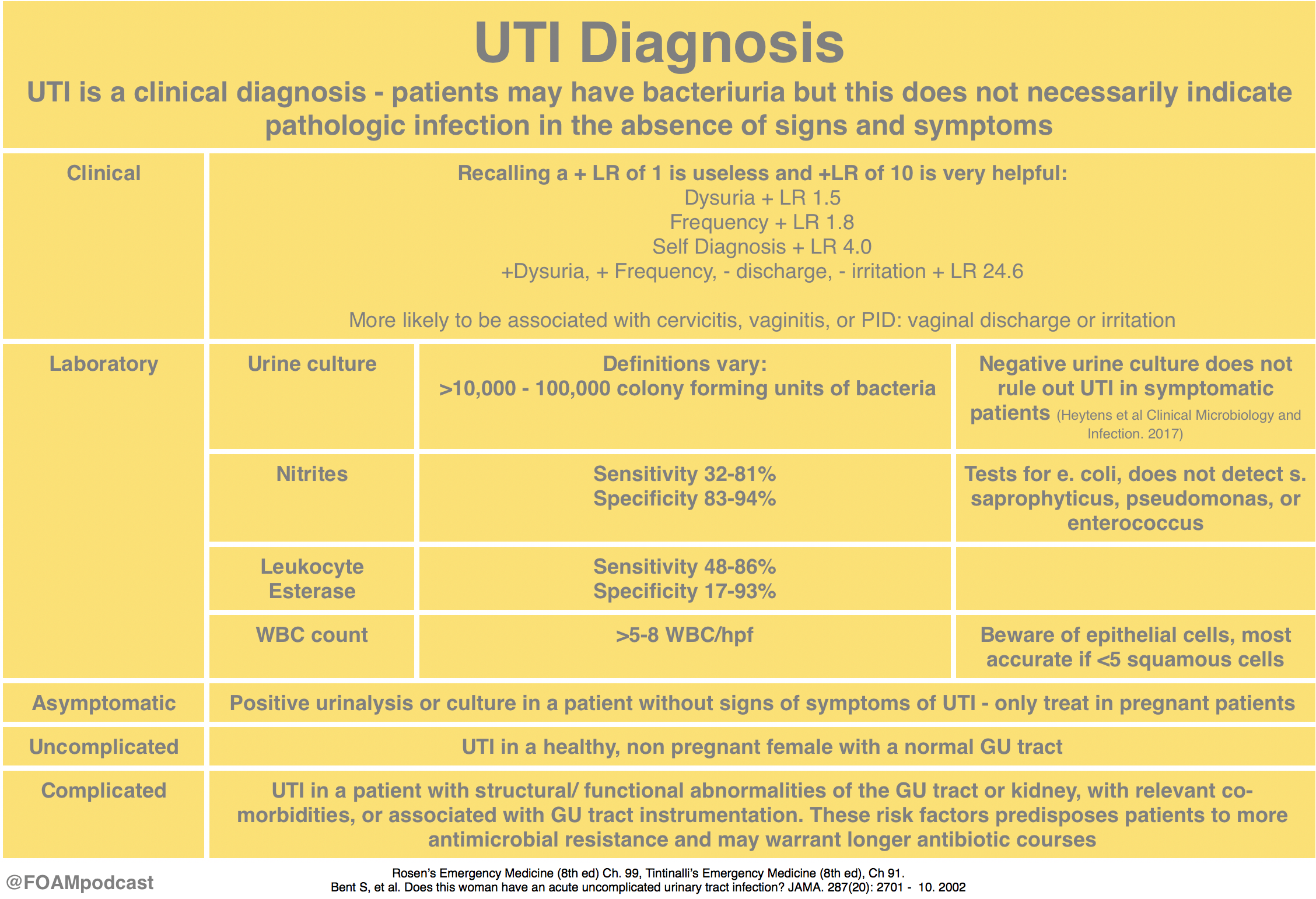
  • UTI is a clinical diagnosis, a dirty urine does not mean the patient has a UTI
  • Urinalyses are more complicated to interpret than we probably understand

The Core Content

Rosen’s Emergency Medicine (8th ed), Chapter 99; Tintialli’s Emergency Medicine (8th ed), Chapter 91; IDSA Guidelines for Treatment and Asymptomatic Bacteriuria

UTI diagnosis

Asymptomatic bacteriuria

UTI Treatment

Rosh Review Emergency Board Review Questions

A 6-year-old girl presents with 4 days of lower abdominal pain. The patient complains of dysuria. On exam, the patient is afebrile and has mild tenderness to palpation in the suprapubic area. No costovertebral tenderness is elicited on exam. A clean-catch urine sample is sent for urinalysis. If positive, which of the following is the most specific to confirm the diagnosis?

A. Glucose

B. Leukocyte esterase

C. Nitrites

D. WBCs (>5 per high power field)

[accordion]
[toggle title=”Answer” state=”closed”]

C. The patient’s presentation is consistent with an uncomplicated urinary tract infection (UTI). The most common cause of a UTI in children >1 year of age is E. coli. Nitrites normally are not found in urine but result when bacteria reduce urinary nitrates to nitrites. Many Gram-negative and some Gram-positive organisms are capable of this conversion, and a positive dipstick nitrite test indicates that these organisms are present in significant numbers (i.e., more than 10,000 per mL). This test is specific (92%–100%) but not highly sensitive (19%–48%). A positive result is helpful, but a negative result does not rule out UTI. The nitrite dipstick reagent is sensitive to air exposure, so containers should be closed immediately after removing a strip. After 1 week of exposure, 33% of strips give false-positive results, and after 2 weeks, 75% give false-positive results. Non-nitrate-reducing organisms also may cause false-negative results, and patients who consume a low-nitrate diet may have false-negative results.

Glucose (A) normally is filtered by the glomerulus, but it is almost completely reabsorbed in the proximal tubule. Glycosuria occurs when the filtered load of glucose exceeds the ability of the tubule to reabsorb it (i.e., 180–200 mg per dL). Etiologies include diabetes mellitus, Cushing’s syndrome, liver and pancreatic disease, and Fanconi’s syndrome. Leukocyte esterase (B) is produced by neutrophils and may signal pyuria associated with UTI. It has a sensitivity of 72%­–97% and specificity of 41%–86%. Leukocyte casts in the urinary sediment can help localize the area of inflammation to the kidney. Organisms such as Chlamydia and Ureaplasma urealyticum should be considered in patients with pyuria and negative cultures. Other causes of sterile pyuria include balanitis, urethritis, tuberculosis, bladder tumors, viral infections, nephrolithiasis, foreign bodies, exercise, glomerulonephritis, and corticosteroid and cyclophosphamide use. Leukocytes (D) may be seen under low- and high-power magnification. Men normally have fewer than 2 white blood cells (WBCs) per HPF; women normally have fewer than 5 WBCs per HPF; >5 WBCs/HPF is associated with a 90%–96% sensitivity and 47%–50% specificity.

[/toggle]
[/accordion]

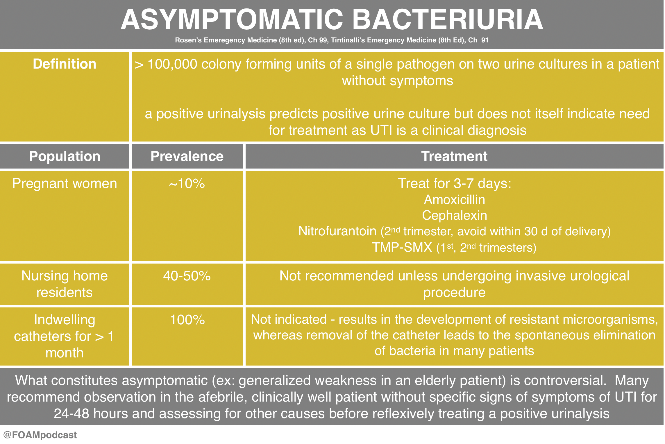
A 24-year old woman presents with URI symptoms. She is 32 weeks pregnant. As part of her work-up, you order a urinalysis, which shows 2+ bacteria with no WBCs. Two days later, the lab calls you and informs you that the urine culture is positive. You call the patient back and she denies symptoms of urinary tract infection. With regards to the urine culture results, what treatment is indicated?

A. Cephalexin 500 mg QID for 7 days

B. Ciprofloxacin 500 mg QID for 7 days

C. No treatment is necessary

D. Trimethoprim-sulfamethoxazole 1 DS tablet BID for 3 days

[accordion]
[toggle title=”Answer” state=”closed”]

A. The patient has asymptomatic bacteriuria of pregnancy confirmed by a positive urine culture and should be treated with an oral antibiotic that is known to be safe in pregnancy, such as cephalexin 500 mg QID for 7 days. Asymptomatic bacteriuria is common in the general population and in most scenarios does not require therapy. However due to the high risk of complication seen during pregnancy, it should be treated with antibiotics. It is seen in 2-10% of pregnant women and is commonly due to E. coli. Pregnant women have an increased risk of developing urinary tract infections due to the pressure that the enlarged uterus exerts on the ureters and bladder, incomplete emptying during voiding and impaired ureteral peristalsis from progesterone-induced relaxation of the ureteral smooth muscle. Complications of untreated asymptomatic bacteriuria include development of a lower urinary tract infection, pyelonephritis, renal abscess, renal failure, bacteremia, sepsis, intrauterine growth retardation, premature labor and neonatal death. Treatment options generally include cephalosporins, such as cephalexin, amoxicillin (or amoxicillin-clavulanate) and nitrofurantoin. All of which are recognized as Category B by the Food and Drug Administration; meaning that animal studies have failed to show a risk to the fetus. Treatment duration should be for 7-10 days.

Ciprofloxacin (B) and trimethoprim-sulfamethoxazole (D) are Category C and D, respectively, and therefore should be avoided in pregnancy when possible. Because there is increased risk for complication during pregnancy, antibiotic treatment (C) is recommended.

[/toggle]
[/accordion]

References:

Gupta K et al. International Clinical Practice Guidelines for the Treatment of Acute Uncomplicated Cystitis and Pyelonephritis in Women: A 2010 Update by the Infectious Diseases Society of America and the European Society for Microbiology and Infectious Diseases.  Infect Dis (2011) 52 (5): e103-e120.

Nicolle L et al. Infectious Diseases Society of America Guidelines for the Diagnosis and Treatment of Asymptomatic Bacteriuria in Adults.   Infect Dis (2005) 40 (5): 643-654.

Rosen’s Emergency Medicine, 8th ed. Chapter 99.

Tintinalli’s Emergency Medicine, 8th ed.  Chapter 91.

Episode 68 – Ischemic Electrocardiograms

ITUNES OR LISTEN HERE

The Free Open Access Medical Education (FOAM)

In the United States, electrocardiograms (ECGs) performed in triage must often be signed by an attending emergency physician shortly after they are performed.  This creates a significant number of interruptions which are not only inconvenient but also distracting for physicians taking care of patients.  A recent study in Academic Emergency Medicine by Hughes and colleagues looks at the potential clinical relevance of having emergency physicians sign ECGs read “normal” by the computer software.  FOAM reviews of this article can be found here:

Triage ECGs

The Core Content

Rosen’s Emergency Medicine (8th ed), Chapter 78 and the 2013 AHA Guidelines

ST Elevation MI (STEMI) 

This is the time-dependent infarction with current goals to obtain reperfusion within 90 minutes (or 60 minutes at some hospitals).

STEMI

However, there are more kinds of ischemia than just STEMI and many of these can be subtle. Close examination of even normal ECGs is necessary.

T wave Ischemia

STEMI mimics

Rosh Review Emergency Board Review Questions

A 45-year-old man presents with chest pain. Which of the following features most strongly predicts acute coronary syndrome as the cause of his chest pain

A. History of diabetes mellitus, hypertension, and tobacco use

B. Pleuritic pain

C. Pressure-like pain

D. Radiation to the right arm

[accordion]
[toggle title=”Answer” state=”closed”]

D.

Chest pain is one of the most common presenting complaints to emergency departments. The causes of chest pain are varied and range from benign (e.g. muscle strain, costochondritis, pleurisy) to life-threatening (e.g. acute coronary syndrome, aortic dissection, pulmonary embolism). The emergency provider’s job is to carefully assess the patient with chest pain and rule out any life-threatening etiologies. As such, acute coronary syndrome must be considered on the differential of every patient presenting with chest pain. Acute coronary syndrome most commonly occurs when an atherosclerotic plaque is disrupted or eroded, resulting in platelet aggregation and thrombus formation at the site, resulting in diminished or absent flow within the affected vessel. Historical features are extremely important in helping categorize patients as low, moderate, or high risk for acute coronary syndrome, and performing a detailed history regarding chest pain complaints is critical. Low risk features include pleuritic, positional, reproducible, and sharp or stabbing pain. In addition, non-exertional pain and pain localized to a small inframammary area have a low likelihood of being cardiac in etiology. High risk features include chest pressure (positive likelihood ratio [LR+] 1.3), pain similar to prior cardiac pain (LR+ 1.8), and associated vomiting or diaphoresis (LR+ 1.9 and 2.0, respectively). The highest risk features include radiation to the right arm or shoulder (LR+ 4.7), left arm (LR+ 2.3), or both arms or shoulders (LR+ 4.1), and exertional chest pain (LR+ 2.4). Of the above choices, radiation to the right arm is the strongest predictor of a cardiac etiology of chest pain.

History of diabetes, hypertension, and tobacco use (A) are traditional cardiac risk factors that predict the lifetime risk of developing coronary artery disease. However, these risks factors are less helpful than historical features in predicting whether an acute presentation of chest pain is due to acute coronary syndrome. Pleuritic pain (B) is not a strong predictor of acute coronary syndrome. Pressure-like pain (C) is a high-risk feature but is less predictive or acute coronary syndrome than pain that radiates to the right arm.

[/toggle]
[/accordion]

References:

  1. Hughes KE, Lewis SM, Katz L, Jones J. Safety of Computer Interpretation of Normal Triage ECGs. Acad Emerg Med. 2016
  2. O’gara PT, Kushner FG, Ascheim DD, et al. 2013 ACCF/AHA guideline for the management of ST-elevation myocardial infarction: a report of the American College of Cardiology Foundation/American Heart Association Task Force on Practice Guidelines. J Am Coll Cardiol. 2013;61(4):e78-140.

Episode 67 – Serious Pediatric Fever

iTunes or Listen Here

We cover an episode of The Skeptic’s Guide to Emergency Medicine that covers a validation study of the Step by Step approach to pediatric fever.  This approach to infants with a fever <3 months old is alluring as it does not necessitate a lumbar puncture.  This algorithm had a better sensitivity and negative predictive value than the Rochester criteria.   The approach did miss some infants with a serious bacterial infection and these tended to be those between 21 and 28 days old and those with fever onset <2 hours prior to arrival.

Step by Step for Pedi Fever

 Core Content

We cover Chapter 116 in Tintinalli’s Emergency Medicine (8th ed) and Rosen’s on pediatric fever.

Infants <3 months old with fever algorithms

Screen Shot 2017-03-25 at 3.06.45 PM

Rosh Review Emergency Board Review Questions

A 25-day-old female presents with fever and cough. Mom denies any symptoms at home. The patient’s 2-year-old brother had a cough and rhinorrhea 1 week prior. On exam, the patient’s temperature is 38.7°C with clear lungs, a benign abdomen, and normal tympanic membranes bilaterally. What is the appropriate workup for this patient?

  1. CBC, chest X-ray
  2. CBC, chest X-ray, urinalysis
  3. CBC, chest X-ray, urinalysis, blood cultures
  4. CBC, chest X-ray, urinalysis, blood cultures, lumbar puncture

[accordion]
[toggle title=”Answer” state=”closed”]

4.  Neonates with fever aged 28 days or younger may have few clues on history and physical examination to guide therapy. Therefore, a high index of suspicion is necessary to detect the febrile neonate with a serious bacterial infection. Obtaining the pertinent medical history from the mother regarding the pregnancy, delivery, and early neonatal life of the febrile neonate is essential. Typically, infections in the 1st week of life are secondary to vertical transmission, and those infections after the 1st week are usually community acquired or hospital acquired. Bacterial meningitis is more common in the 1st month of life than at any other time. An estimated 5%–10% of neonates with early onset group B streptococcal (GBS) sepsis have concurrent meningitis. Therefore, febrile infants (temperature >38°C) younger than 28 days should receive a full sepsis workupCBC, chest X-ray (A), urinalysis (B), and blood cultures (C) are a partial workup for neonatal fever.

[/toggle]
[/accordion]

A two-day-old boy presents to the ED with fever for the past four hours. His birth history includes a normal spontaneous vaginal delivery at term. Parents report noticing that the child “felt warm,” and that he was having copious nasal secretions while feeding. On physical examination, the child appears lethargic, has mottled extremities, and is hot to the touch. Breath sounds are clear bilaterally, and there are no rashes. His vital signs are T 102.9°F, BP 74/48 mm Hg, HR 170 beats per minute, and RR 40 breaths per minute. Which of the following groupings of organisms should your antibiotic choices cover when treating this febrile neonate?

  1. Listeria monocytogenes, Group B streptococcus, Escherichia coli
  2. Mycoplasma pneumoniae, Neisseria meningitidis, Streptococcus pneumoniae
  3. Neisseria meningitidis, Listeria monocytogenes, Streptococcus pneumoniae
  4. Streptococcus pneumoniae, Neisseria meningitidis, Haemophilus influenzae
    [accordion]
    [toggle title=”Answer” state=”closed”]

Answer 1. The febrile neonate is a child 28 days and younger who presents with a fever. These children are at very high risk of serious bacterial infections, including urinary tract infection, pneumonia, meningitis, and bacteremia. Risk factors for serious bacterial infection in a neonate include prematurity, low birth weight, premature or prolonged rupture of membranes, meconium aspiration, or maternal group B streptococcus infection. The evaluation of a neonate with a fever includes CBC, urinalysis, blood culture, urine culture, and a lumbar puncture in order to obtain CSF for cell count, Gram stain, and culture. If the child has respiratory symptoms, a chest X-ray should be performed. If the child has diarrhea, stool testing should also be performed. The most common pathogens involved in serious bacterial infections, including meningitis and bacteremia, in neonates are Listeria monocytogenes, Group B streptococcus, and Escherichia coli. These children can become critically ill very rapidly; therefore, initial management should include a fluid bolus of 20 mL/kg and broad-spectrum antibiotics to cover the most common pathogens in this age group. The most appropriate antibiotics to use in neonates with a fever are ampicillin and cefotaxime. Ampicillin will cover Listeria monocytogenes while cefotaxime will cover Group B streptococcus and Escherichia coli. If there is a history of maternal infection with herpes simplex virus, acyclovir should be added to the empiric broad-spectrum treatment. These patients universally need to be admitted to the hospital for IV antibiotics and observation until all cultures have returned.  Mycoplasma pneumoniae, Neisseria meningitidis, Streptococcus pneumoniae (B) are common pathogens seen in adolescents and young adults. Mycoplasma pneumoniae is a common cause of atypical pneumonia in this age group. Streptococcus pneumoniae is a common bacterial cause of pneumonia, bacteremia, and meningitis while Neisseria meningitidis is primarily a cause of meningitis. Neisseria meningitidis, Listeria monocytogenes, Streptococcus pneumoniae (C) are the primary pathogens causing serious bacterial infections in adults over the age of 65. Listeria monocytogenes is a pathogen that is seen in infants and then later reemerges as a prominent pathogen in older adults. Streptococcus pneumoniae, Neisseria meningitidis, Haemophilus influenzae (D) are the most common pathogens causing serious bacterial infections in children ages one to five years. There has been a significant decline in the incidence of Haemophilus influenzae type B in recent years due to childhood vaccination programs.

[/toggle]
[/accordion]

Episode 66 – Back Pain and Spinal Epidural Abscess

iTunes or Listen Here

  • We cover a post from the fantastic St. Emlyn’s team which breaks down a recently published article in Annals of Emergency Medicine comparing diazepam with placebo in patients with acute low back pain.  We recommend you read the full text of the article by Friedman et al.

diazepam

Core Content

We delve into core content on red flags in back pain and spinal epidural abscess using Rosen’s Emergency Medicine and Tintinalli’s Emergency Medicine Chapter 279 as a guide.

red flags

SEA

Rosh Review Emergency Board Review Questions

  • A 13-year-old boy on chemotherapy for acute lymphoblastic leukemia presents with progressive lower back pain for 2 weeks. Per mom, he has had subjective fevers and a “bulge in his lower back” that is warm to touch. He is currently afebrile and has no focal neurologic deficits. An MRI is obtained as seen in the image above. Which of the following regarding this patient’s condition is true?

A. An appropriate antibiotic regimen is cefepime and metronidazole

B. Direct extension of skin and soft tissue infections is the most common cause

C. Erythrocyte sedimentation rate is a sensitive screening tool

D. Most patients present with back pain, fever, and a focal neurologic deficit

[accordion]
[toggle title=”Answer” state=”closed”]

C.  Erythrocyte sedimentation rate is a sensitive screening toolThe patient has a posterior epidural abscess with adjacent osteomyelitis and myositis. The most common cause of an epidural abscess is hematogenous spread of infection, not direct extension from skin or soft tissue infection. Major risk factors include diabetes, intravenous drug abuse, chronic renal failure, alcoholism, and immunosuppression. The most common organism involved is Staphylococcus aureus. Other organisms include streptococci, anaerobes, gram-negative bacilli, and Pseudomonas aeruginosa. Patients present with localized back pain with tenderness to percussion. Fevers are common (reported in up to 75% of patients). MRI is the diagnostic modality of choice, but erythrocyte sedimentation rate is a sensitive marker and may be used in conjunction with plain radiographs to screen for infectious spinal disease.Vancomycin is an important component of the antibiotic regimen because it covers methicillin-resistant Staphylococcus aureus (MRSA) and is usually combined with a 3rd-generation cephalosporin (A). Cefepime is a 4th-generation cephalosporin with pseudomonas coverage. Metronidazole or clindamycin can be added for anaerobic coverage. While the classic triad consists of back pain, fever, and neurologic deficits (D), only a small proportion of patients actually have all three components at presentation. The most common cause of an epidural abscess is hematogenous spread of infection, not direct extension from skin or soft tissue infection (B).

[/toggle]
[/accordion]

An 8-year-old previously healthy boy presents with back pain and fever for 4 days. He complains of pain in the low back, which is increased with bending or twisting. The pain radiates down into his left leg. He denies trauma. Physical exam is remarkable only for tenderness to palpation over the lumbar spine. What management is indicated

A. Ibuprofen and follow up with his pediatrician

B. MRI of the lumbar spine

C. Plain radiographs of the lumbar spine

D. Urinalysis

[accordion]
[toggle title=”Answer” state=”closed”]

B.

MRI of the lumbar spine. This patient presents with symptoms consistent with discitis and should have an MRI for diagnosis. Discitis is a rare infection of the nucleus pulposus and can involve the cartilaginous endplate and vertebral body. It can occur spontaneously or after surgical procedures. Typically, patients present with severe pain localized to the spinal level of involvement. Any movement of the spine exacerbates the pain. Children often present with sudden onset of back pain and refusal to walk. The lumbar spine is most commonly involved and the average age of patients is 7 years. Radicular symptoms are common (present in 50-90% of cases) and most patients will present with fever (90%). Neurologic deficits are uncommon. Serious pathology should be suspected in any pediatric patient presenting with back pain. Neurologic symptoms are rarely present. MRI is the best study for diagnosis and can also rule out other critical diagnoses including epidural abscess. Laboratory studies are non-specific and insensitive but typically will have an elevated erythrocyte sedimentation rate. White blood cell counts are frequently within the normal range.

[/toggle]
[/accordion]

Episode 65 – Contrast-Induced Nephropathy and Genitourinary Trauma

iTunes or Listen Here

We cover Free Open Access Medical Education (FOAM) on Contrast-Induced Nephropathy (CIN).  A large retrospective study by Hinson and colleagues in Annals of Emergency Medicine, reignited enthusiasm in the FOAM world about the questionable entity (and clinical significance) of CIN. The data on CIN is somewhat difficult to parse out as the data consists entirely of retrospective cohort and case-control studies. The highest risk of CIN seems to come from large volume contrast procedures such as percutaneous coronary angiography. Studies looking at the risk of CIN after contrast-enhanced CT scan have been less conclusive.

The American College of Radiology (ACR) Manual on Contrast Induced Nephropathy

CIN

This Emergency Medicine Literature of Note post covers the AMACING  trial, which looks at intravenous fluid administration (0.9% NaCl) versus usual care in patients “at risk” for CIN undergoing contrast-enhanced CT scan.   The study found a difference of -0.1% (95% CI -2.25 to 2.06), which was below the non-inferiority margin. Prior literature shows similar results, with no clear-cut efficacy from prevention strategies for CIN.

This post by Dr. Joel Topf (nephrologist @kidneyboy) on the Precious Bodily Fluids blog discusses a nephrologist’s take on CIN.

EM topics post on fluids in CIN.

Core Content

We then delve into core content on genitourinary trauma using Rosen’s Chapter 47 and Tintinalli’s Chapter265 as a guide.

Screen Shot 2017-02-28 at 1.43.40 PM Screen Shot 2017-02-28 at 1.44.33 PM

 Rosh Review Emergency Board Review Questions

An 18-year-old man involved in a motor vehicle collision is transferred from a rural facility after being diagnosed with a pelvic fracture. After your initial assessment and stabilization, a secondary survey is performed. On examination, he is noted to have blood at his urethral meatus as well as a scrotal hematoma. Which of the following is the most appropriate next step?

A. Retrograde cystogram

B. Retrograde urethrogram

C. Suprapubic catheter placement

D. Transurethral urinary catheter placement

[accordion]
[toggle title=”Answer” state=”closed”]

B. The patient should undergo a retrograde urethrogram to rule out an underlying urethral injury. In patients with a pelvic fracture and signs of urethral injury, a retrograde urethrogram should be performed in the supine position prior to urethral instrumentation. If a partial disruption is identified, one attempt to place a 12- or 14-French Foley or coude catheter can be made. If unsuccessful or a complete tear is diagnosed, then a suprapubic catheter will need to be placed. Pelvic fractures with displacement of the pubic symphysis can cause laceration or avulsion of the prostatic urethra. The three classic findings of urethral injury include blood at the urethral meatus, a high-riding prostate and a scrotal or perineal hematoma. Failure to recognize a urethral injury can lead to urethral stricture formation and urinary incontinence.

Urethral manipulation can convert a partial urethral tear to a complete tear, therefore transurethral urinary catheter placement (D) should be avoided until urethral integrity is known. Suprapubic catheter placement (C) may ultimately be necessary if complete urethral injury is diagnosed, however in this case a retrograde urethrogram should be performed prior to proceeding with suprapubic placement. The patient may also have a bladder injury and may require a retrograde cystogram (A), however urethral integrity must be evaluated by a retrograde urethrogram prior to performing a cystogram

[/accordion]

A 29-year-old man presents to the ED after a MVC. A pelvic fracture is identified on radiography. His vital signs are stable. The decision is made to place a Foley catheter, but blood is noted at the urethral meatus. Which of the following is an appropriate next step?

A. Consult a urologist

B. Obtain a CT scan to evaluate for urethral injury

C. Perform a retrograde urethrogram

D. Place a condom catheter

[accordion]
[toggle title=”Answer” state=”closed”]

C. Perform a retrograde urethrogram. In general, a Foley catheter should not be placed in the setting of suspected urethral injury. In such cases, it is recommended that further testing be performed to evaluate for urethral injury. A retrograde urethrogram should be performed. If there is no contrast extravasation, then a Foley catheter can safely be inserted.  A urologist should be consulted (A) if a urethral injury is confirmed by the retrograde urethrogram. A CT scan (B) is a poor study to identify urethral injuries. A condom catheter (D) does not allow for accurate urine output measurements and may delay identification of a urethral injury.

[/accordion]

References:

  1. Hinson JS, Ehmann MR, Fine DM, et al. Risk of Acute Kidney Injury After Intravenous Contrast Media Administration. Ann Emerg Med. 2017
  2. ACR Manual on Contrast Media.  v10.2. 2016. p33-40
  3. “Genitourinary System.”  Rosen’s Emergency Medicine, 8th ed. Chapter 47, 479-499.e1
  4. “Genitourinary Trauma.” Tintinalli’s Emergency Medicine: A Comprehensive Review.  8th ed.  Chapter 265

Episode 64 – Lumbar Puncture and Central Nervous System Infections

iTunes or Listen Here

Ultrasound is gaining popularity in assisting lumbar punctures (LPs). We review this technique as well as Free Open Access Medical Education (FOAM) from the following sites:  5 Min SonoSinai EM, and PEM pearls from ALiEM.

US guided LP

  • This technique is likely most helpful in difficult patients. A recent study demonstrated 27% absolute increase in first attempt success using ultrasound in infants <6 months old undergoing LP; however, the success rate in both arms was abysmal with only 57% success rate in the ultrasound arm.
  • Core Content

We then delve into core content on meningitis, encephalitis, and antiNMDA receptor encephalitis using Rosen’s Chapter 109 and Tintinalli’s Chapters 117 and 174 as a guide.

 Rosh Review Emergency Board Review Questions

A 40-year-old man with HIV presents with two weeks of progressive headache, malaise, and fever. On examination, he has mild nuchal rigidity, confusion, and a temperature of 38.2oC. Cerebrospinal fluid analysis shows a white blood cell count 360 cells/mL with a monocyte predominance, glucose 28 mg/dL, and protein 220 mg/dL. What is the treatment of choice?

A. Acyclovir

B. Amphotericin B

C. Ceftriaxone

D. Vancomycin

[accordion]
[toggle title=”Answer” state=”closed”]

B. Amphotericin B . Cryptococcal meningoencephalitis is an opportunistic infection that occurs primarily in patients with advanced AIDS, although it can be seen in immunocompromised transplant patients as well. The majority of HIV-related cases occur when the CD4 count is < 100 cells/mm3. Patients present with progressive headache, nausea, malaise, and fever over the course of 1 – 2 weeks. Examination findings are typical for meningitis and include altered mental status, photophobia, and fever. Meningismus is less commonly seen. Presentations can be subtle and a high index of suspicion is needed for diagnosis. A CT scan of the brain is indicated if there are signs of increased intracranial pressure or focal neurologic deficits. Lumbar puncture should be performed with careful measurement of the opening pressure. Cerebrospinal fluid will show a mildly elevated white blood cell count with a monocyte predominance, decreased glucose, and mildly elevated protein. However, in some cases the cerebrospinal fluid will only show minor, if any, abnormalities. Cryptococcal antigen testing of the CSF is nearly 100% sensitive and specific. India ink staining will show budding organisms. Treatment of choice is intravenous amphotericin B in addition to oral flucytosine for 14 days followed by an 8 week course of oral fluconazole.  Acyclovir (A) is used in the treatment of herpes meningoencephalitis. Ceftriaxone (C) and vancomycin (D) are indicated in the treatment of bacterial meningitis.

[/toggle]
[/accordion]

A previously healthy 18-year-old woman presents to the emergency department with complaints of fever, headache, and neck stiffness. She is accompanied by her sister, who expresses concern because the patient seems suddenly confused and cannot remember what she did yesterday. After you administer empiric intravenous antibiotics, which of the following is the next best step?

A. Chest X-ray

B. Complete blood count with differential

C. Lumbar puncture

D. Urinalysis

[accordion]
[toggle title=”Answer” state=”closed”] C. 
 Lumbar puncture.  Meningitis is an inflammation of the tissues surrounding the brain and spinal cord (meninges) and may be of infectious (bacterial, viral, or fungal) and various other etiologies. The classic clinical manifestations include nuchal rigidity, fever and altered mental status. Patients often present with headache as well. All patients with suspected meningitis should have lumbar puncture (LP) to evaluate the cerebrospinal fluid (CSF) unless this procedure is contraindicated. There are no absolute contraindications to LP. Relative contraindications include patients with evidence of increased intracranial pressure, thrombocytopenia, bleeding diathesis or spinal epidural abscess. Acute bacterial meningitis is a medical emergency and left untreated or treated late is almost universally fatal. Treatment involves addressing systemic complications and initiating empiric antibiotic therapy as soon as possible.

Up to half of patients with pneumococcal meningitis may have evidence of pneumonia on chest X-ray (A), but this is not part of the initial workup of patients with suspected meningitis. Complete blood count with differential (B) is often ordered in the workup for bacterial meningitis and generally shows increased white blood cell count, but is not as important to order initially as an LP. Urinalysis (D) is generally not a helpful test in the diagnosis of meningitis and therefore not recommended in the workup for patients with suspected bacterial meningitis.

[/toggle]
[/accordion]

References:

  1. Attia J, Hatala R, Cook DJ, Wong JG. The rational clinical examination. Does this adult patient have acute meningitis?. JAMA. 1999;282(2):175-81.
  2. Nakao JH, Jafri FN, Shah K, Newman DH. Jolt accentuation of headache and other clinical signs: poor predictors of meningitis in adults. Am J Emerg Med. 2014;32(1):24-8.
  3. Neal JT, Kaplan SL, Woodford AL, Desai K, Zorc JJ, Chen AE. The Effect of Bedside Ultrasonographic Skin Marking on Infant Lumbar Puncture Success: A Randomized Controlled Trial. Ann Emerg Med. 2016.

Episode 63 – Bradycardia

iTunes or Listen Here

Emergent transvenous pacing is a crucial skill for emergency physicians.  It may be daunting due to the pacing box and various catheters. We have found that  routinely rehearsing the procedure, reviewing where pacing equipment is in our departments, and where

In this episode we review the following videos:

  • Practical Pointers for Pacemaker Placement by Dr. Jason Nomura
  • Transvenous pacing video by Dr. Al Sachetti
  • Pacing 101 (Transcutaneous is Just Stupid) by Dr. Joe Bellezo on the Ultrasound Podcast
    • This podcast lays down the argument that transcutaneous pacing is stupid.  Transcutaneous pacing is difficult – patients are diaphoretic, capture rates may be 40%, and it takes a significant amount of energy. Further, it hurts.
      • However, in our opinion, there may be a role.  In this talk, Dr. Bellezo quotes a 1981 study, in which emergent pacers were placed in “6 minutes.”  Review of this study finds that this was actually 6 minutes, 45 seconds (closer to 7 minutes) [1].  This likely does not reflect the majority of emergency providers experience, certainly not ours, where the range is more often 15-30 minutes for the procedure.  Thus, transcutaneous pacing may be a temporizing measure as one locates the ultrasound, gathers the supplies, and prepares for transvenous pacing in the unstable patient.

transvenous pacing

pacing

Core Content

We delve into core content on bradycardias and heart blocks using Rosen’s Emergency Medicine (8th edition) Chapter 79 “Dysrthymias” and Tintinalli’s Emergency Medicine (8th edition) Chapter 18  “Cardiac Rhythm Disturbances” as a guide.

bradycardias

bradycardia

 Rosh Review Emergency Board Review Questions

Question 1a.

A 71-year-old woman presents after a fall at home. Her electrocardiogram is shown below.

content_ecg_-3rd_degree_heart_block

[accordion]
[toggle title=”Answer” state=”closed”]

Third Degree Heart block.

[/toggle]
[/accordion]

Question 2.

A. Administration of epinephrine

B. Defibrillation

C. Observation

  • D. Placement of transcutaneous pacer pads

[accordion]
[toggle title=”Answer” state=”closed”]

D. The patient has third degree heart block. In third degree atrioventricular (AV) block, also known as complete heart block, there is no conduction through the AV node, and an escape pacemaker is responsible for the ventricular rate. On electrocardiogram, P waves occur at regular intervals, and QRS complexes occur at regular intervals, but there is no association between the P waves and QRS complexes. When the block occurs in the AV node, a junctional escape pacemaker takes over with a rate of 40-60 beats/minute, and the QRS complex is narrow. If the block occurs at the infranodal level, a ventricular escape pacemaker paces at a rate of 40 beats/minute or less. Infranodal blocks results in a wide QRS complex. Patients with third degree heart block require cardiac pacing, as the slow escape rhythm is rarely adequate to maintain cardiac output and tissue perfusion. Transcutaneous pacing should be initiated while arrangements for transvenous pacing are made. Third degree blocks are commonly associated with cardiac ischemia or infarction. A nodal third degree block (narrow QRS complex) is a complication of acute inferior wall myocardial infarction, and may last for several days. Extensive acute anterior wall infarction is associated with infranodal third degree blocks (wide complex QRS), indicating damage to the infranodal conduction system. When a third degree heart block is seen with acute myocardial infarction, mortality is increased.

Administration of epinephrine (A) is incorrect. Defibrillation (B) the treatment for cardiac arrest from ventricular fibrillation or pulseless ventricular tachycardia.  Observation (C) is incorrect since the patient’s slow heart rate is likely not adequate to maintain cardiac output.

[/toggle]
[/accordion]

Question 3.

A 56-year-man presents to the ED with right arm pain and some chest discomfort. The day prior to arrival, he tried using heavier weights at the gym. He has a history of hypertension, hyperlipidemia, and COPD. In the ED, his vital signs are BP 136/90, HR 60, RR 16, and oxygen saturation 97% on room air. His rhythm strip is seen below. Which is the most appropriate management for this rhythm?

content_ecg_-_second_degree_heart_block_type_ii

A. Aspirin

B. Cardioversion

C. Observation

D. Temporary pacing

[accordion]
[toggle title=”Answer” state=”closed”]

D. This ECG demonstrates type II second-degree heart block. Second-degree heart block is defined by one or more impulses not reaching the ventricles and is classified as type I and type II. Type II second-degree heart block, also known as Mobitz II, is caused by an infranodal conduction abnormality, most commonly in the bundle of His or the purkinje fibers. ECG findings demonstrate random dropped QRS complexes without any changes in the PR interval. Type II second-degree heart block carries a worse prognosis than type I second-degree heart block and necessitates treatment. Unlike type I, atropine has no effect on the His-Purkinje system and may worsen conduction. Temporary pacing is critical in this case because this rhythm can devolve to complete heart block. In the ED, transcutaneous or transvenous pacing should be instituted if the patient is symptomatic and there should be immediate consultation with a cardiologist. Patients with Mobitz II in the setting of an acute myocardial infarction should be treated with temporary pacing and revascularization; following revascularization most conduction abnormalities will improve or resolve and will not require permanent pacing.

[/toggle]

[/accordion]

Question 4.

A man who presents with syncope is placed on the cardiac monitor. On the monitor you note a repeating trend of 6 P waves, 5 of which are followed by a narrow QRS complex and 1 of which is not followed by a QRS complex. The PR interval during this trend progressively increases. Which of the following is the most likely diagnosis?

A. First-degree AV block

B. Third-degree AV block

C. Type I second-degree AV block

D. Type II second-degree AV block

[accordion]
[toggle title=”Answer” state=”closed”]

C. A key distinction between first-degree and second-degree heart block is that in first-degree block the P wave is always followed by a QRS complex. In other words, the ratio of P waves to QRS complexes is 1:1, or, the electrical signal from the atria always passes to the ventricles. In second-degree AV block, the electrical impulse sometimes gets to the ventricles. There are two main types of second-degree AV block. In Mobitz type I, or Wenckebach, second-degree block, there is a progressive beat-to-beat lengthening of the PR interval until a P wave does not conduct through the AV node. The absent conduction and resultant “missing” QRS complex is called a “dropped” QRS, which represents an absent beat of ventricular contraction. First-degree AV block (A) has a 1:1 ratio of P waves to QRS complexes. Mobitz type II second-degree heart block (D) is characterized by a nonconducted P wave which is not preceded by progressive PR interval prolongation. AV dissociation, or third-degree AV block (B), occurs when none of the P waves conduct through the AV node. This complete AV block occurs with separate atrial and ventricular rates. There is no discrete correlation or trend between P waves and QRS complexes.

[/toggle]

[/accordion]

References:

  1. Lang R, David D, Klein HO, et al. The use of the balloon-tipped floating catheter in temporary transvenous cardiac pacing. Pacing Clin Electrophysiol. 1981;4(5):491-6.
  2. Roberts J and Hedges J. “Emergency Cardiac Pacing.” Roberts and Hedges’ Clinical Procedures in Emergency Medicine

Suviving Sepsis Campaign Guidelines 2017

iTunes or Listen Here

The new Surviving Sepsis Campaign Guidelines are out in Critical Care Medicine.  The biggest change is the change in the definition of sepsis, which now uses the language of Sepsis 3.0, introduced in 2016. Other significant changes include dropping protocolized care (ex: early goal directed therapy), and recommending against combination antibiotic therapy (double coverage) for a single pathogen [1,2].

Bonus discussion on new validation study of qSOFA

Screen Shot 2017-01-19 at 4.02.07 PM

References:

  1. Opal SM, Rubenfeld GD, Poll T Van Der, Vincent J, Angus DC. The Third International Consensus Definitions for Sepsis and Septic Shock (Sepsis-3). 2016;315(8):801–10.
  2. Rhodes A, Evans LE, Alhazzani W, et al. Surviving Sepsis Campaign: International Guidelines for Management of Sepsis and Septic Shock: 2016. Crit Care Med [Internet] 2017;1.
  3. Keh D, Trips E, Marx G, et al. Effect of Hydrocortisone on Development of Shock Among Patients With Severe Sepsis: The HYPRESS Randomized Clinical Trial. JAMA. 2016;316(17):1775-1785.
  4. Seymour CW, Liu VX, Iwashyna TJ, et al. Assessment of Clinical Criteria for Sepsis. Jama [Internet] 2016;315(8):762.
  5. Freund Y, LeMachatti N, Krastinova E.Prognostic Accuracy of Sepsis-3 Criteria for In-Hospital Mortality Among Patients W.ith Suspected Infection Presenting to the Emergency Department. JAMA. 2017;317(3):267-268.

Roundup: Favorite Literature of 2016

(iTUNES OR LISTEN HERE)

Article highlights of 2016

Sepsis 3.0

Singer M, Deutschman CS, Seymour CW, et al: The Sepsis Definitions Task Force The Third International Consensus Definitions for Sepsis and Septic Shock (Sepsis-3).

  • This iteration of sepsis discarded systemic inflammatory response syndrome (SIRS), noting it’s poor discriminatory ability.  Further, these authors sought to define sepsis as a dysregulated response to infection, not just a normal response to infection (i.e. fever and tachycardia are normal responses to infection and therefore are, alone, not evidence of sepsis).

Sepsis 3.0

The quick SOFA score (qSOFA) also came out in hopes it “provides simple bedside criteria to identify adult patients with suspected infection who are likely to have poor outcomes.”  It is not part of the sepsis definition  but may help in identifying those that will die or have a 3 day ICU stay.  Studies looking at the performance of qSOFA applied retrospectively to data sets have not been promising [1,2].

Back Up Head Elevated Intubation

Khandelwal et al. Head-elevated Patient Positioning Decreases Complications of Emergent Tracheal Intubation in the Ward and Intensive Care Unit. Anesthesia and Analgesia. Apr 2016.

back up head elevated intubation

Ketorolac Dose

Motov, S, Yasavolian, M, Likourezos, A, et al. Comparison of Intravenous Ketorolac at Three Single-Dose Regimens for Treating Acute Pain in the Emergency Department. .Ann Emerg Med. 2016 Dec 16.

ketorolac

Pulmonary Embolism in Syncope 

Prandoni et al. Prevalence of Pulmonary Embolism among Patients Hospitalized for Syncope.   N Engl J Med 2016; 375:1524-1531. This trial is probably one of the most over-reacted to trials of 2016.  News outlets, both by lay media and for health professionals overstated the claims of this trial.

image

What the trial actually did…

PESIT

Out of Hospital Cardiac Arrest Prognostication

Jabre et al. Early Identification of Patients With Out-of-Hospital Cardiac Arrest With No Chance of Survival and Consideration for Organ Donation.   Intern Med. 2016 Dec 6;165(11):770-778

OHCA

ALTE (Apparent Life Threatening Event) is out, replaced with a new definition and classification, BRUE (Brief Resolved Unexplained Event).

Tieder JS, Bonkowsky JL, Etzel RA, Franklin WH, Gremse DA, Herman B et al. Subcommittee on Apparent Life Threatening Events. Brief Resolved Unexplained Events (Formerly Apparent Life-Threatening Events) and Evaluation of Lower-Risk Infants: Executive Summary.  Pediatrics. 2016

Screen Shot 2016-05-04 at 9.33.37 AM

References:

  1. Churpek MM, Snyder A, Han X et al. qSOFA, SIRS, and Early Warning Scores for Detecting Clinical Deterioration in Infected Patients Outside the ICU. Am J Respir Crit Care Med. 2016. [article]
  2. Brabrand M, Havshøj U, Graham CA. Validation of the qSOFA score for identification of septic patients: A retrospective study. European Journal of Internal Medicine. 36:e35-e36. 2016. [article]