Episode 58 – Ophthalmology

The Free Open Access Medical Education (FOAM) 

Listen Here or iTunes

We cover an incredible ophthalmology resource, OphthoBook.com, by Dr. Tim Roots.  This resource has a free book and excellent free video lectures.  Specifically, we detail a hilarious video on eye exam tricks, especially targeting individuals who “can’t see.”

[table id=3 /]

  • Core Content
  • We previously reviewed eye trauma in this podcast. In this episode, we review ophthalmology basics using Tiintinalli’s Emergency Medicine Chapter 241.
  • screen-shot-2016-10-09-at-5-22-35-pm

When doing the pupillary exam, it is important to assess for an afferent pupillary defect (APD). Ophthalmologists will want “there is or is NOT an APD” when you consult them for essentially any reason.  Normal pupils constrict when the light is shown in either pupil (direct and consensual constriction). To assess for an APD, perform the “swinging light test.”

screen-shot-2016-10-10-at-9-56-49-am

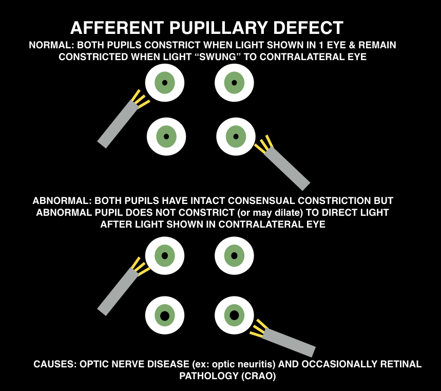
Causes: optic nerve pathology (ex: optic neuritis) or occsaionally, retinal pathology (CRAO)

screen-shot-2016-10-09-at-8-34-25-pm

Peer reviewed by Michael Westafer, MD ophthalmologist and glaucoma fellow at Cleveland Clinic.

Generously Donated Rosh Review Questions

A 72-year-old man presents with a painful red eye and visual loss worsening over the last 24 hours. He recently had cataract surgery. Examination of the eye reveals the image above. Which of the following is the most likely?

A. Endophthalmitis

B. Hyphema

C. Uveitis

D. Vitreous hemorrhage

[accordion]
[toggle title=”Answer” state=”closed”]

A. Endophthalmitis is an infection involving the anterior, posterior and vitreous chambers of the eye. It results from trauma (blunt globe rupture, penetrating injury, foreign bodies) and alsoiatrogenically after ocular surgery like cataract repair. Patients complain of severe pain in the eye and visual impairment or loss. Examination of the eye reveals decreased visual acuity, injected conjunctiva, chemosis and haziness of the infected chambers. Infections are treated with both systemic and intraocular antibiotics.

A hyphema (B) is blood in the anterior chamber usually caused by trauma. When the patient is in an upright position, blood will layer along the inferior aspect of the anterior chamber. As the hyphema increases in size, it elevates intraocular pressure. In some cases admission is warranted for patients with large hyphemas (>50%), decreased vision, sickle cell disease and elevated intraocular pressure. Treatment is aimed at decreasing pressure with topical (beta-blocker, alpha agonist or carbonic anhydrase inhibitors) and systemic therapy (carbonic anhydrase inhibitor, mannitol). Uveitis (C) occurs after blunt trauma in which the iris and ciliary body are inflamed causing ciliary spasm. Patients complain of significant photophobia with significant eye pain. Examination of the eye reveals perilimbal conjunctival injection (also called ciliary flush) and a small, poorly dilating pupil. Photophobia occurs with light shone on both the affected and unaffected eye. On slit lamp, cells (white and red) and flare (protein) are noted in the anterior chamber. Treatment is with a topical cycloplegic agent to minimize spasm. Vitreous hemorrhage (D) occurs as a result of injuries to the retina, uveal tract and their associated vascular structures. Common associated conditions include diabetic retinopathy, retinal vein occlusion and trauma. Patients complain of decreased visual acuity and floaters. The condition is not typically painful. Diagnosis is made with ocular ultrasound showing blood products in the posterior chamber.

[/toggle]
[/accordion]

What is a dependent pocket of pus seen in the anterior chamber called?

  • [accordion]
  • [toggle title=”Answer” state=”closed”]
  • Hypopyon.
  • [/toggle]
  • [/accordion]

A 51-year-old man walks into a movie theater and experiences acute onset of right eye pain associated with nausea, vomiting, and cloudy vision. Which of the following is expected during the ophthalmologic exam?

A. Cherry-red spot in the macular area

B. Deep anterior chamber

C. Intraocular pressure >21 mm Hg

D. Miotic pupil

[accordion]
[toggle title=”Answer” state=”closed”]

The patient has acute angle-closure glaucoma. This condition results in optic nerve damage from increased intraocular pressure. In patients with a narrow anterior chamber angle, reduced illumination (like entering a dark movie theater) causes mydriasis. Subsequently, folds of the peripheral iris can block the angle, which prevents aqueous humor outflow. This leads to a rapid elevation of intraocular pressure causing ocular pain, a hazy cornea, ciliary flush, a firm globe, and optic nerve damage if the pressure is not promptly relieved. An intraocular pressure >21 mm Hg is considered elevated. Pressures can elevate quickly to >60 mm Hg. The higher the pressure, the quicker damage occurs to the optic nerve and the poorer the prognosis. The diagnosis is often delayed due to the associated symptoms of nausea, vomiting, and abdominal pain. Treatment involves reducing aqueous humor production with IV acetazolamide, topical beta-blockers (timolol), and topical alpha-agonists (apraclonidine). Topical miotic agents (pilocarpine) are used to reverse the angle closure. Topical steroidshelp to reduce inflammation. Hyperosmotic agents (mannitol, glycerol) can also be administered for further reduction in intraocular pressure.

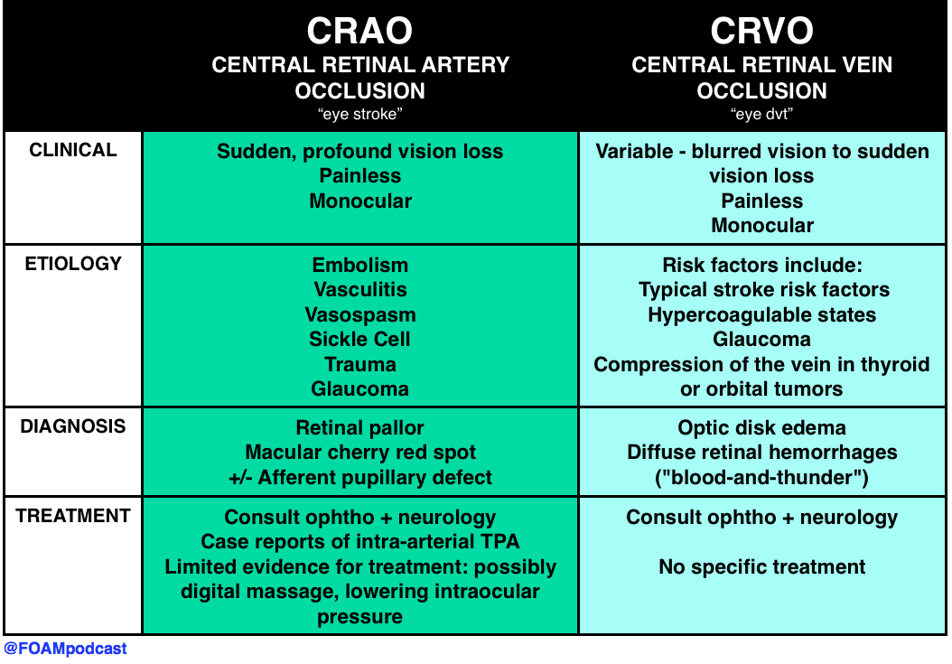
The funduscopic finding of a pale retina with a cherry-red spot in the macular area (A) is consistent with central retinal artery occlusion. This condition is associated with sudden unilateral vision loss that is painless. It is caused by a thrombotic plaque or embolus of the central retinal artery. A deep anterior chamber (B) is protective against acute angle-closure glaucoma. Individuals with a narrow chamber are at increased risk. The pupil in acute angle-closure glaucoma is most commonly fixed and mid-dilated, rather than miotic (D). Miotic pupils are associated with opiate use, cholinergic toxicity, and pontine strokes.

[/toggle]
[/accordion]

References:

“Eye Emergencies.”  Chapter 241.  Tintinalli’s Emergency Medicine: A Comprehensive Review. 8th ed.

Episode 57 – Lithium Toxicity & Rhabdomyolysis

The Free Open Access Medical Education (FOAM) 

Listen Here or iTunes

We cover a post from Dr. Josh Farkas on PULMcrit on lithium toxicity. The key message from the post is: a single serum lithium level doesn’t necessitate dialysis, despite a recommendation from the EXTRIP working group  to initiate dialysis in patients with a lithium level > 5 mEq/L [1].  Dr. Farkas advocates for aggressive management in asymptomatic patients with chronic lithium toxicity and patients without impaired renal function.

Lithium neurotoxicity does not correlate with serum lithium levels; it depends on the concentration in the CNS.

  • The argument is that the neurologic effects, among the most worrisome sequelae of lithium toxicity, results from lithium crossing the blood-brain-barrier (BBB) into the central nervous system (CNS). Thus, in an acute ingestion, patients may have a higher serum lithium level, but given the acuity, the lithium may not cross into the CNS.  Conversely, a patient with chronic toxicity may have a mildly elevated serum lithium level but the lithium has had time to cross the BBB into the CNS so more neurotoxicity can result from lower serum lithium concentrations.

Differentiating between acute, chronic, and acute on chronic toxicity is important in management.

  • Acute – expect the levels will rise rapidly as the bolus of lithium is absorbed.
  • Chronic – expect the levels will not rise rapidly given a lack of lithium bolus.
  • Acute on chronic – again, expect levels to rise rapidly and may see more neurotoxicity as some lithium may have passed through the BBB into the CNS.

screen-shot-2016-09-30-at-6-19-25-pm

screen-shot-2016-09-30-at-6-02-28-pm

Core Content

We review rhabdomyolysis using Rosen’s Emergency Medicine (8e)  Chapter 160 and Tintinalli’s Emergency Medicine (8e), Chapter 181.

screen-shot-2016-10-01-at-12-05-27-pm

Generously Donated Rosh Review Questions

A 17-year-old man presents to the ED from a correctional facility complaining of general malaise with nausea and vomiting one day after a weightlifting competition. Vital signs are T 37.2°C, BP 100/65 mm Hg, HR 125, and RR 22. Physical exam reveals an uncomfortable, fatigued male who has diffuse muscle soreness. Urinalysis shows 3+ blood without red blood cells. What is the most important next test to direct the acute management of this patient?

A. Creatine kinase assay

B. Electrocardiogram

C. Electrolyte panel with blood urea nitrogen and creatinine

D.Microscopic urinalysis

[accordion]
[toggle title=”Answer” state=”closed”]

B. Electrocardiogram. The patient presents with exercised-induced rhabdomyolysis. Although the causes of rhabdomyolysis are manifold, potential complications are independent of etiology. Of all the complications, hyperkalemia is the most concerning and, undiagnosed, can lead to sudden cardiac death. ECG changes indicative of hyperkalemia are thus critical to identify early in the course of management. Hyperkalemia results from impaired calcium transport with increased intracellular calcium accumulation, cellular necrosis, and expulsion of intracellular contents (including potassium) into the bloodstream. Rhabdomyolysis can also lead to acute tubular necrosis and kidney failure, which will exacerbate developing hyperkalemia by decreasing renal potassium clearance. Hyperkalemia is an immediate life-threatening condition that develops shortly after muscle injury. Absent point-of-care electrolyte analyzers, the most rapid way to screen for hyperkalemia is through an electrocardiogram.

Rhabdomyolysis is not defined by a specific creatine kinase (A) level. But, in general, a serum CK >5 times the upper limit of normal (a threshold that may differ by lab) is considered indicative of rhabdomyolysis. Although kidney injury can occur at any level, higher CKs correlate with an increased likelihood for the development of acute renal failure. In the absence of cerebral or myocardial infarction, a CK >5 times is diagnostic for serious muscle injury. Urinalysis (D) typically shows brownish discoloration with “large” blood on dipstick but few, if any, red blood cells on microscopic evaluation. This occurs because most dipstick tests cannot distinguish myoglobinuria from hematuria or hemoglobinuria. Protein, brown casts, and renal tubular epithelial cells may also be present. Measures of renal function and an electrolyte panel (C) should be obtained in all patients with suspected rhabdomyolysis. But, as mentioned, waiting for results may lead to a delay in the identification of life-threatening complications. In addition to hyperkalemia, hyperphosphatemia and hypocalcemia may also be seen. Additional (though less worrisome) laboratory abnormalities include elevated uric acid and low albumin.

[/toggle]
[/accordion]

One Step Further Question: What is the late complication of rhabdomyolysis associated with thrombocytopenia, hypofibrinogenemia, and an elevated D-dimer?

[accordion]
[toggle title=”Answer” state=”closed”]

Disseminated intravascular coagulopathy may occur and is a result of muscle necrosis with liberation of activating substances from injured cells.

[/toggle]
[/accordion]


  • References:
  • 1. Decker BS, Goldfarb DS, Dargan PI et al. Extracorporeal Treatment for Lithium Poisoning: Systematic Review and Recommendations from the EXTRIP Workgroup. Clinical Journal of the American Society of Nephrology. 10(5):875-887. 2015.
  • 2. “Chapter 160. Lithium.”  Rosen’s Emergency Medicine.  8th edition.
  • 3. “Chapter 181. Lithium.” Tintinalli’s Emergency Medicine: A Comprehensive Review. 8th edition.
  • 4. Grellar H. “Chapter 72. Lithium.” Goldfrank’s Toxicology. 10th edition.
  • 5.  “Chapter 127. Rhabdomyolysis. Rosen’s Emergency Medicine.  8th edition.
  • 6. “Chapter 89. Rhabdomyolysis.” Tintinalli’s Emergency Medicine: A Comprehensive Review. 8th edition.

Episode 56 – Altitude

iTunes or Listen Here

The Free Open Access Medical Education (FOAM)

We review a FOAM post by Dr. Matthew MacPartlin on Rollcage Medic on flying after a pneumothorax.

Increases in altitude are accompanied by decreases in pressure that cause the volume of gas to expand (Boyle’s law – pressure and gas volume are inversely associated). As such, gas filled spaces, such as a pneumothorax, may expand during ascent, particularly many thousand meters, as in is the case of aircraft.

  • British Thoracic Society (2011) – No commercial flights until full resolution of pneumothorax, confirmed by chest x-ray. They ideally recommend waiting 7 days after resolution of spontaneous pneumothorax and 14 days after resolution of traumatic pneumothorax. The risk of pneumothorax recurrence drops after one year [1].
    • An observational paper by Sacco and colleagues report the experience of patients flying after chest tube removal, but before the 7-14 day waiting period and found, in their experience, this was safe [2]

Core Content

Tintinalli (8e) Ch 221, Rosen’s Emergency Medicine (8e) Ch 144.

screen-shot-2016-09-09-at-6-22-50-am

Generously Donated Rosh Review Questions

A 20-year-old woman is climbing Mt. Kilimanjaro when she begins developing a headache followed by vomiting. As you begin to assess her, she has a grand mal seizure. Which of the following treatments should immediately be started?

A. Descent and acetazolamide

B. Ibuprofen and dexamethasone

C. Supplemental oxygen and acetazolamide

D. Supplemental oxygen, descent, and dexamethasone

[accordion]
[toggle title=”Answer” state=”closed”]

This patient has developed high-altitude cerebral edema (HACE), a rare but potentially life-threatening form of high-altitude illness. Most cases of HACE are described in ascension past 12 000 feet although it may happen at altitudes as low as 8 200 feet. In HACE, mild altered mental status can rapidly progress to coma in as little as 12 hours. HACE is characterized by global cerebral dysfunction evidenced by headache, fatigue, vomiting, ataxia, confusion, generalized seizures, slurred speech, and focal neurologic deficits. It is a clinical diagnosis although imaging will show cerebral edema. Treatment should start with high-flow oxygen, dexamethasone, and immediate descent. Additionally, if hyperbaric treatment is available, it should be initiated. Ibuprofen (B) and other NSAIDs can be used for prophylaxis prior to ascent as can acetazolamide (A and C).

[/toggle]
[/accordion]

A 32-year-old man complains of dyspnea on exertion and a cough with clear, watery sputum. He has been climbing Mt. Kilimanjaro for 2 days. Other than descent, what treatment can be started immediately?

A. Acetazolamide

B. Albuterol

C. Furosemide

D. Portable hyperbaric chamber therapy

[accordion]
[toggle title=”Answer” state=”closed”]

The patient is suffering from high-altitude pulmonary edema (HAPE) and should be treated with descent from altitude and hyperbaric oxygen therapy. HAPE is the most common fatal manifestation of high-altitude illness. It typically does not develop until the climber has passed 10,000 feet of elevation but it can happen at lower altitudes with heavy activity. The symptoms of HAPE usually begin 2-4 days after arrival at high altitude. Typically, patients experience marked dyspnea on exertion, fatigue with minimal effort, dry cough and difficulty with recovering from exertion. As HAPE progresses, patients will have a cough productive of copious clear secretions and have rales on examination. In severe cases, hemoptysis can develop. Although symptoms may mimic pneumonia or acute cardiogenic pulmonary edema, HAPE should be suspected in the correct clinical scenario. Rapid identification and management is central to preventing morbidity and mortality. The first and most important step in management is descent of the patient. Moderate decreases in altitude (1500 – 3000 feet) can rapidly resolve symptoms. If a hyperbaric chamber is available, it should be employed as simulated descent is just as effective and may be more logistically feasible.

Furosemide (C) is a loop diuretic, which decreases intravascular volume. There is a delay in onset of action and in recent years it has been replaced by pulmonary vasodilators like nifedipine. Additionally, the dehydration associated with furosemide makes it potentially dangerous in these patients. Moreover, HAPE is caused by hypoxia-induced pulmonary vasoconstriction and not from contractility problems of the heart. Acetazolamide (A) is a carbonic anhydrase inhibitor and can be used to treat mild altitude related symptoms like acute mountain sickness (AMS) or to prevent the development of high-altitude illnesses but it is not an appropriate treatment in HAPE. Albuterol (B) plays no role in the treatment of HAPE as bronchospasm is not the problem

[/toggle]
[/accordion]

References

  1. Ahmedzai S, Balfour-Lynn IM, Bewick T et al. Managing passengers with stable respiratory disease planning air travel: British Thoracic Society recommendations. Thorax. 66(Suppl 1):i1-i30. 2011. [article]
  2. Sacco F, Calero KR. Safety of early air travel after treatment of traumatic pneumothorax.  Int J Circumpolar Health. 2014; 73.
  3. Cheatham ML, Safcsak K. Air travel following traumatic pneumothorax: when is it safe? Am Surg 1999; 65:1160–1164
  4. Yaron M, Paterson RD, Davis CB. “High Altitude Medicine.” Chapter 144. Rosen’s Emergency Medicine (8e).
  5. Hackett PH, Davis CB. “High Altitude Disorders.” Chapter 221. Tintinalli’s Emergency Medicine: A Comprehensive Study Guide.

Episode 55 – Petechiae & Purpura

iTunes or Listen Here

We cover several excellent post on rashes, including:

@FOAMpodcast
@FOAMpodcast

Core Content

Next, we dive into core content on platelet problems including problems caused by drugs, immune thrombocytopenic purpura (ITP/idiopathic thrombocytopenic purpura) and thrombotic thrombocytopenic purpura (TTP) using Tintinalli Chapter 233 (8th ed) and Rosen’s Chapter 122 (7th ed) as a guide.

Screen Shot 2016-08-24 at 9.46.51 AM

A previously healthy 3-year-old boy presents to the emergency room with bruising to his arms, legs, trunk, and face. The mother reports that the boy was well appearing yesterday and noted the bruising upon waking up this morning. She also noticed that his gums were bleeding while brushing his teeth before coming to the hospital. Mom denies any trauma but notes her son had a viral illness three weeks ago that kept him out of daycare. A complete blood count is notable for a white blood cell count of 7,000, hemoglobin of 12, and platelets of 10,000. Which of the following is the most likely diagnosis?

A. Idiopathic thrombocytopenia purpura

B. Leukemia

C. Thrombotic thrombocytopenic purpura

D. Wiskott-Aldrich syndrome

[accordion]
[toggle title=”Answer” state=”closed”]

A. Idiopathic thrombocytopenic purpura (ITP) is the most common cause of acute onset thrombocytopenia in an otherwise healthy child. It is most often seen in children aged 2-6 yearsand usually occurs 1 to 4 weeks after a viral illness. ITP is an autoimmune process where antibodies bind to the platelet surface. The platelet-antibody complex is then ingested by splenic macrophages and destroyed. The common physical exam findings are petechiae and purpura. If the child has hepatosplenomegaly, bone or joint pain, or lymphadenopathy, leukemia or other diagnoses should be suspected. In adolescents with new-onset ITP, systemic erythematous lupus should be suspected. Seventy to eighty percent of children will have spontaneous resolution of the symptoms. Treatment depends on the severity of the illness and includes such interventions as platelet transfusion for bleeding patients, steroids, and IVIG. Patients with ITP who have persistent or severe headache should undergo a head CT scan to rule out intracranial hemorrhage.


Leukemia (B) is rarely associated with thrombocytopenia alone and is typically seen with other cell lines being low with immature cells. In addition, physical examination may be remarkable for hepatosplenomegaly or lymphadenopathy. Children with leukemia often complain of bone pain. Leukemia is diagnosed by bone marrow biopsy and treated with chemotherapy. Thrombotic thrombocytopenic purpura (TTP) (C) is characterized by fever, micoangiopathic hemolytic anemia (MAHA), thrombocytopenia, abnormal renal function, and CNS changes. The microvascular thrombi are responsible for the clinical signs and symptoms. Lab findings associated with MAHA include anemia, schistocytes, spherocytes, and elevated reticulocytes. TTP is treated with plasmapheresis. Recurrent TTP is seen in patients with ADAMTS-13 deficiency. Wiskott-Aldrich syndrome (D) is an example of congenital thrombocytopenia. It is characterized by thrombocytopenia, small platelets, recurrent infections, and eczema. This disorder is X-linked.

[/toggle]
[/accordion]

A 9-year-old boy presents to the ED with bilateral knee pain, low-grade fever, nausea, vomiting, and diarrhea for the past 4 days. His vital signs are blood pressure of 116/80 mm Hg, heart rate of 98 beats per minute, respiratory rate of 14 breaths per minute, and a temperature of 38.1°C. On examination, you note the rash seen above. Urinalysis is positive for hematuria. Which of the following statements is the most accurate?

A. Despite plasma exchange, most patients progress to chronic renal impairment

B. Long-term prednisone therapy improves 5-year survival to greater than 50%

C. The disease is self-limited; most cases resolve within 6-8 weeks

D. Without treatment, the disease carries a mortality rate of 80% at one year

[accordion]
[toggle title=”Answer” state=”closed”]

C. The patient has Henoch-Schönlein purpura (HSP). This small-vessel vasculitis predominantly occurs in small children. Most cases follow an upper respiratory tract infection. HSP classically presents with fever, abdominal pain, arthritis, hematuria, and a pathognomonic round, palpable, symmetrical rash that appears on the dependent areas of the legs and buttocks. NSAIDs, dapsone, and prednisone have all been shown to relieve symptoms. The course of disease is typically self-limited. Most cases resolve within 6 to 8 weeks, with a recurrence rate of up to 33%.


Plasma exchange (A) has been found to be successful in the treatment of microscopic polyangiitis. In HSP, most patients do not progress to chronic renal impairment. Prednisone (B) therapy has increased the 5-year survival rate to greater than 50% in Churg-Strauss Syndrome, which is typically associated with fever, weight loss, malaise, and pulmonary symptoms, but it does not affect survival in HSP. Granulomatosis with polyangiitis (D), not HSP, once carried a 1-year mortality rate of 80%, however, the combination of cyclophosphamide and corticosteroids has been successful in inducing remission in more than 90% of patients.

[/toggle]
[/accordion]

Episode 54 – The Pericardium

iTunes or Listen Here 

We cover ultrasound guided pericardiocentesis using the posts from EMin5, CoreEM, and the Ultrasound Podcast.

Historically, pericardiocentesis is taught using a landmark based method; however, use of ultrasound guidance may increase success.  Experts recommend an approach wherever the largest pocket of fluid exists and each location has particular downsides to be aware of.

Screen Shot 2016-07-27 at 10.10.18 AM

Core Content

We delve into core content on the pericardium using Rosen’s (8th ed) Chapter 82 and Tintinalli (8th ed) Chapter 55.

Screen Shot 2016-07-27 at 10.34.13 AM

Generously Donated Rosh Review Questions

1.A 25-year-old man presents to the ED with chest pain, shortness of breath, and fever. Vital signs include BP 98/50 mm Hg, HR 136 beats/minute, RR 26 breaths/minute, and T 102.4°F. On auscultation, you hear rales to the mid-thorax bilaterally. Bedside cardiac ultrasound shows global hypokinesis and a small pericardial effusion. Which of the following organisms is the most common cause of this condition worldwide?

A. Coxsackievirus B

B. Mycobacterium tuberculosis

C. Plasmodium falciparum

D. Trypanosoma cruzi

  • [accordion]
    [toggle title=”Answers” state=”closed”]D. Trypanosoma cruz.  This patient presents with signs and symptoms of myocarditis accompanied by pericarditis. Myocardial injury results from inflammation of the myocardium. The most common etiology worldwide is Chagas disease, caused by the protozoan Trypanosoma cruzi. The protozoan is spread by the reduviid bug, also known as the kissing bug as it feeds on the faces of those affected. Unfortunately, in many patients, the cause of myocarditis is idiopathic. Other noninfectious causes include connective tissue disorders such as scleroderma, toxins such as chemotherapy, cocaine, and heavy metals, and peripartum myocarditis. Symptoms often include a viral prodrome with fever, myalgias, and generalized weakness. Patients may present with chest pain, symptoms of acute heart failure, tachycardia, dysrhythmias, syncope, cardiogenic shock, or even sudden cardiac death. Diagnosis can be very difficult and patients often present to the ED multiple times prior to being diagnosed. An ECG may show global or segmental ST elevation, nonspecific ST segment and T wave changes, dysrhythmias, or conduction delays. Troponin and creatinine phosphokinase are often elevated. Echocardiography classically shows global hypokinesis. Management is primarily supportive; however, patients with new left bundle branch block or low ejection fraction may require a left ventricular assist device as a bridge to cardiac transplantation in some cases as these are poor prognostic indicators. The most common long-term sequelae of myocarditis is dilated cardiomyopathy.[/toggle]
    [/accordion]

2.A 56-year-old woman with a history of lymphoma presents to the Emergency Department at the recommendation of her primary care physician. During a routine visit, she had a chest X-ray that showed a “big heart.” She denies chest pain, shortness of breath, leg swelling, cough, orthopnea, or lightheadedness. Her vital signs include temperature 98.6 ºF, HR 88 beats/minute and regular, RR 14 breaths/minute, BP 121/89 mm Hg, and oxygen saturation 98% on room air. Her cardiac and neck exams are within normal limits. A bedside ultrasound reveals a small pericardial effusion. Which of the following is the next best step in management?

A.Lower extremity ultrasound

B. Pericardiocentesis

C. Reassurance and close follow up

D. Thoracic Surgery consultation

  • [accordion]

[toggle title=”Answers” state=”closed”] C.  Reassurance and close follow up. The patient likely has a malignant pericardial effusion secondary to her known malignancy. Pericardial effusions are accumulations of fluid in the pericardial space that occur rapidly or gradually. Rapid accumulation of pericardial effusion can produce tamponade physiology and hypotension. This requires pericardiocentesis for emergent decompression of the effusion. Pericardial effusions that develop gradually often occur secondary to cancer (e.g. lymphoma, lung cancer, breast cancer, melanoma) or as the result of cancer treatment (e.g. radiation). Clinical signs or symptoms are determined by the rate of fluid accumulation. Asymptomatic pericardial effusions require no immediate treatment. Echocardiography is the diagnostic tool of choice. Chest X-ray may show a large cardiac silhouette indicating gradual fluid accumulation within a stretched pericardium. Malignant pericardial effusions can be managed in a variety of ways, including systemic or intrapericardial chemotherapy, or a pericardial window with pericardial resection. Lower extremity venous ultrasound (A) is an imaging modality to evaluating and diagnosing a deep venous thrombosis (DVT). This patient has no clinical features suggesting a DVT. Pericardiocentesis (B) is indicated in patients with symptomatic pericardial effusions or those who are experiencing tamponade physiology with hypotension. Thoracic surgery consultation (D) is not indicated since the patient is asymptomatic and hemodynamically stable.

[/toggle]
[/accordion]

3. A 4-year-old girl is brought to the ER by her parents due to lethargy. A week prior the girl had a cough and colds. Later symptoms progressed to include fever and malaise. She has been less active with decreased appetite. A few hours prior to arrival in the ER, she has been having difficulty of breathing. On exam, temperature is 38.3°C, respiratory rate of 35, heart rate of 126, blood pressure of 90/60, clear breath sounds, hepatomegaly, and poor pulses. Which of the following is the most likely diagnosis?

A. Bronchiolitis

B. Dysrhythmia

C. Myocarditis

D.  Pneumonia

  • [accordion]
    [toggle title=”Answers” state=”closed”]The girl demonstrates signs and symptoms that are suspicious for myocarditis which is a condition that results from inflammation of the heart muscle. Majority of children present with acute or fulminant disease. Myocarditis can be caused by infectious, toxic, or autoimmune conditions. Common causes of viral myocarditis include enterovirus (coxsackie group B), adenovirus, parvovirus B19, Epstein-Barr virus, cytomegalovirus, and human herpes 6 (HHV-6). The presentation of the disease is variable and patients can present with broad symptoms that range from subclinical disease to cardiogenic shock, arrhythmias, and sudden death. There is usually a history of a recent respiratory or gastrointestinal illness within the previous weeks. There is a prodrome of fever, myalgia, and malaise several days prior to the onset of symptoms of heart dysfunction. Then patients present with heart failure symptoms that include dyspnea at rest, exercise intolerance, syncope, tachypnea, tachycardia, and hepatomegaly. Testing is focused on determining the severity of cardiac dysfunction and these include electrocardiography (ECG), cardiac biomarkers, chest radiography, and echocardiography. Confirmation of myocarditis is generally made by cardiac magnetic resonance imaging or endomyocardial biopsy.

Dysrhythmia (B) usually presents with palpitations, syncope, chest pain. In the vignette, the girl’s symptoms are more consistent with a myocarditis. A primary dysrhythmia resulting in myocardial injury is differentiated from myocarditis by an endomyocardial biopsy. Bronchiolitis (A) is typically a disease in children younger than two years of age. It is diagnosed clinically with the characteristic findings of a viral upper respiratory prodrome followed by increased respiratory effort. Pneumonia (D) usually presents with respiratory complaints, particularly cough, tachypnea, retractions, and abnormal lung examination which were not present in the vignette.

[/toggle]
[/accordion]

Episode 53 – Intracranial Hemorrhage

iTunesor Listen Here

We cover a post by Dr. Rory Spiegel, EMNerd: The Case of Differing Perspectives, on the results of the ATACH-2 trial on blood pressure control in intracranial hemorrhage (ICH). This study sought to determine the safety and efficacy of the intensive blood pressure lowering.

Population: adults (>18 y/o) with ICH on CT scan, GCS ≥ 5 and <4.5 hours since symptom onset (changed mid-study)

Intervention: Reduce and maintain the hourly minimum systolic blood pressure in the range of 110 to 139 mm Hg throughout the period of 24 hours after randomization (“intensive treatment”). Preferred agents for blood pressure control in order of preference were 1. nicardipine 2. labetalol (diltiazem or urapidil if not available)

  • Mean minimum in hours 0-2: 128.9±16 mm Hg

Control: Reduce and maintain the hourly minimum systolic blood pressure in the range of 140 to 179 mm Hg throughout the period of 24 hours after randomization.

  • Mean minimum in hours 0-2: 141.1±14.8 mm Hg

Outcome: The primary outcome was the proportion of patients who had moderately severe or severe disability (modified Rankin scale score (mRS) 4-5) or those who had died (mRS 6; hereafter referred to as “death or disability”) at 3 months.

  • death or disability: Intensive treatment = 186 participants (38.7%) vs Standard treatment = 181 (37.7%)

Bottom Line:  INTENSIVE blood pressure lowering <140mmHg is not supported by the literature. AHA and Rosens/Tintinalli recommend goal of 160-180 mmHg or diastolic <130 mmHg.

We also discuss the history of blood pressure control in ICH and the pendulum swing on this in recent years, using an episode of the Skeptic’s Guide to Emergency Medicine, Episode 73. This episode covers the Interact-2 trial.

Core Content

We delve into core content on ICH using Rosen’s (8th ed) and Tintinalli (8th ed)

Screen Shot 2016-07-18 at 10.59.54 AM

The use of platelets in patients with ICH on antiplatelet agents (aspirin, clopidogrel, etc) has been controversial and both Rosen’s and Tintinalli acknowledge that there is no good quality evidence that platelets are beneficial in this population.   The PATCH trial, recently published in the Lancet, found no benefit to platelet transfusion in patients with ICH who had taken antiplatelet agents in the 7 days prior to their ICH. See this review from Dr. Salim Rezaie of Rebel EM.

Population: patients ≥18years of age with non-traumatic ICH confirmed by CT and GCS ≥ 8 who took antiplatelet agent for at least 7 days prior (excluded thrombocytopenia/known coagulopathy)

Intervention: platelet transfusion

Control: standard care without platelet confusion

Outcome: primary outcome was the difference in functional outcome at 3 months after randomisation scored with the mRS

  • Odds of a shift towards death or dependence at 3 months: adjusted OR 2.05 (95% CI 1·18–3·56; p = 0.0114). This favors the control group.
  • Secondary outcome of survival: 68% in platelet transfusion group vs 77% in standard group (p = 0.15)

Note:  Like many stroke trials, the planned statistical analysis plan changed after the data came in. Initially authors planned a fixed dichotomous analysis (yes or no, did they have an mRS of 4-6 at 3 months). They changed this to an ordinal logistic regression analysis of the shift of all categories of the mRS at 3 months as this has greater “statistical efficiency”.

Generously Donated Rosh Review Questions

Question 1.  A patient presents with nausea, vomiting, right-sided hemiplegia and non-occipital headache. His gaze is deviated to the left, but he denies loss of sensation. Thirty minutes later, he becomes stuporous and progresses into coma. The pupils are now fixed and dilated. Abnormal posturing is absent. A brain CT scan is ordered. You would expect to find intracerebral hemorrhage in which of the following sites

A. Left pons

B. Left putamen

C. Right cerebellum

D. Right thalamus

[accordion]
[toggle title=”Answer” state=”closed”]

A large putamen hemorrhage results in a rapid progression of hemiplegia, nausea, vomiting and headache over 30 minutes, which is quickly followed by ipsilateral deviation of the eyes,stupor, coma and mydriatic pupils (“blown-pupil”, associated with brainstem compression and occulomotor nerve palsy). Acute management includes controlling intracranial pressure and hypertension. Surgical removal of clots is seldom successful, however, some neurologic function may be salvaged in those without coma or those with lobar clots.

Pontine hemorrhages (A) are characterized by total paralysis, rapid coma, decerebrate rigidity (abnormal posturing marked by shoulder adduction, elbow extension, wrist pronation and flexion, digit flexion, leg extension and plantarflexion) and small (miotic), not mydriatic, reactive pupils. Cerebellar insults (C) are evidenced by vomiting, occipital headache, inability to stand, vertigo, and eye deviation to the opposite side of the lesion. In this scenario, the eye deviation makes sense, but not the presenting symptoms.Thalamic lesions (D) are marked by complete hemisensory loss. Also common is hemiparesis (bleeding extension into the internal capsule) and aphasia.

Putamen Hemorrhage: Contralateral hemiparesis/hemiplegia, Contralateral sensory loss, Homonymous hemianopia

[/toggle]
[/accordion]

References:

  1. Qureshi AI, Palesch YY, Barsan WG, et al. Intensive Blood-Pressure Lowering in Patients with Acute Cerebral Hemorrhage. N Engl J Med [Internet] 2016;NEJMoa1603460. Available from: http://www.nejm.org/doi/10.1056/NEJMoa1603460
  2. Anderson C, Heeley E, Huang Y, et al. Rapid Blood-Pressure Lowering in Patients with Acute Intracerebral Hemorrhage. N Engl J Med [Internet] 2013;368(25):2355–65. Available from: http://www.nejm.org/doi/abs/10.1056/NEJMoa1214609
  3. “Spontaneous Subarachnoid and Intercerebral Hemorrhage.”  Chapter 166.  Tintialli’s Emergency Medicine: A Comprehensive Review. 8th ed.
  4. “Stroke.” Chapter 101. Rosen’s Emergency Medicine. 8th ed.
  5. Baharoglu MI, Cordonnier C, Salman RA-S, et al. Platelet transfusion versus standard care after acute stroke due to spontaneous cerebral haemorrhage associated with antiplatelet therapy (PATCH): a randomised, open-label, phase 3 trial. Lancet [Internet] 2016;6736(16):1–9. Available from: http://linkinghub.elsevier.com/retrieve/pii/S0140673616303920

Just In Time – A FOAM Resource Review

iTunes or Listen Here

Just in time for the new interns, we review our favorite resources to use on shift.

Free Open Access Medical Education (FOAM) exists in forms that are suitable for self-study or function as resources and those that are easy to use resources to consult on shift, Just In Time (JIT) Resources.

Our Favorite Free JIT Resources

[table id=1 /]

Episode 52 – The Esophagus

ITUNES OR LISTEN HERE

The Free Open Access Medical Education (FOAM)

We cover a Scancrit post on the Back Up Head Elevated (BUHE) intubation position.  This post details a multicenter retrospective observational study by Khandelwal et al in Anesthesia & Analgesia

FullSizeRender (4)

Population: 528 adults undergoing emergent intubation

Intervention: Head up at least 30 degrees during intubation

Control: Supine intubation

Primary Outcome: Occurrence of intubation related complication (difficult intubation: >3 attempts or prolonged intubation, hypoxemia, esophageal intubation, or pulmonary aspiration) – 22.6% in supine group vs 9.3% in the head elevated position.  Absolute difference of 13.3%

Limitations: Did not look at emergency department intubation.  More experienced intubators used the BUHE positioning, which could confound the reduction in intubation related complications [1].

BUHE or Head Elevated Laryngoscopy Position (HELP) has also been found to

  • Improve laryngeal view [2,3]
  • Prolong safe apnea time [4]

Note: in patients with possible spinal injuries, one may use reverse trendelenberg (or forego the back up head elevated position)

Core Content

We delve into core content on the esophagus using Rosen’s (8th ed) Chapter 71 and Chapter 77 in Tintinalli (8th ed)

DYSPHAGIA

Emergent conditions may include stroke (most common cause), myasthenia/botulism/or other neuromuscular problems (may also have concomitant respiratory failure). Many causes do not need emergent workup.

@FOAMpodcast
@FOAMpodcast

ESOPHAGEAL FOOD BOLUS

Screen Shot 2016-06-27 at 5.29.03 AM

Generously Donated Rosh Review Questions

An 87-year-old woman presents to the ED after her caregiver witnessed the patient having difficulty swallowing over the past 2 days. The patient is having difficulty with both solids and liquids. She requires multiple swallowing attempts and occasionally has a mild choking episode. She has no other complaints. Your exam is unremarkable. Which of the following is the most likely cause of her condition?

A. Achalasia

B. Cerebrovascular accident

C. Esophageal neoplasm

D. Foreign body

[accordion]
[toggle title=”Answer” state=”closed”]

B. Cerebrovascular accident.  Dysphagia can be divided into two categories: transfer and transport. Transfer dysphagia occurs early in swallowing and is often described by the patient as difficulty with initiation of swallowing. Transport dysphagia occurs due to impaired movement of the bolus down the esophagus and through the lower sphincter. This patient is experiencing a transfer dysphagia. This condition is most commonly due to neuromuscular disorders that result in misdirection of the food bolus and requires repeated swallowing attempts. A cerebrovascular accident (stroke)that causes muscle weakness of the oropharyngeal muscles is frequently the underlying cause. Achalasia (A) is the most common motility disorder producing dysphagia. It is typically seen in patients between 20 and 40 years of age and is associated with esophageal spasm, chest pain, and odynophagia. Esophageal neoplasm (C) usually leads to dysphagia over a period of months and progresses from symptoms with solids to liquids. It is also associated with weight loss and bleeding. Foreign bodies (D) such as a food bolus can lead to dysphagia, but patients are typically unable to tolerate secretions and are often observed drooling. These patients do not have difficulty in initiating swallowing.

[/toggle]
[/accordion]

A 33-year-old man presents with dysphagia to both solids and liquids, with solids being worse than liquids. He describes a sensation of the food getting stuck in his chest. Occasionally, he needs to raise his arms above his head to help food pass into his stomach. His primary care doctor has been treating him for GERD over the previous six months, but his symptoms are getting worse. Which of the following is the most likely diagnosis?

A. Achalasia

B. Diffuse esophageal spasm

C. Schatzki ring

D. Zenker’s diverticulum

[accordion]

[toggle title=”Answer” state=”closed”]

Achalasia This patient most likely has achalasia, which is an esophageal dysmotility disorder due to failure of the lower esophageal sphincter to relax. Dysphagia is the most common symptom. While all patients have dysphagia to solids, only two-thirds have liquid dysphagia. By standing after eating, straightening one’s back, or raising the arms above the head, the esophageal pressure increases, which can help emptying into the stomach. Symptoms usually begin with mild dysphagia in patients who are 20 to 40 years old; symptoms are usually progressive. Diffuse esophageal spasm (B) is a hypermotility disorder causing strong, uncoordinated peristaltic contractions that do not propel food effectively to the stomach. Dysphagia, regurgitation of food, and chest pain are commonly present. The dysphagia is intermittent and does not progress over time. A Schatzki ring (C) is a fibrous band-like structure in the distal esophagus that is the most common cause of dysphagia with solids. Patients typically do not experience difficulty with liquids. Zenker’s diverticulum (D) is an acquired disease that is due to an out-pouching in the mucosa of the pharynx. It typically occurs in individuals older than 50 years and can cause regurgitation, cough, and halitosis from food that becomes stuck in the diverticulum.

[/toggle]
[/accordion]

[accordion]
[toggle title=”What are three common treatments for achalasia?” state=”Closed”]

Nitroglycerin to reduce lower esophageal sphincter tone, endoscopic injection of botulinum toxin into the muscle of the sphincter, and surgical myotomy.  Diffuse esophageal spasm (B) is a hypermotility disorder causing strong, uncoordinated peristaltic contractions that do not propel food effectively to the stomach. Dysphagia, regurgitation of food, and chest pain are commonly present. The dysphagia is intermittent and does not progress over time. A Schatzki ring (C) is a fibrous band-like structure in the distal esophagus that is the most common cause of dysphagia with solids. Patients typically do not experience difficulty with liquids. Zenker’s diverticulum (D) is an acquired disease that is due to an out-pouching in the mucosa of the pharynx. It typically occurs in individuals older than 50 years and can cause regurgitation, cough, and halitosis from food that becomes stuck in the diverticulum.

[/toggle]
[/accordion]

References

  1. Khandelwal et al. Head-elevated Patient Positioning Decreases Complications of Emergent Tracheal Intubation in the Ward and Intensive Care Unit. Anesthesia and Analgesia. Apr 2016.
  2. Lee et al. Laryngeal exposure during laryngoscopy is better in the 25 degree back-up position than in the supine position. British Journal of Anaesthesia. July 2007.
  3. Levitan RM, Mechem CC, Ochroch EA, Shofer FS, Hollander JE. Head-elevated laryngoscopy position: improving laryngeal exposure during laryngoscopy by increasing head elevation. Annals of emergency medicine. 41(3):322-30. 2003.
  4. Ramkumar V, Umesh G, Philip FA. Preoxygenation with 20º head-up tilt provides longer duration of non-hypoxic apnea than conventional preoxygenation in non-obese healthy adults. Journal of anesthesia. 25(2):189-94. 2011. [pubmed]
  5. Bodkin RP, Weant KA, Baker Justice S, Spencer MT, Acquisto NM. Effectiveness of glucagon in relieving esophageal foreign body impaction: a multicenter study. The American Journal of Emergency Medicine. 34(6):1049-1052. 2016.
  6. Weant KA, Weant MP. Safety and efficacy of glucagon for the relief of acute esophageal food impaction. American journal of health-system pharmacy : AJHP : official journal of the American Society of Health-System Pharmacists. 69(7):573-7. 2012
  7. ASGE Standards of Practice Committee, Ikenberry SO, Jue TL, Anderson MA, Appalaneni V, Banerjee S, Ben-Menachem T, Decker GA, Fanelli RD, Fisher LR, Fukami N, Harrison ME, et al. Management of ingested foreign bodies and food impactions. Gastrointest Endosc. 2011 Jun;73(6):1085-91

FOAMcastini – The Aorta and No Analgesia Will #!&?% You Up

ITUNES OR LISTEN HERE

We cover pearls from smaccDUB (Social Media and Critical Care Conference in Dublin, Ireland), Day 3. We are here thanks to the Rosh Review.

Dr. Scott Weingart – “Post-Intubation Sedation

  • Analgesia first. Try a hydromorphone 1mg push while you’re waiting for the fentanyl drip. The endotracheal tube is uncomfortable.
  • Minimize sedation. There’s this principle: eCASH: early Comfort using Analgesia, minimal Sedatives and maximal Humane care [1]. 
  • Sedation: go for dexmedetomidine if you have it (but it’s expensive) or propofol. This is supported by the Society of Critical Care Medicine (SCCM) Pain, Agitation, and Delirium guidelines [2].
  • Be careful with rocuronium.  The long duration of rocuronium means that you can’t assess for pain or discomfort so you must be responsible and get these

Dr. David Carr – “The Aorta Will #!&?% You Up”

Screen Shot 2016-06-16 at 6.57.56 PM

Dr. Kathleen Thomas – “Oh Sh**! They’re bombing the hospital!”

We should not need a website entitled STOPBOMBINGHOSPITALS.ORG but, unfortunately, over the past 4 years, 400 hospitals have been bombed. This passionate, wrenching talk is a “must see” and “must listen” when the free talks are released on the SMACC podcast over the course of the next year.

References

  1. Vincent J, Shehabi Y, Walsh TS et al. Comfort and patient-centred care without excessive sedation: the eCASH concept. Intensive Care Med. 42(6):962-971. 2016. [article]
  2. Barr J, Fraser GL, Puntillo K et al. Clinical Practice Guidelines for the Management of Pain, Agitation, and Delirium in Adult Patients in the Intensive Care Unit. Critical Care Medicine. 41(1):263-306. 2013. [article]
  3. Watt JM, Amini A, Traylor BR, Amini R, Sakles JC, Patanwala AE. Effect of paralytic type on time to post-intubation sedative use in the emergency department. Emergency medicine journal : EMJ. 30(11):893-5. 2013. [pubmed]
  4. Imamura H, Sekiguchi Y, Iwashita T et al. Painless Acute Aortic Dissection. Circ J. 75(1):59-66. 2011. [article]
  5. Diercks DB, et al. Clinical policy: critical issues in the evaluation and management of adult patients with suspected acute nontraumatic thoracic aortic dissection. Ann Emerg Med. 2015 Jan;65(1):32-42.e12. PMID: 25529153.
  6. Hagan PG, Nienaber CA, Isselbacher EM. The International Registry of Acute Aortic Dissection (IRAD): new insights into an old disease. JAMA. 283(7):897-903. 2000. [pubmed]

FOAMcastini – Undifferentiated Agitation and Cured Pork

ITUNES OR LISTEN HERE

We cover pearls from smaccDUB (Social Media and Critical Care Conference in Dublin, Ireland), Day 2. We are here thanks to the Rosh Review.

Dr. Reuben Strayer (@emupdates) – “Disruption, Danger, and Droperidol: Emergency Management of the Agitated Patient”  

Dr. Strayer presented a brilliant talk on dealing with the quintessential Emergency Medicine patient – the undifferentiated acutely agitated patient. These patients are high risk and require emergent stabilization and resuscitation.  

Dr. Reuben Strayer's (@emupdates) algorithm for agitation in the ED
Dr. Reuben Strayer’s (@emupdates) algorithm for agitation in the ED
  • Oh, and, droperidol is not dangerous [1]. See this post on the controversial black box warning.

Dr. Haney Mallemat – “The PEA Paradox”

  • The typical way we think about PEA, the “H’s and T’s,” is overly complicated. Further, we are horrendous at pulse palpation (see this for more) [2,3], and so what we think is PEA may not actually be PEA.  Dr. Mallemat proposed something along the lines of the following the following.

Screen Shot 2016-06-15 at 6.43.52 PM

  • For some FOAM commentary on the limitations of this approach, see this post by Dr. Rory Spiegel [4].

Dr. Michele Dominico – “How Usual Resuscitative Maneuvers Can Kill Paediatric Cardiac Patients”

Interventions we jump to in sick patients – oxygenation, ventilation, vasopressors – these can kill pediatric patients with cardiac pathology. She gave examples of some high yield pearls in these already terrifying patients.

Screen Shot 2016-06-15 at 6.59.08 PM

EM Literature update by Drs. Ashley Shreves and Ryan Radecki

 Ridiculous Research Pearls from Drs. Ashley Shreves and Ryan Radecki

  • Perception of dyspnea and pulmonary function tests change with stress – and rollercoaster rides.  Rietveld S, van Beest I. Rollercoaster asthma: when positive emotional stress interferes with dyspnea perception. Behaviour research and therapy. 45(5):977-87. 2007. [pubmed]
  • Cured pork for epistaxis? Possibly. Researchers will try everything, especially if it involves bacon. Humphreys I, Saraiya S, Belenky W, Dworkin J. Nasal packing with strips of cured pork as treatment for uncontrollable epistaxis in a patient with Glanzmann thrombasthenia. The Annals of otology, rhinology, and laryngology. 120(11):732-6. 2011. [pubmed]

References:

  1. Calver L, Page CB, Downes MA et al. The Safety and Effectiveness of Droperidol for Sedation of Acute Behavioral Disturbance in the Emergency Department. Annals of Emergency Medicine. 66(3):230-238.e1. 2015. [article]
  2. Tibballs J, Weeranatna C. The influence of time on the accuracy of healthcare personnel to diagnose paediatric cardiac arrest by pulse palpation. Resuscitation. 81(6):671-5. 2010. [pubmed]
  3. Eberle B, Dick WF, Schneider T, Wisser G, Doetsch S, Tzanova I. Checking the carotid pulse check: diagnostic accuracy of first responders in patients with and without a pulse. Resuscitation. 33(2):107-16. 1996. [pubmed]
  4. Bergum D, Skjeflo GW, Nordseth T. ECG patterns in early pulseless electrical activity-Associations with aetiology and survival of in-hospital cardiac arrest. Resuscitation. 104:34-9. 2016.