Episode 33 – Hemoptysis

(ITUNES OR Listen Here)

The Free Open Access Medical Education (FOAM)

Dr. Ryan Radecki of Emergency Medicine Literature of Note reviews Gestational Age D-Dimers covering an article by Murphy and colleagues in BJOG.  

The paper: The authors took blood samples from 760 healthy pregnant patients at one point during their pregnancy. They propose a continuous increase for a normal d-dimer cut off throughout gestation.

  • 1-12 weeks:    n=33, 81% with normal d-dimer
  • 19-21 weeks:  n=53, 32% with normal d-dimer
  • 28-36 weeks: n=8, 6% with normal d-dimer
  • 39-40 weeks: 0, 0% normal d-dimer
  • Postpartum day 2: n=12, 8% with normal d-dimer

Dr. Radecki’s “Take Home:

  • Dr. Kline has advocated for the following d-dimer cut offs in pregnancy: 1st trimester 750 ng/mL, 2nd trimester 1000 ng/mL, and 3rd trimester 1250 ng/mL(based on a standard cut-off of 500 ng/mL) and this may be reasonable but is not rooted in robust evidence.

Interestingly, this post was followed by another post covering an article by Hutchinson et al from Am J Roentgenol showing that of 174 CTPAs initially read as positive, 45 were read as negative by chest radiologist upon blinded retrospective review.  That means 25.9% of this cohort diagnosed with PE apparently had negative CT scans.

Core Content – Hemoptysis

Tintinalli (7e) Chapter 66;  Rosen’s Emergency Medicine (8e) Chapter 24

Etiology: Most common causes are bronchitis (often blood tinged sputum), infection (abscess, pneumonia, tuberculosis), neoplasm (lung cancer).  Other causes include iatrogenic causes (bronchoscopy, biopsy, aspirated foreign body), anticoagulation, and autoimmune diseases such as granulomatous polyangiitis (Wegener’s), lupus, and Goodpasture’s.

Workup:

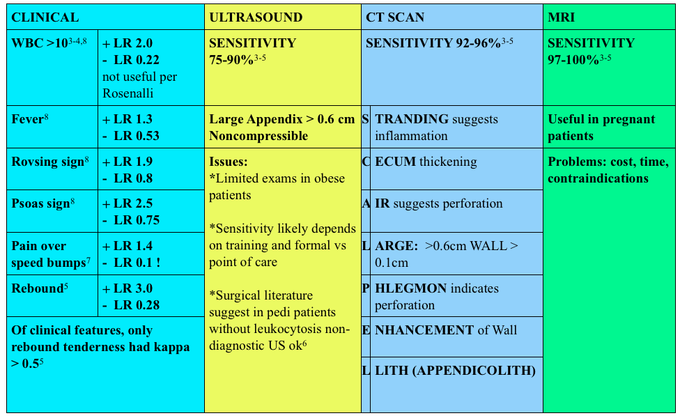
Hemoptysis Workup

Generously Donated Rosh Review Questions 

Question 1. A 50-year-old man, nonsmoker, presents to the ED with a 2-day history of cough now associated with frank hemoptysis. He denies any constitutional symptoms. Vital signs are BP 125/70, HR 80, RR 16, and pulse oximetry is 98% on room air. On exam, his lung fields are clear; the remainder of the exam is unremarkable. A chest radiograph is performed, which is normal. [polldaddy poll=9039260]

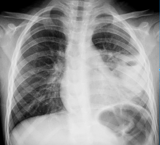
Question 2. A 55-year-old man, smoker, presents to the ED with hemoptysis and dyspnea for 4 weeks. His VS are T 37°C, BP 146/76 mm Hg, HR 85 bpm, RR 20 per minute, and oxygen saturation 96% on RA. His lung exam reveals distant breath sounds on the left side. His chest X-ray is shown below. [polldaddy poll=9039262]
Rosh Review
Rosh Review

Answers

1.C. The patient is hemodynamically stable with a normal chest radiograph, so he does not require ICU admission (A). Patients with massive hemoptysis require ICU admission. The decision to perform a bronchoscopy (B) in this patient will be left up to the pulmonologist. Given the overall clinical picture, urgent bronchoscopy is not required in this case. With massive hemoptysis, an emergent bronchoscopy is indicated. Bronchitis (D) typically presents with the abrupt onset of cough with blood-streaked purulent sputum. The patient in the clinical scenario has persistent frank hemoptysis, which mandates further investigation. In a patient who does not smoke, is under the age of 40, and has a normal chest radiograph and scant hemoptysis, treatment for bronchitis can be initiated with outpatient follow-up.

2. B. Although bronchitis (A) is the most common cause of hemoptysis (responsible for 15%-30% of cases), patients present with cough as the dominant symptom and have abnormal lung exams and normal chest x-rays. The cough may be productive of sputum. The diagnosis of pneumonia (C) requires focal findings on physical exam or infiltrates on radiographic imaging and is typically accompanied by a fever. Patients with lung cancer are at increased risk for pulmonary embolism (D). This patient’s Wells score is 2 (one point each for hemoptysis and malignancy), which makes the likelihood of PE 16% in an ED population. Given the lung mass seen on chest x-ray, lung cancer is more likely than PE.

References:
Episode 32 – Appendicitis

(ITUNES OR Listen Here)

The Free Open Access Medical Education (FOAM)

This week we cover a post from Dr. Rory Spiegel, author of EMnerd, on initial nonoperative management of acute appendicitis based on an article by Salminen et al in JAMA 2015.

  • 530 patients with CT confirmed acute, uncomplicated appendicitis were randomized to operative intervention (n=273 receiving open laparotomies) or non-operative intervention (n=257 receiving antibiotics).
  • 27.3% (n=70, CI 22-33.2%) of patients who received medical management (ertapenem x 3 days then 5 days of levofloxacin) had an appendectomy by the 1 year mark
    • 7 patients (2.7%) in medical management group had complicated appendicitis at one year, 0 had abscesses
    • 45 patients (20.5%) in the operative group had surgical complications

This is a non-inferiority study where the intent  is to demonstrate that an experimental treatment (antibiotics alone) is not substantially worse than a control treatment (immediate surgery). The authors set the non-inferiority margin at 24%, which means that a failure rate (appendectomy by 1 year) >24% would render medical management inferior.

Authors Conclusion: “Among patients with CT-proven, uncomplicated appendicitis, antibiotic treatment did not meet the prespecified criterion for noninferiority compared with appendectomy.”

Spiegel’s Conclusion: “there is a great deal to be determined before this non-invasive strategy can be considered mainstream practice…in what was once considered an exclusively surgical disease, the majority of patients can effectively be managed conservatively. Despite not meeting their own high standards for non-inferiority, the authors demonstrated that for most patients with acute appendicitis, when treated conservatively with antibiotics we can avoid surgical intervention without complications of delays to definitive care.”

More FOAM on non-operative treatment of appendicitis: The SGEM

Core Content
Tintinalli (7e) Chapters 84, 124;  Rosen’s Emergency Medicine (8e) Chapter 93

Appendicitis

Diagnosis:

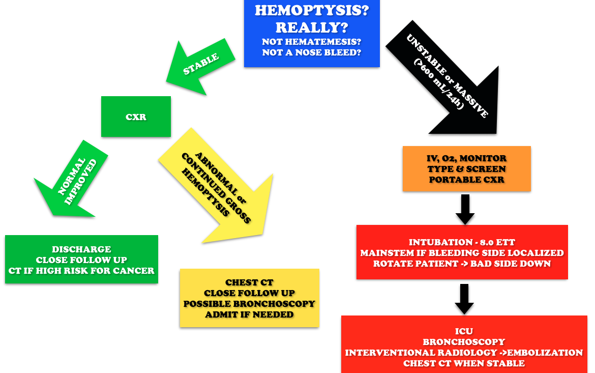
Diagnosing Acute Appendicitis

Use of contrast enhanced CT scans controversial.  Rosenalli and the American College of radiology concur that oral contrast is probably not needed but does increase the emergency department length of stay [3-5].

Treatment:

  • Surgical consult
  • Antibiotics:
    • Broad spectrum beta-lactams: ampicillin-sulbactam 3g IV (75 mg/kg IV in peds)  piperacillin-tazobactam 4.5g IV, cefoxitin 2g IV (40 mg/kg IV in peds)  OR metronidazole 500 mg IV + ciprofloxacin 400 mg IV

Other things to consider in special populations in right lower quadrant:

Pelvic Inflammatory Disease and Tubo-Ovarian Abscess in women

  • Symptoms: vaginal discharge, adnexal or uterine tenderness, lower abdominal pain, cervical motion tenderness, fever
  • Cause: chlamydia and neisseria gonorrhoea most commonly
  • Treatment: ceftriaxone 250 mg IM + doxycycline 100 mg BID x 14 days

Typhlitis (neutropenic enterocolitis) –

  • Symptoms: crampy abdominal pain (often RLQ), abdominal distension, fever, diarrhea, bloody diarrhea
  • Diagnosis: CT scan
  • Treatment: NPO, IV fluids, broad spectrum antibiotics (piperacillin-tazobactam, meropenem, metronidazole) + surgical consult if needed
  • Complications: perforation, gastrointestinal bleeding, sepsis

Generously Donated Rosh Review Questions 

1. A 22-year-old man presents with abdominal pain followed by vomiting for 1 day. His examination is significant for right lower quadrant tenderness to palpation. He has a negative Rovsing sign. [polldaddy poll=9026936]

2. A 22-year-old woman presents with lower abdominal pain and vaginal discharge. She is sexually active with men with inconsistent barrier protection. Her vitals are normal other than temperature of 101°F. On examination, there is yellow cervical discharge, no cervical motion tenderness, but uterine and left adnexal tenderness. An ultrasound does not show any evidence of tubo-ovarian abscess. [polldaddy poll=9026939]

Answers.

1. B. Sensitivity or the true positive rate measures the proportion of actual positives that are correctly identified as such. It is determined by dividing the number of true positives of the test by the number of true positives + false negatives. Tests with a high sensitivity are good for ruling out disease as the test has very few false negatives. A test with high sensitivity is advantageous as a screening tool as it misses very few people with the disease. The onset of pain before vomiting has been found to be as high as 100% sensitive in diagnosing acute appendicitis.Rovsing’s sign (D) (indirect tenderness) describes pain felt in the right lower quadrant upon palpation of the left lower quadrant. This sign signifies the presence of peritoneal irritation and has a sensitivity of 58%. Right lower quadrant pain (C) has a sensitivity of 81% and fever (A) has a sensitivity of 67%.

2.This patient presents with signs and symptoms consistent with pelvic inflammatory disease (PID) and should be treated with ceftriaxone 250 mg IM and 2 weeks of doxycycline. PID is an ascending infection beginning in the cervix and vagina and ascending to the upper genital tract. Neisseria gonorrhoeae and Chlamydia trachomatis are most commonly implicated. It can present with a myriad of symptoms although lower abdominal pain is the most common. Other symptoms include fever, cervical or vaginal discharge and dyspareunia. Pelvic examination reveals cervical motion tenderness (CMT), adnexal tenderness and vaginal or cervical discharge. Inadequately treated PID can lead to tubo-ovarian abscess, chronic dyspareunia and infertility. Due to the variable presentation and serious sequelae, the CDC recommends empiric treatment of all sexually active women who present with pelvic or abdominal pain and have any one of the following: 1) CMT, 2) adnexal tenderness or 3) uterine tenderness. Treatment should cover the most common organisms and typically consists of a third generation cephalosporin (ceftriaxone) and a prolonged course of doxycycline. Patients with systemic manifestations or difficulty tolerating PO should be admitted for management.Ceftriaxone and azithromycin (A) are used in the treatment of cervicitis or urethritis. Clindamycin (C) and metronidazole (D) are used in the treatment of bacterial vaginosis.
References:

1.Salminen P, Paajanen H, Rautio T, et al. Antibiotic Therapy vs Appendectomy for Treatment of Uncomplicated Acute Appendicitis: The APPAC Randomized Clinical Trial. JAMA. 2015;313(23):2340

2. Horst JA, Trehan I, Warner BW et al. Can Children With Uncomplicated Acute Appendicitis Be Treated With Antibiotics Instead of an Appendectomy? Ann Emerg Med. 2015;66:(2)119-22

3.”Acute Appendicitis.” Tintinalli’s Emergency Medicine: A Comprehensive Study Guide.  7th ed. pp 574-581.

4. “Acute Appendicitis” Rosen’s Emergency Medicine. 8th ed. pp. 1225-1232.e2

5. ACR Appropriateness Criteria.  American College of Radiology. 2013.

6. Cohen B, Bowling J, Midulla P, et al. The non-diagnostic ultrasound in appendicitis: is a non-visualized appendix the same as a negative study? J Pediatr Surg. 2015;50:(6)923-7

7. Ashdown HF, D’Souza N, Karim D, Stevens RJ, Huang A, Harnden A. Pain over speed bumps in diagnosis of acute appendicitis: diagnostic accuracy study. BMJ. 2012;345:e8012.

8. Bundy DG, Byerley JS, Liles EA. Does This Child Have Appendicitis? 2009;298(4):438–451.