Vaccine-Induced Thrombotic Thrombocytopenia

Apple Podcasts , SpotifyListen Here

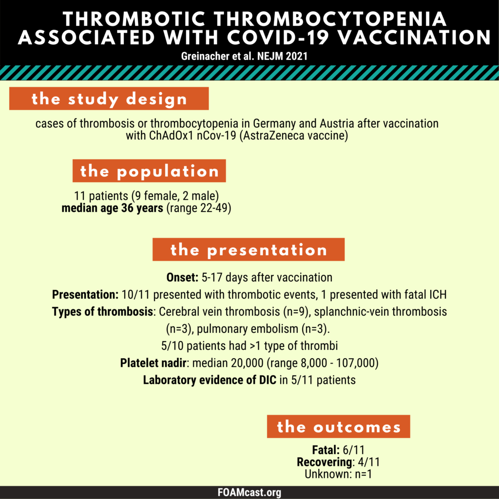
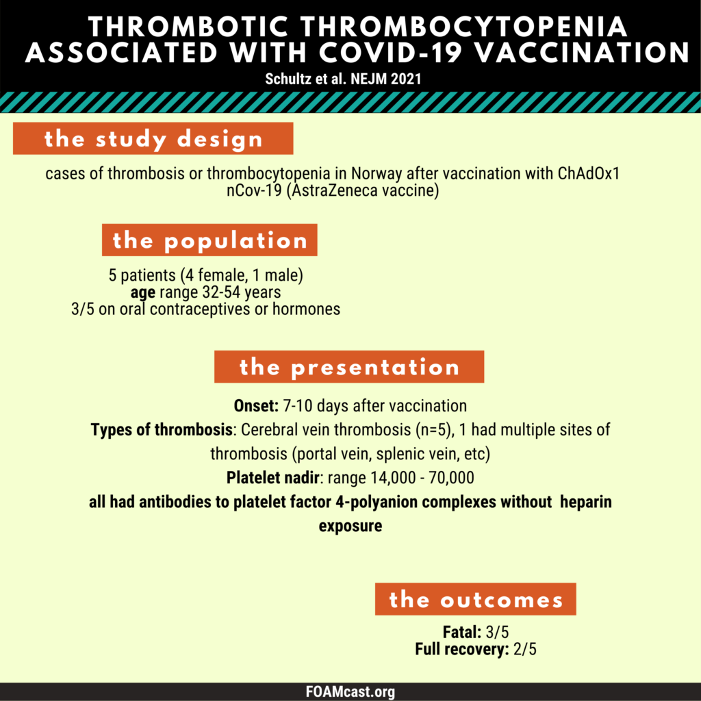
In this episode, we discuss thrombotic thrombocytopenia associated with the viral vector COVID-19 vaccines (VITT: Vaccine-Induced Thrombotic Thrombocytopenia) – a very very rare, but real entity. Information is still emerging so numbers will likely change in the coming days. For more on cerebral vein thrombosis in general, check out this podcast.

Greinacher et al NEJM 2021
Schultz et al. NEJM 2021
ACIP Presentation by Jannsen 4/14/21, Muir et al NEJM 2021

International Society of Thrombosis and Hemostasis Statement

United Kingdom Guidance On Thrombosis Following Vaccinations

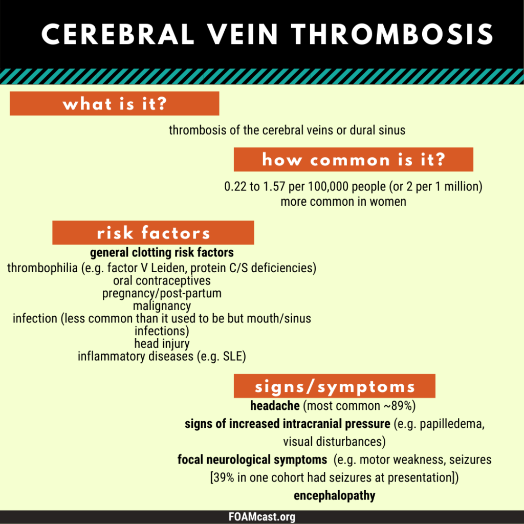
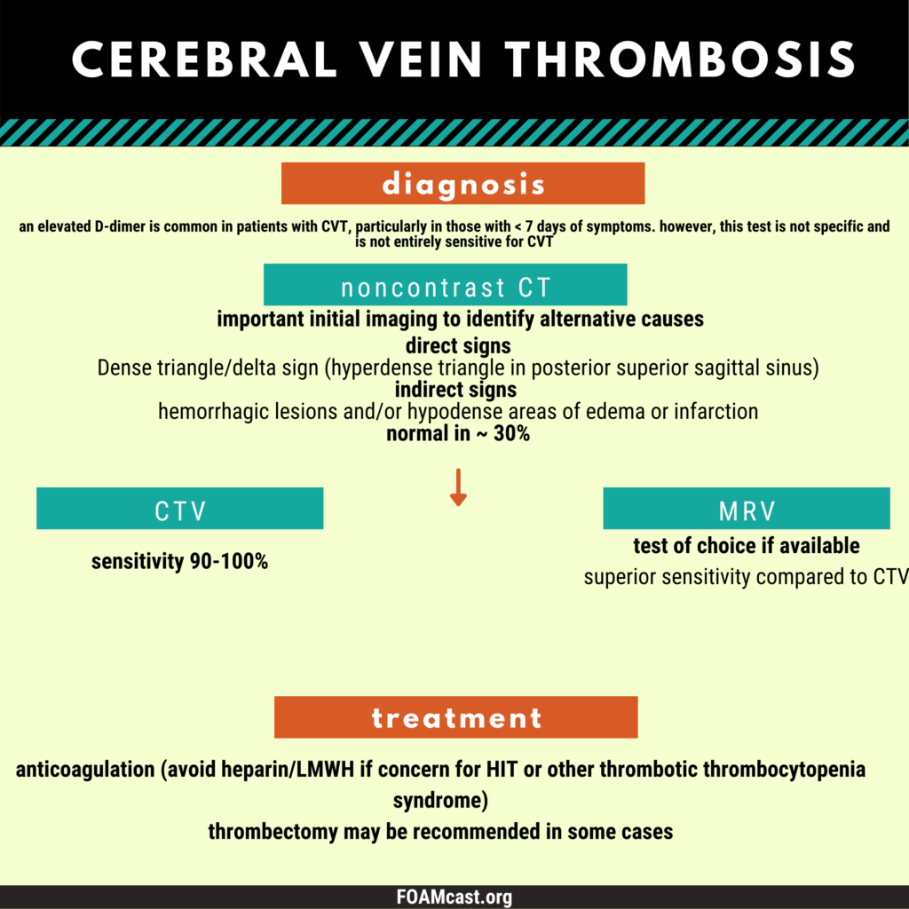
Cerebral Vein Thrombosis

Apple Podcasts , SpotifyListen Here

If you’re looking for vaccine-induced thrombotic thrombocytopenia (CVT/CVST with thrombocytopenia following AstraZeneca or J&J COVID-19 vaccine), check out this podcast.

Multicenter Cohort Study: Ferro JM et al. Prognosis of cerebral vein and dural sinus thrombosis: results of the International Study on Cerebral Vein and Dural Sinus Thrombosis (ISCVT). Stroke. 2004 Mar;35(3):664-70. Epub 2004 Feb 19. PMID: 14976332.

Utility of D-dimer:

  1. Dentali F et al. D-dimer testing in the diagnosis of cerebral vein thrombosis: a systematic review and a meta-analysis of the literature. J Thromb Haemost. 2012 Apr;10(4):582-9.PMID: 22257124.
  2. Meng R et al. Evaluation of plasma D-dimer plus fibrinogen in predicting acute CVST. Int J Stroke. 2014 Feb;9(2):166-73. Epub 2013 Mar 19. PMID: 23506130

Psychogenic Non-epileptic Attacks (PNEA)

iTunes or Listen Here

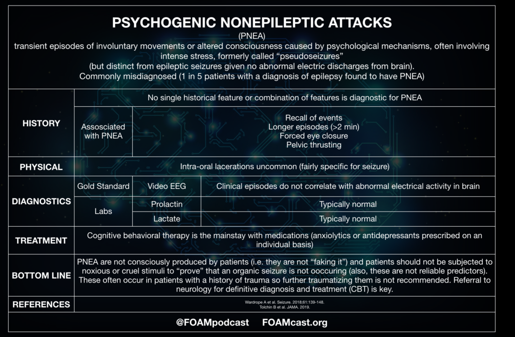
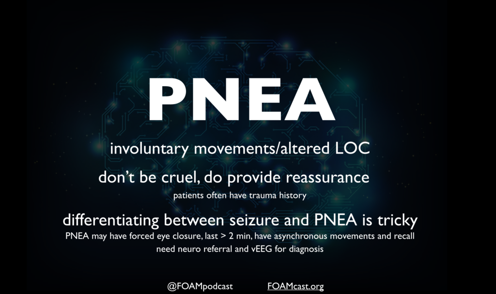
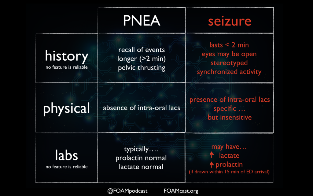
Psychogenic non-epileptic attacks (PNEA) have been formally referred to as pseudoseizures or psychogenic non-epileptic seizures. Recently, this entity has been rebranded to PNEA to reflect that there is no actual seizure activity and potentially prevent confusion and mislabeling among patients.

A 10-year-old girl presents with episodes of brief staring spells that she is unaware of. The patient’s mother states that the onset and termination of the spells are abrupt. The patient is otherwise healthy. She has lots of friends at school and has excellent academic performance. Physical examination and laboratory studies are normal at this time. Which of the following is the most likely diagnosis?

  1. Absence seizure
  2. Attention deficit hyperactivity disorder
  3. Autism spectrum disorder
  4. Complex partial seizure

[accordion]
[toggle title=”Answer” state=”closed”]

  1. Absence seizure is a type of generalized seizure characterized by loss of consciousness sometimes accompanied by myotonic, atonicclonic, or tonic components, autonomic components (e.g., enuresis), or automatisms. The onset of absence seizure commonly occurs in early childhood with a predilection to the female sex. It usually terminates by the age of 20 years or may transform into another form of generalized seizure. Absence seizure may be typical or atypical. The onset and termination of a typical absence seizure are abrupt while they may be gradual in an atypical absence seizure. Additionally, atypical seizure commonly occurs in patients with multiple seizure types and demonstrates more marked changes in tone. Patients with an atypical absence seizure may also have a developmental delay or mental retardation. Patients with absence seizure may miss a few words or break off mid-sentence during an attack. The attack period is so brief that the patient is unaware of it. Physical examination may be normal. Having the child hyperventilate for three to five minutes may precipitate a staring attack. Diagnosis of typical absence seizure is with electroencephalography that shows bursts of bilateral synchronous and symmetric 3-Hertz spike-wave activity. A slower spike-wave discharge is noted in an atypical absence seizure. Treatment is with antiepileptic medications including ethosuximide and valproic acid. All patients with absence seizure should be referred to a neurologist for management. Drug levels of valproic acid should be monitored.While attention deficit hyperactivity disorder (B) has a childhood onset, it is marked by symptoms of inattentiveness, hyperactivity, and impulsivity that are not present in the vignette above. Autism spectrum disorder (C) may also have an early onset. However, patients suffer from pervasive difficulties with social communication and repetitive interests and behavior that are absent in the vignette above. In complex partial seizure (D), the duration of seizure usually lasts > 30 seconds, automatisms are present, and the termination of the seizure is gradual.

[/toggle]
[/accordion]

References:

  1. Lafrance WC, Baker GA, Duncan R, Goldstein LH, Reuber M. Minimum requirements for the diagnosis of psychogenic nonepileptic seizures: a staged approach: a report from the International League Against Epilepsy Nonepileptic Seizures Task Force. Epilepsia. 2013;54(11):2005-18.
  2. Wardrope A, Newberry E, Reuber M. Diagnostic criteria to aid the differential diagnosis of patients presenting with transient loss of consciousness: A systematic review. Seizure. 2018;61:139-148.
  3. Chen et al. Value of witness observations in the differential diagnosis of transient loss of consciousness.  Neurology. 2019 Feb 26;92(9):e895-e904. doi: 10.1212/WNL.0000000000007017. Epub 2019 Jan 25.
  4. Shmuely S, Bauer PR, Van zwet EW, Van dijk JG, Thijs RD. Differentiating motor phenomena in tilt-induced syncope and convulsive seizures. Neurology. 2018;90(15):e1339-e1346.
  5. Doğan EA, Ünal A, Ünal A, Erdoğan Ç. Clinical utility of serum lactate levels for differential diagnosis of generalized tonic-clonic seizures from psychogenic nonepileptic seizures and syncope. Epilepsy Behav. 2017;75:13-17.

Episode 66 – Back Pain and Spinal Epidural Abscess

iTunes or Listen Here

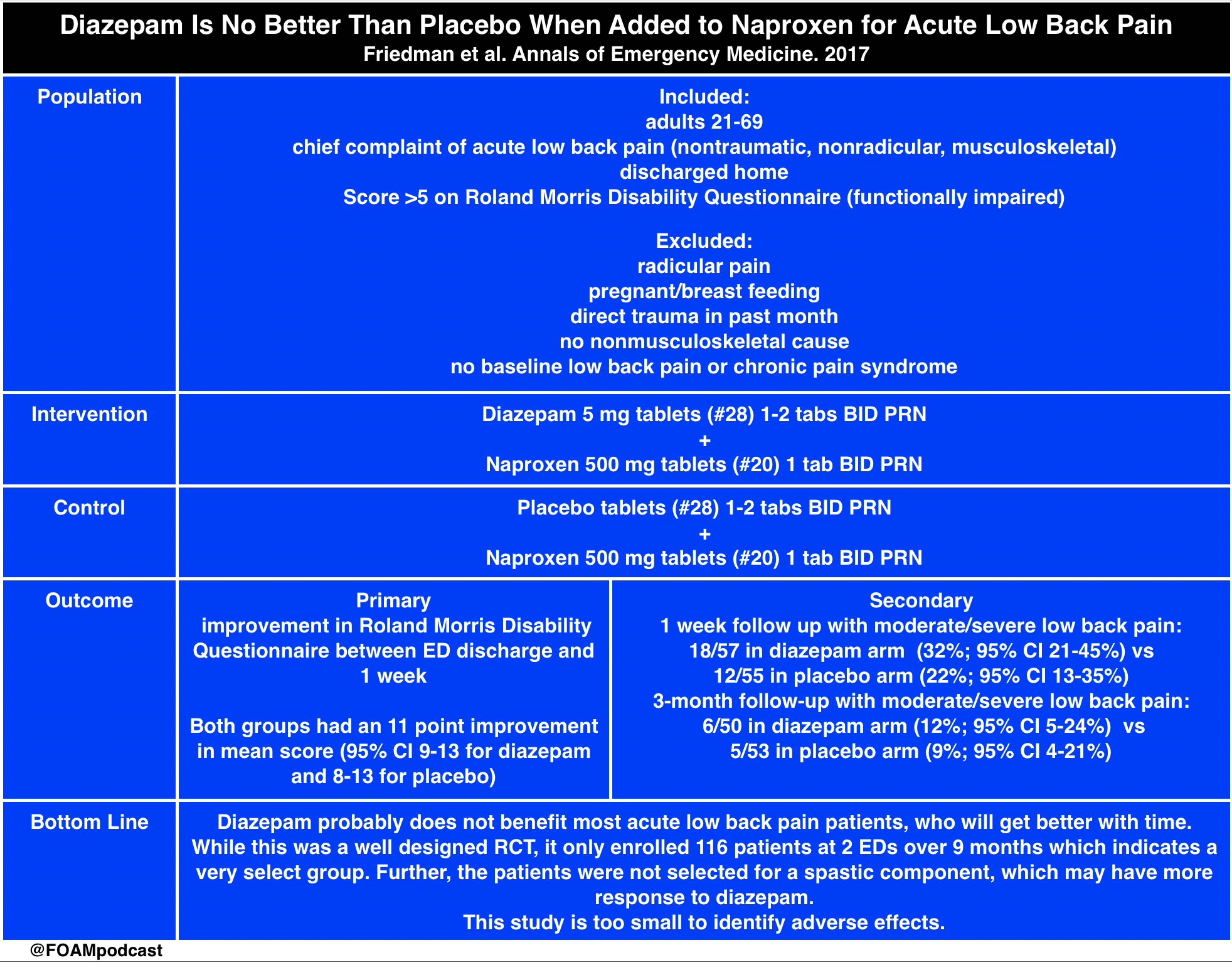
  • We cover a post from the fantastic St. Emlyn’s team which breaks down a recently published article in Annals of Emergency Medicine comparing diazepam with placebo in patients with acute low back pain.  We recommend you read the full text of the article by Friedman et al.

diazepam

Core Content

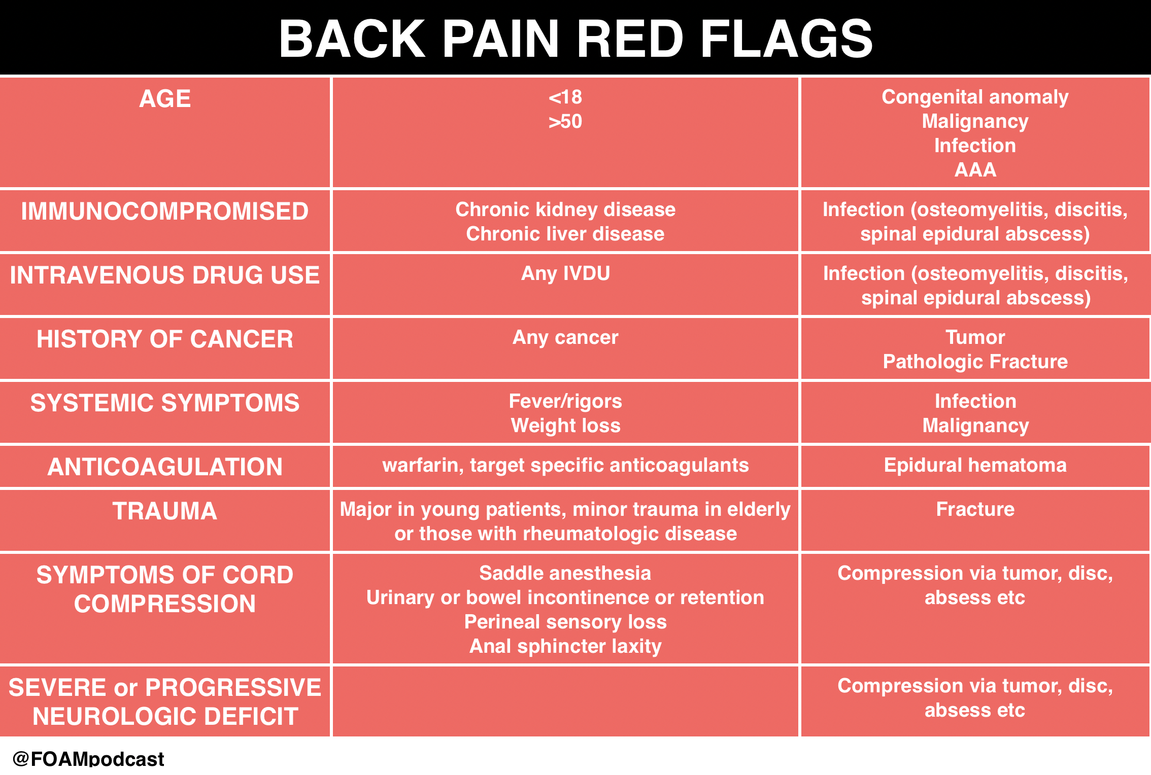
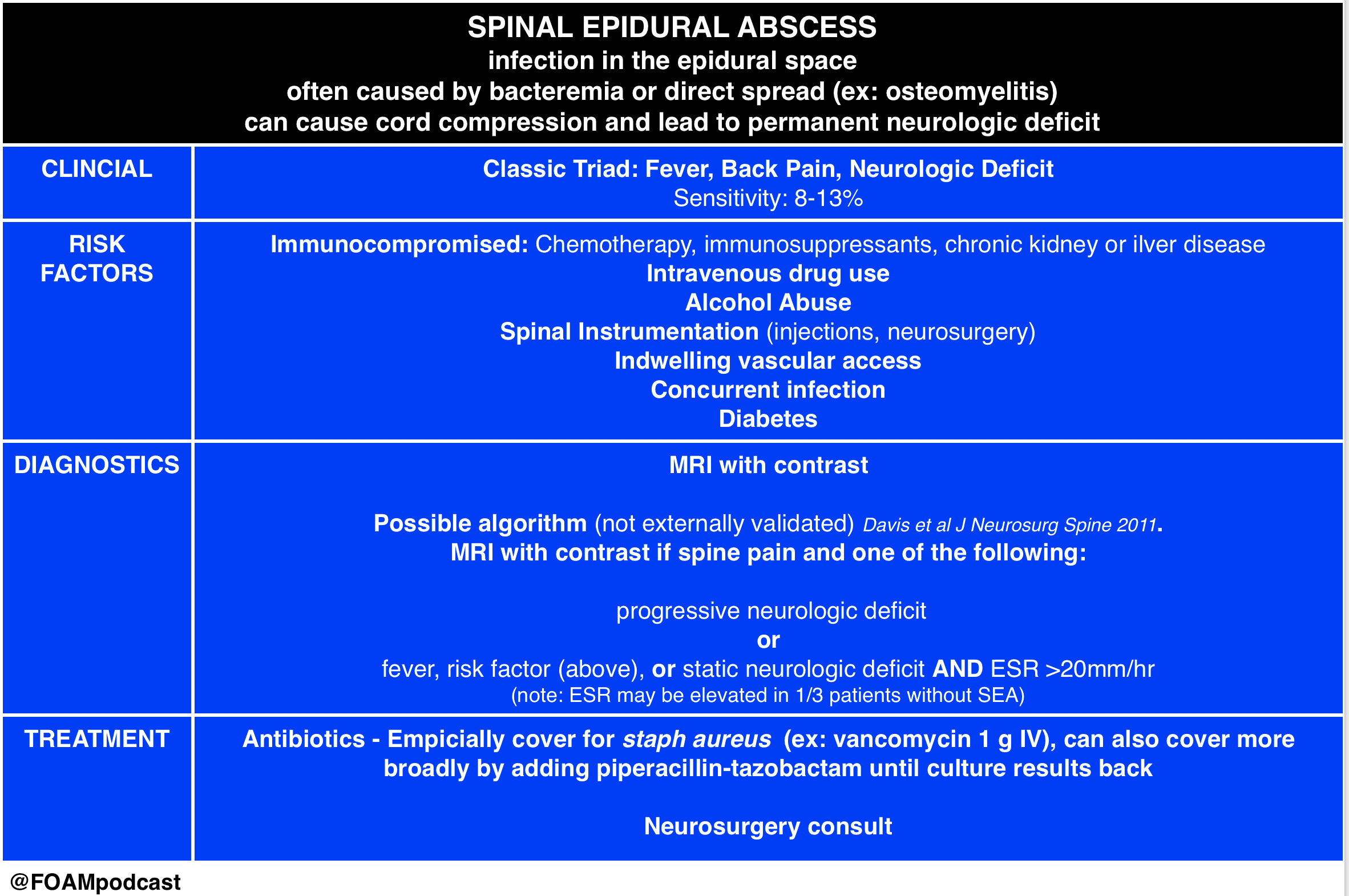
We delve into core content on red flags in back pain and spinal epidural abscess using Rosen’s Emergency Medicine and Tintinalli’s Emergency Medicine Chapter 279 as a guide.

red flags

SEA

Rosh Review Emergency Board Review Questions

  • A 13-year-old boy on chemotherapy for acute lymphoblastic leukemia presents with progressive lower back pain for 2 weeks. Per mom, he has had subjective fevers and a “bulge in his lower back” that is warm to touch. He is currently afebrile and has no focal neurologic deficits. An MRI is obtained as seen in the image above. Which of the following regarding this patient’s condition is true?

A. An appropriate antibiotic regimen is cefepime and metronidazole

B. Direct extension of skin and soft tissue infections is the most common cause

C. Erythrocyte sedimentation rate is a sensitive screening tool

D. Most patients present with back pain, fever, and a focal neurologic deficit

[accordion]
[toggle title=”Answer” state=”closed”]

C.  Erythrocyte sedimentation rate is a sensitive screening toolThe patient has a posterior epidural abscess with adjacent osteomyelitis and myositis. The most common cause of an epidural abscess is hematogenous spread of infection, not direct extension from skin or soft tissue infection. Major risk factors include diabetes, intravenous drug abuse, chronic renal failure, alcoholism, and immunosuppression. The most common organism involved is Staphylococcus aureus. Other organisms include streptococci, anaerobes, gram-negative bacilli, and Pseudomonas aeruginosa. Patients present with localized back pain with tenderness to percussion. Fevers are common (reported in up to 75% of patients). MRI is the diagnostic modality of choice, but erythrocyte sedimentation rate is a sensitive marker and may be used in conjunction with plain radiographs to screen for infectious spinal disease.Vancomycin is an important component of the antibiotic regimen because it covers methicillin-resistant Staphylococcus aureus (MRSA) and is usually combined with a 3rd-generation cephalosporin (A). Cefepime is a 4th-generation cephalosporin with pseudomonas coverage. Metronidazole or clindamycin can be added for anaerobic coverage. While the classic triad consists of back pain, fever, and neurologic deficits (D), only a small proportion of patients actually have all three components at presentation. The most common cause of an epidural abscess is hematogenous spread of infection, not direct extension from skin or soft tissue infection (B).

[/toggle]
[/accordion]

An 8-year-old previously healthy boy presents with back pain and fever for 4 days. He complains of pain in the low back, which is increased with bending or twisting. The pain radiates down into his left leg. He denies trauma. Physical exam is remarkable only for tenderness to palpation over the lumbar spine. What management is indicated

A. Ibuprofen and follow up with his pediatrician

B. MRI of the lumbar spine

C. Plain radiographs of the lumbar spine

D. Urinalysis

[accordion]
[toggle title=”Answer” state=”closed”]

B.

MRI of the lumbar spine. This patient presents with symptoms consistent with discitis and should have an MRI for diagnosis. Discitis is a rare infection of the nucleus pulposus and can involve the cartilaginous endplate and vertebral body. It can occur spontaneously or after surgical procedures. Typically, patients present with severe pain localized to the spinal level of involvement. Any movement of the spine exacerbates the pain. Children often present with sudden onset of back pain and refusal to walk. The lumbar spine is most commonly involved and the average age of patients is 7 years. Radicular symptoms are common (present in 50-90% of cases) and most patients will present with fever (90%). Neurologic deficits are uncommon. Serious pathology should be suspected in any pediatric patient presenting with back pain. Neurologic symptoms are rarely present. MRI is the best study for diagnosis and can also rule out other critical diagnoses including epidural abscess. Laboratory studies are non-specific and insensitive but typically will have an elevated erythrocyte sedimentation rate. White blood cell counts are frequently within the normal range.

[/toggle]
[/accordion]

Episode 64 – Lumbar Puncture and Central Nervous System Infections

iTunes or Listen Here

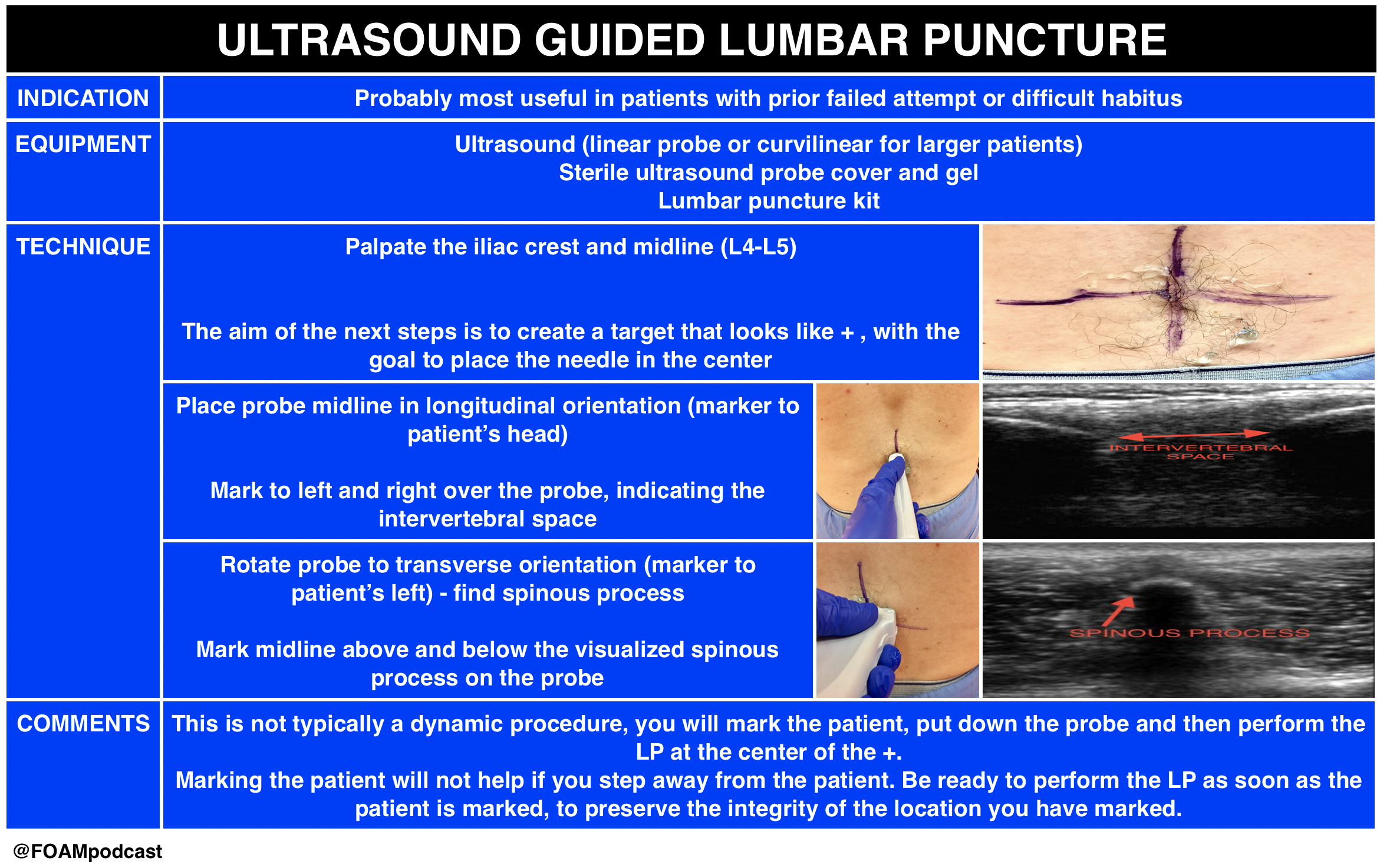
Ultrasound is gaining popularity in assisting lumbar punctures (LPs). We review this technique as well as Free Open Access Medical Education (FOAM) from the following sites:  5 Min SonoSinai EM, and PEM pearls from ALiEM.

US guided LP

  • This technique is likely most helpful in difficult patients. A recent study demonstrated 27% absolute increase in first attempt success using ultrasound in infants <6 months old undergoing LP; however, the success rate in both arms was abysmal with only 57% success rate in the ultrasound arm.
  • Core Content

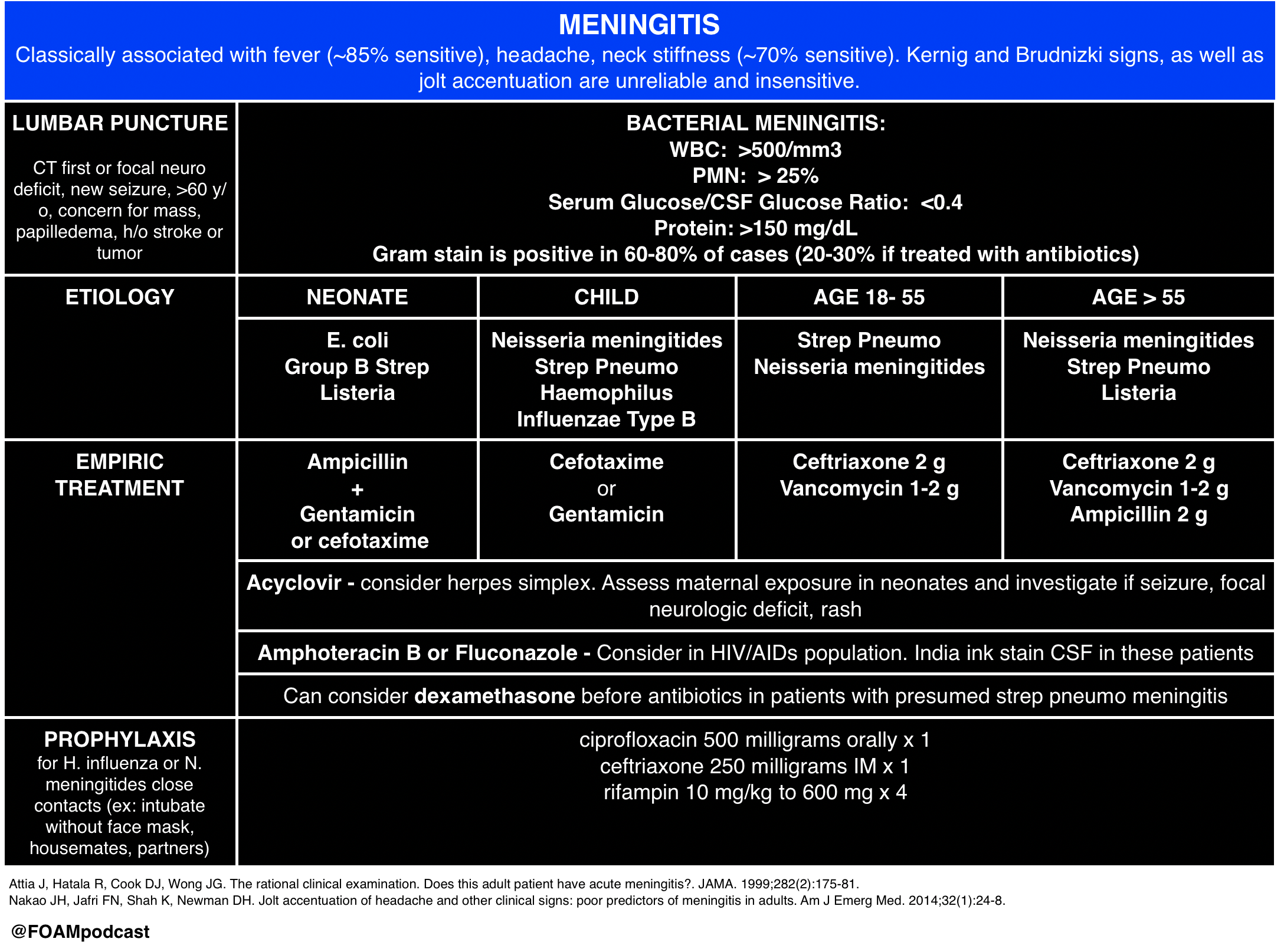
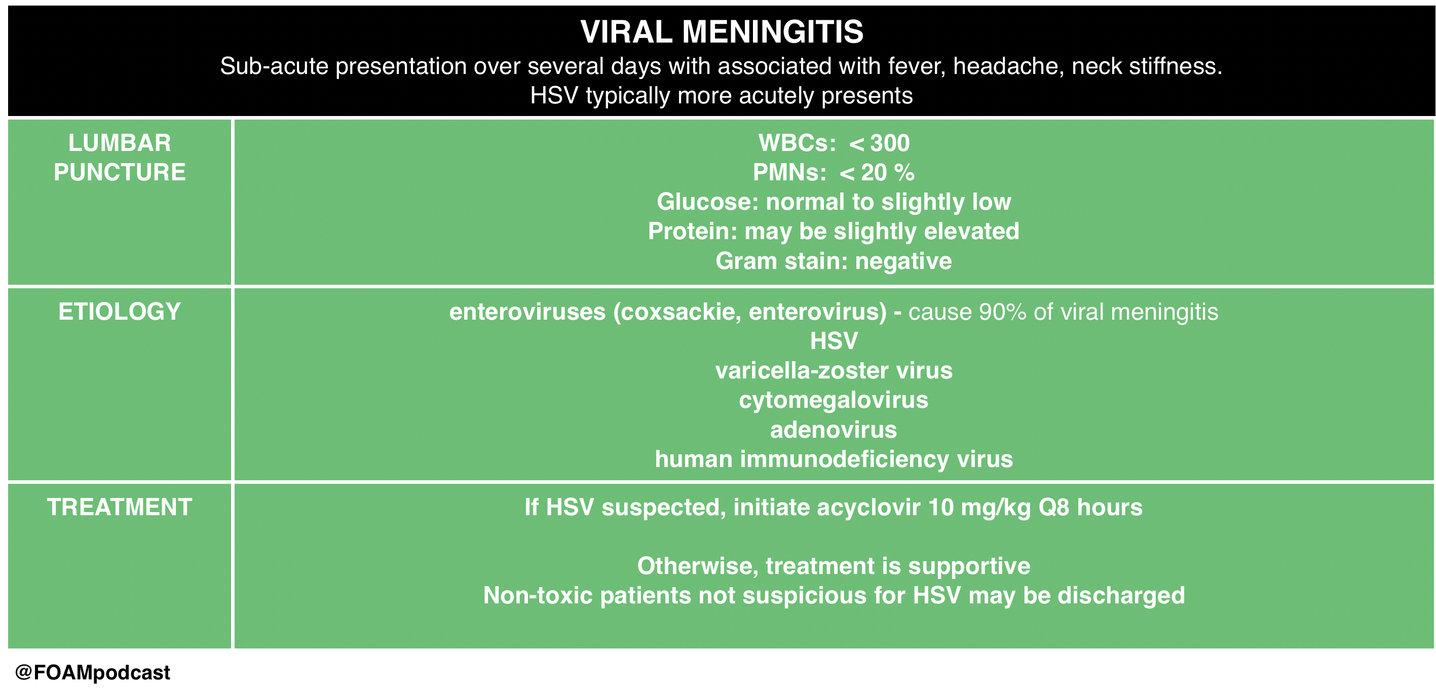
We then delve into core content on meningitis, encephalitis, and antiNMDA receptor encephalitis using Rosen’s Chapter 109 and Tintinalli’s Chapters 117 and 174 as a guide.

 Rosh Review Emergency Board Review Questions

A 40-year-old man with HIV presents with two weeks of progressive headache, malaise, and fever. On examination, he has mild nuchal rigidity, confusion, and a temperature of 38.2oC. Cerebrospinal fluid analysis shows a white blood cell count 360 cells/mL with a monocyte predominance, glucose 28 mg/dL, and protein 220 mg/dL. What is the treatment of choice?

A. Acyclovir

B. Amphotericin B

C. Ceftriaxone

D. Vancomycin

[accordion]
[toggle title=”Answer” state=”closed”]

B. Amphotericin B . Cryptococcal meningoencephalitis is an opportunistic infection that occurs primarily in patients with advanced AIDS, although it can be seen in immunocompromised transplant patients as well. The majority of HIV-related cases occur when the CD4 count is < 100 cells/mm3. Patients present with progressive headache, nausea, malaise, and fever over the course of 1 – 2 weeks. Examination findings are typical for meningitis and include altered mental status, photophobia, and fever. Meningismus is less commonly seen. Presentations can be subtle and a high index of suspicion is needed for diagnosis. A CT scan of the brain is indicated if there are signs of increased intracranial pressure or focal neurologic deficits. Lumbar puncture should be performed with careful measurement of the opening pressure. Cerebrospinal fluid will show a mildly elevated white blood cell count with a monocyte predominance, decreased glucose, and mildly elevated protein. However, in some cases the cerebrospinal fluid will only show minor, if any, abnormalities. Cryptococcal antigen testing of the CSF is nearly 100% sensitive and specific. India ink staining will show budding organisms. Treatment of choice is intravenous amphotericin B in addition to oral flucytosine for 14 days followed by an 8 week course of oral fluconazole.  Acyclovir (A) is used in the treatment of herpes meningoencephalitis. Ceftriaxone (C) and vancomycin (D) are indicated in the treatment of bacterial meningitis.

[/toggle]
[/accordion]

A previously healthy 18-year-old woman presents to the emergency department with complaints of fever, headache, and neck stiffness. She is accompanied by her sister, who expresses concern because the patient seems suddenly confused and cannot remember what she did yesterday. After you administer empiric intravenous antibiotics, which of the following is the next best step?

A. Chest X-ray

B. Complete blood count with differential

C. Lumbar puncture

D. Urinalysis

[accordion]
[toggle title=”Answer” state=”closed”] C. 
 Lumbar puncture.  Meningitis is an inflammation of the tissues surrounding the brain and spinal cord (meninges) and may be of infectious (bacterial, viral, or fungal) and various other etiologies. The classic clinical manifestations include nuchal rigidity, fever and altered mental status. Patients often present with headache as well. All patients with suspected meningitis should have lumbar puncture (LP) to evaluate the cerebrospinal fluid (CSF) unless this procedure is contraindicated. There are no absolute contraindications to LP. Relative contraindications include patients with evidence of increased intracranial pressure, thrombocytopenia, bleeding diathesis or spinal epidural abscess. Acute bacterial meningitis is a medical emergency and left untreated or treated late is almost universally fatal. Treatment involves addressing systemic complications and initiating empiric antibiotic therapy as soon as possible.

Up to half of patients with pneumococcal meningitis may have evidence of pneumonia on chest X-ray (A), but this is not part of the initial workup of patients with suspected meningitis. Complete blood count with differential (B) is often ordered in the workup for bacterial meningitis and generally shows increased white blood cell count, but is not as important to order initially as an LP. Urinalysis (D) is generally not a helpful test in the diagnosis of meningitis and therefore not recommended in the workup for patients with suspected bacterial meningitis.

[/toggle]
[/accordion]

References:

  1. Attia J, Hatala R, Cook DJ, Wong JG. The rational clinical examination. Does this adult patient have acute meningitis?. JAMA. 1999;282(2):175-81.
  2. Nakao JH, Jafri FN, Shah K, Newman DH. Jolt accentuation of headache and other clinical signs: poor predictors of meningitis in adults. Am J Emerg Med. 2014;32(1):24-8.
  3. Neal JT, Kaplan SL, Woodford AL, Desai K, Zorc JJ, Chen AE. The Effect of Bedside Ultrasonographic Skin Marking on Infant Lumbar Puncture Success: A Randomized Controlled Trial. Ann Emerg Med. 2016.

Bell’s Palsy and Burns

(ITUNES OR LISTEN HERE)

We are in Las Vegas at ACEP 2016 thanks to Annals of Emergency Medicine and ACEPnow and discuss high yield and cutting edge lectures each day.

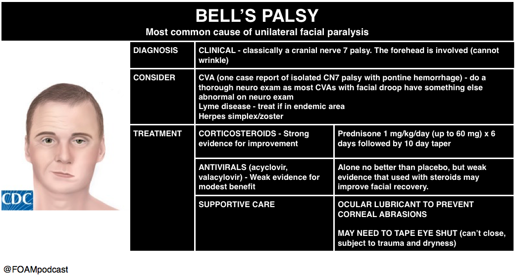
Dr. Megan Osborn – Bell’s Palsy or Stroke?

Traditional teaching: we can differentiate Bell’s palsy (lower motor neuron) from a stroke (upper motor neuron by assessing forehead involvement.  If the patient can wrinkle their forehead? Think stroke.  Dr. Megan Osborn tackled the question: does this actually work all the time in her talk in the New Speakers Forum.

screen-shot-2016-10-19-at-8-19-31-am

Dr. Toree McGowan – Burns

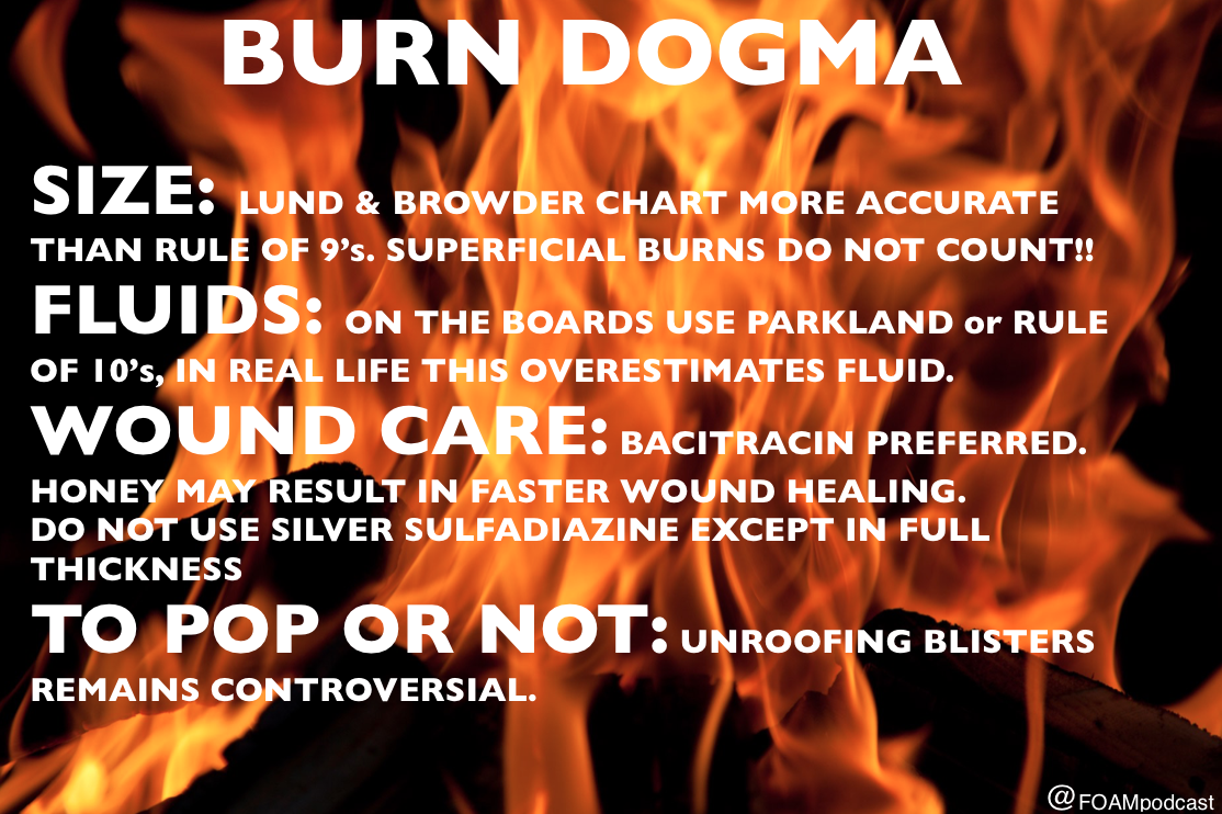
Check out this podcast for more on burns

screen-shot-2016-10-19-at-8-33-56-am

  1. Sherman SC, Thompson TM, Thompson TT. Pontine hemorrhage presenting as an isolated facial nerve palsy. Annals of emergency medicine. 46(1):64-6. 2005. [pubmed]
  2. AAN Bell’s Palsy Guideline Update
  3. Fahimi J, Navi BB, Kamel H. Potential Misdiagnoses of Bell’s Palsy in the Emergency Department. Annals of Emergency Medicine. 63(4):428-434. 2014. [article]
  4. Madhok VB, Gagyor I, Daly F. Corticosteroids for Bell’s palsy (idiopathic facial paralysis). The Cochrane database of systematic reviews. 7:CD001942. 2016. [pubmed]
  5. Gagyor I, Madhok VB, Daly F. Antiviral treatment for Bell’s palsy (idiopathic facial paralysis). The Cochrane database of systematic reviews. 2015. [pubmed]
  6. Wasiak J, Cleland H, Campbell F. Dressings for superficial and partial thickness burns. The Cochrane database of systematic reviews. 2008. [pubmed]
  7. Ringh M, Rosenqvist M, Hollenberg J et al. Mobile-Phone Dispatch of Laypersons for CPR in Out-of-Hospital Cardiac Arrest. N Engl J Med. 372(24):2316-2325. 2015. [article]

Episode 53 – Intracranial Hemorrhage

iTunesor Listen Here

We cover a post by Dr. Rory Spiegel, EMNerd: The Case of Differing Perspectives, on the results of the ATACH-2 trial on blood pressure control in intracranial hemorrhage (ICH). This study sought to determine the safety and efficacy of the intensive blood pressure lowering.

Population: adults (>18 y/o) with ICH on CT scan, GCS ≥ 5 and <4.5 hours since symptom onset (changed mid-study)

Intervention: Reduce and maintain the hourly minimum systolic blood pressure in the range of 110 to 139 mm Hg throughout the period of 24 hours after randomization (“intensive treatment”). Preferred agents for blood pressure control in order of preference were 1. nicardipine 2. labetalol (diltiazem or urapidil if not available)

  • Mean minimum in hours 0-2: 128.9±16 mm Hg

Control: Reduce and maintain the hourly minimum systolic blood pressure in the range of 140 to 179 mm Hg throughout the period of 24 hours after randomization.

  • Mean minimum in hours 0-2: 141.1±14.8 mm Hg

Outcome: The primary outcome was the proportion of patients who had moderately severe or severe disability (modified Rankin scale score (mRS) 4-5) or those who had died (mRS 6; hereafter referred to as “death or disability”) at 3 months.

  • death or disability: Intensive treatment = 186 participants (38.7%) vs Standard treatment = 181 (37.7%)

Bottom Line:  INTENSIVE blood pressure lowering <140mmHg is not supported by the literature. AHA and Rosens/Tintinalli recommend goal of 160-180 mmHg or diastolic <130 mmHg.

We also discuss the history of blood pressure control in ICH and the pendulum swing on this in recent years, using an episode of the Skeptic’s Guide to Emergency Medicine, Episode 73. This episode covers the Interact-2 trial.

Core Content

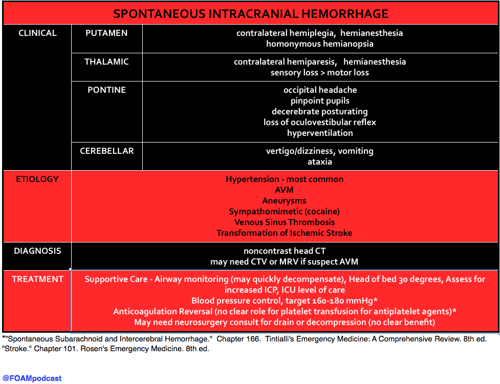
We delve into core content on ICH using Rosen’s (8th ed) and Tintinalli (8th ed)

Screen Shot 2016-07-18 at 10.59.54 AM

The use of platelets in patients with ICH on antiplatelet agents (aspirin, clopidogrel, etc) has been controversial and both Rosen’s and Tintinalli acknowledge that there is no good quality evidence that platelets are beneficial in this population.   The PATCH trial, recently published in the Lancet, found no benefit to platelet transfusion in patients with ICH who had taken antiplatelet agents in the 7 days prior to their ICH. See this review from Dr. Salim Rezaie of Rebel EM.

Population: patients ≥18years of age with non-traumatic ICH confirmed by CT and GCS ≥ 8 who took antiplatelet agent for at least 7 days prior (excluded thrombocytopenia/known coagulopathy)

Intervention: platelet transfusion

Control: standard care without platelet confusion

Outcome: primary outcome was the difference in functional outcome at 3 months after randomisation scored with the mRS

  • Odds of a shift towards death or dependence at 3 months: adjusted OR 2.05 (95% CI 1·18–3·56; p = 0.0114). This favors the control group.
  • Secondary outcome of survival: 68% in platelet transfusion group vs 77% in standard group (p = 0.15)

Note:  Like many stroke trials, the planned statistical analysis plan changed after the data came in. Initially authors planned a fixed dichotomous analysis (yes or no, did they have an mRS of 4-6 at 3 months). They changed this to an ordinal logistic regression analysis of the shift of all categories of the mRS at 3 months as this has greater “statistical efficiency”.

Generously Donated Rosh Review Questions

Question 1.  A patient presents with nausea, vomiting, right-sided hemiplegia and non-occipital headache. His gaze is deviated to the left, but he denies loss of sensation. Thirty minutes later, he becomes stuporous and progresses into coma. The pupils are now fixed and dilated. Abnormal posturing is absent. A brain CT scan is ordered. You would expect to find intracerebral hemorrhage in which of the following sites

A. Left pons

B. Left putamen

C. Right cerebellum

D. Right thalamus

[accordion]
[toggle title=”Answer” state=”closed”]

A large putamen hemorrhage results in a rapid progression of hemiplegia, nausea, vomiting and headache over 30 minutes, which is quickly followed by ipsilateral deviation of the eyes,stupor, coma and mydriatic pupils (“blown-pupil”, associated with brainstem compression and occulomotor nerve palsy). Acute management includes controlling intracranial pressure and hypertension. Surgical removal of clots is seldom successful, however, some neurologic function may be salvaged in those without coma or those with lobar clots.

Pontine hemorrhages (A) are characterized by total paralysis, rapid coma, decerebrate rigidity (abnormal posturing marked by shoulder adduction, elbow extension, wrist pronation and flexion, digit flexion, leg extension and plantarflexion) and small (miotic), not mydriatic, reactive pupils. Cerebellar insults (C) are evidenced by vomiting, occipital headache, inability to stand, vertigo, and eye deviation to the opposite side of the lesion. In this scenario, the eye deviation makes sense, but not the presenting symptoms.Thalamic lesions (D) are marked by complete hemisensory loss. Also common is hemiparesis (bleeding extension into the internal capsule) and aphasia.

Putamen Hemorrhage: Contralateral hemiparesis/hemiplegia, Contralateral sensory loss, Homonymous hemianopia

[/toggle]
[/accordion]

References:

  1. Qureshi AI, Palesch YY, Barsan WG, et al. Intensive Blood-Pressure Lowering in Patients with Acute Cerebral Hemorrhage. N Engl J Med [Internet] 2016;NEJMoa1603460. Available from: http://www.nejm.org/doi/10.1056/NEJMoa1603460
  2. Anderson C, Heeley E, Huang Y, et al. Rapid Blood-Pressure Lowering in Patients with Acute Intracerebral Hemorrhage. N Engl J Med [Internet] 2013;368(25):2355–65. Available from: http://www.nejm.org/doi/abs/10.1056/NEJMoa1214609
  3. “Spontaneous Subarachnoid and Intercerebral Hemorrhage.”  Chapter 166.  Tintialli’s Emergency Medicine: A Comprehensive Review. 8th ed.
  4. “Stroke.” Chapter 101. Rosen’s Emergency Medicine. 8th ed.
  5. Baharoglu MI, Cordonnier C, Salman RA-S, et al. Platelet transfusion versus standard care after acute stroke due to spontaneous cerebral haemorrhage associated with antiplatelet therapy (PATCH): a randomised, open-label, phase 3 trial. Lancet [Internet] 2016;6736(16):1–9. Available from: http://linkinghub.elsevier.com/retrieve/pii/S0140673616303920

Episode 50 – Seizures

(ITUNES OR LISTEN HERE)

The Free Open Access Medical Education (FOAM)

We cover Special Seizures from EMin5.com by Dr. Anna Pickens. This is a short video summarizing important diagnoses to consider when a seizure doesn’t stop after the first or second round of benzodiazepines.

[expand title=”Female“]

Consider eclampsia, which typically occurs >20 weeks gestation to 6 weeks post-partum. Magnesium 4-6 grams IV, followed by infusion. Watch for apnea, check reflexes. [/expand]


[expand title=”Possible history of alcohol abuse“]

Consider alcohol withdrawal seizure. These patients respond to benzodiazepines and will typically NOT respond to antiepileptics such as phenytoin. They just need larger doses of benzodiazepines, so ramp up the dosages of those (10-100 mg of diazepam) and may require phenobarbital or propofol. [/expand]


[expand title=”Possible isoniazid exposure“]

Isoniazid toxicity can cause seizures, coma, metabolic acidosis, often within only 30 minutes of ingestion. The treatment here is pyridoxine (vitamin B6). These patients will need large doses and many recommend empirically giving 5 GRAMS intravenously (note: this is takes many vials as the typical dose of this medication is 50-100 MILLIGRAMS). This is a great review by First10inEM [/expand]


[expand title=”Consider hyponatremia“]

Treat with 100 mL of 3% hypertonic saline over 10-15 minutes. Can repeat x 1. Beware of rapidly correcting sodium in these patients due to central pontine myelinolysis/osmotic demyelination syndrome. [/expand]

Core Content

We delve into core content on seizures using Rosen’s (8th edition) Chapter 18 and Chapter 171 in Tintinalli (8th edition)
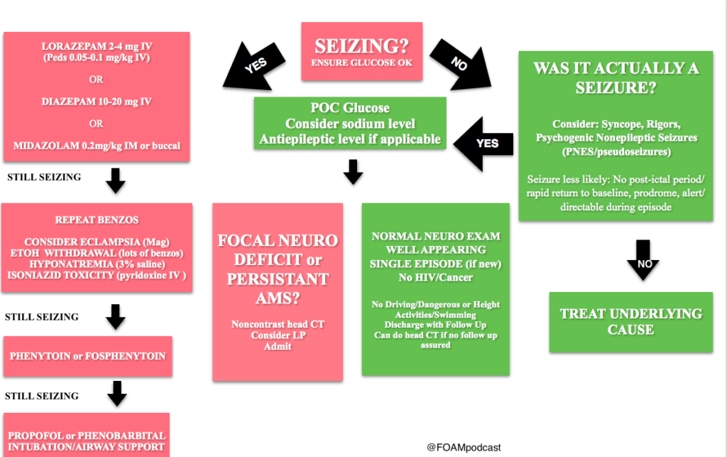
Screen Shot 2016-05-24 at 10.16.02 PM

Febrile Seizures AAP Guidelines

Febrile Seizures

Generously Donated Rosh Review Questions

A fifteen-year-old girl presents for evaluation and clearance for sports play. She has played team sports in the past, and would like to join the swim team this year. She was recently diagnosed with a seizure disorder. Her seizures are usually in the mornings, are generalized tonic-clonic seizures, and last for 1-2 minutes. They occur once per week. She is currently taking topiramate for seizure control. Her physical exam and vital signs are reassuring. Which of the following is the best recommendation for this patient?A. Allow participation in swimming as this is not a contact sport
B. Deny participation in all sports until seizures are better controlled
C. Permit patient to join the swim team as long as she has rectal diazepam with her at all times
D. Refuse participation in swimming as seizures are poorly controlled

[expand title=”Answer”]

D. Refuse participation in swimming as seizures are poorly controlled. Swimming (A) is a danger to children with poorly controlled seizures, as there is a risk that the child will have a seizure during the exercise and could suffer near-drowning or death. Some sporting events are safe for children with epilepsy (B), such as running. While the child participating in non-contact and non-aquatic sports may still have a seizure, the risk of morbidity and mortality to the child and other participants is low. Rectal diazepam (C) is a pharmacologic therapy that can stop seizures once they begin; however, the risk for morbidity and mortality remains high for children with poorly controlled seizures.

[/expand]

A full-term 3-week-old girl is brought in by her parents who report that she has been “acting funny” for 2 hours. They noticed that she has been moving her lips nonstop. She was a full-term, normal, spontaneous vaginal delivery and has been feeding well with adequate wet diapers since hospital discharge. She is afebrile and vital signs are normal. The anterior fontanelle is flat, and red reflexes are present. Heart, lung, and abdominal exams are normal. Her neurologic exam is positive for root, suck, and Moro reflexes, upgoing Babinski reflexes, and rhythmic lip-smacking movements. What is the most appropriate next step to take with this baby?
A. Administer a benzodiazepine
B. Initiate EEG monitoring
C. Perform a CT scan of the brain
D. Provide reassurance that this is normal behavior

[expand title=”Answer”]

This baby is having neonatal seizures, which are often subtle and more likely to be focal than tonic clonic. The most common manifestations are lip smacking, eye deviation, staring, rhythmic blinking, and bicycling movements. The patient should receive a benzodiazepine, such as lorazepam, midazolam, or diazepam, to stop the seizure. This should be followed by a workup that is directed at finding the underlying cause because neonatal seizures are less likely to be idiopathic than seizures in older children. A full septic workup is mandated for neonates with seizures, whether or not they are febrile. This includes CBC, blood culture, chemistry, UA, urine culture, chest x-ray, and CSF analysis. The infant should receive antibiotics and acyclovir and should be admitted to the hospital for further evaluation. Trauma can also cause neonatal seizures. It is important to look for signs of trauma such as bruising, bulging fontanelles, and retinal hemorrhages; the history may also include poor feeding, lethargy, or vomiting. Any infant with suspected head trauma should have a CT scan (C) and undergo a full child-abuse workup. An EEG (B) can be considered after more life-threatening causes of seizures, such as infection or trauma, are ruled out. A newborn (< 1 month old) with abnormal behavior should never be sent home with parental reassurance (D) only. Many new parents mistake normal behaviors for abnormal ones, but any truly abnormal behavior needs further investigation.[/expand]


References:

Kornegay JG. Chapter 171: Seizures Tintinalli’s Emergency Medicine: A Comprehensive Review (8e).

McMullan JT, Davitt AM, Pollack CV. “Seizures.” Rosen’s Emergency Medicine, Chapter 18, 156-161.e1




Episode 41 – Vertigo

(ITUNES OR LISTEN HERE)

The Free Open Access Medical Education (FOAM)

We cover two bits of FOAM, one from Emergency Medicine Literature of note on the use of meclizine for vertigo and an EMcrit episode on the HiNTs exam.

Emergency Medicine Literature of Note – Dr. Ryan Radecki – Treating what you believe is peripheral vertigo?  Using meclizine? So are most people.  But this is not an evidence based practice.  While meclizine is Rosen approved, Tintinalli recommends transdermal scopolamine as the first line treatment [1,2]. Following a recent recall of meclizine (oddly because iron bottles contained meclizine instead of iron), Dr. Radecki probes into why we use meclizine for vertigo.

  • Meclizine is an anti-histamine and has been thought to have anti-emetic properties.
  • A 1968 paper compared 16 anti-emetics/combinations and did NOT conclude that meclizine was the best. In fact, scopolamine and amphetamine performed best. Promethazine (phenergan) is also a good choice based on this paper [3].

EMcrit – Dr. Scott Weingart – The HiNTs exam has taken off, particularly in the FOAM world, as a means of disguising between central and peripheral causes of vertigo. In 2010, an EMcrit episode popularized this in the FOAM world. See this video demonstrating the HiNTs exam.

  • Head Impulse – rapid head rotation by the examiner with the subject’s vision fixed on an object (i.e. examiner’s nose). The examiner rapidly rotates the head towards midline and the patient’s eyes should remain fixed on the target. 
    • Abnormal (loss of fixation on target and movement of eyes away from target followed by correct saccade as patient fixates on target) = peripheral (vestibulo-ocular reflex impaired)
    • Normal = central
  • Nystagmus 
    • Horizontal nystagmus with a unidirectional fast phase (away from affected side) = probably peripheral
      • Patients with horizontal nystagmus may have central pathology but may have direction-changing nystagmus (i.e. fast phase beating in one direction when looking to right and the opposite direction when looking left).
    • Vertical or nystagmus = central pathology
  • Test of Skew Patients should fixate on a target while the provider alternately covers each eye. As the cover is moved from one eye to the other, the uncovered eye must correct for the misalignment and will look up or down to focus back on the target. This slight correction is observed repeatedly as the cover is moved from one eye to the other.
    • Skew deviation/misalignment = probably central, often in posterior fossa abnormalities
    • No skew deviation= peripheral

INFARCT – Impulse Normal, Fast-phase Alternating nystagmus, and Refixation on Cover Test

Issues with HiNTs

  • Can only be performed on patients with continuous vertigo.
  • External validity is a major issue with HiNTs.
    • Providers – Of the 4 studies have examined the operating characteristics of HiNTs, none have used emergency providers and instead have examined how the exam performs in the hands of two neuro-ophthalmologists, neuro-otologists, and neurologists with 4 hours of specialized training in the exam. It’s unclear whether HiNTs would be reliable or valid when performed by emergency providers [4-6].
    • Patients – The patients examined in many of these studies have other indicators of badness on neurologic exam. In one study, patients had to have gait instability and/or truncal ataxia to enroll.  Then, 76/101 (76%) of those patients had a central cause. These patients were sick and not the undifferentiated vertiginous patients we see primarily as emergency providers [4-6].
    • In the words of leading HiNTs expert Dr. Newman-Toker, HiNTs “requires expertise not routinely available in emergency departments.” As such, his team is piloting quantitative video-oculography to aid in diagnosis using HiNTs [7]. An Annals of Emergency Medicine review also warned that HiNTs may not be ready for emergency provider use [9].

More FOAM on HiNTs: EMJclub, EMNerd

Core content 

We delve into core content on vertigo using Rosen’s Medicine (8e) Chapter 19,  and Tintinalli’s Emergency Medicine: A Comprehensive Study Guide  (7e) Chapter 164 “Vertigo and Dizziness.”

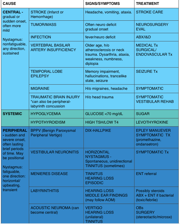
Vertigo is often characterized by the sensation of spinning and falls into the broad and frustrating category of “dizziness.”  Often, when a dizzy patient presents we perseverate on characterizes what the patient means by “dizzy.” However, some argue that this is not an appropriate approach as a study found 50% of patients changed the character of their dizziness when questioned again after 10 minutes [9].  Additionally, the clinical characteristics differentiating peripheral from central causes of vertigo are not entirely reliable. Despite these limitations, it is expected that we are familiar with “classic” presentations.

Screen Shot 2016-01-13 at 8.32.04 AM
*”Classic” presentations

Generously Donated Rosh Review Questions 

  1. A 50-year-old man presents with episodic severe vertigo lasting hours, with associated symptoms of unilateral tinnitus, fluctuating low-frequency hearing loss, and aural fullness. [polldaddy poll=9245427]
  2. A 20-year-old woman presents with an acute onset of dizziness. The patient describes the sensation that the room is spinning when she turns her head to the left and it is accompanied by nausea and vomiting. The symptoms resolve with turning her head away from that side. Examination reveals nystagmus elicited by deviating the eyes to the left and no other neurologic findings. [polldaddy poll=9245971]

Answers

1. B. Meniere’s disease is characterized by episodic severe vertigo lasting hours, with associated symptoms of unilateral tinnitus, fluctuating low-frequency hearing loss, and aural fullness. Typical onset is in the fifth decade of life. The cause is uncertain but is speculated to result from allergic, infectious, or autoimmune injury. The histopathologic finding includes endolymphatic hydrops, which is thought to be caused by either overproduction or underresorption of endolymph in the inner ear. Meniere’s disease is a clinical diagnosis mostly based on history. Testing may be obtained to support the diagnosis and rule out other disorders. Audiometry often demonstrates a low-frequency sensorineural hearing loss. An FTA-ABS test may be obtained to rule out syphilis. Electronystagmography (ENG) may demonstrate a unilateral peripheral vestibular weakness on caloric testing. When the diagnosis is uncertain, a brain MRI with contrast is obtained to evaluate for a retrocochlear lesion. The differential diagnosis of Meniere’s disease includes acute labyrinthitis, neurosyphilis, labyrinthine fistula, autoimmune inner ear disease, vestibular neuronitis, and migraine-associated vertigo.The most common cause of peripheral vestibular vertigo in adults is benign paroxysmal positional vertigo (A). BPPV occurs in all age groups but more often between ages 50 and 70 but is not associated with hearing loss and made worse with movement. In a perilymph fistula (C) rapid changes in air pressure (barotrauma), otologic surgery, violent nose blowing or sneezing, head trauma, or chronic ear disease may cause leakage of perilymph fluid from the inner ear into the middle ear and result in episodes of vertigo. Associated signs and symptoms are variable but can include a sudden pop in the ear followed by hearing loss, vertigo, and sometimes tinnitus. Acute vertigo associated with nausea and vomiting (but without neurologic or audiologic symptoms) that originates in the vestibular nerve is known as vestibular neuronitis (D). Vestibular neuronitis can occur spontaneously or can follow viral illness.

2. B. This patient presents with peripheral vertigo most consistent with benign paroxysmal peripheral vertigo (BPPV) and should be treated with an Epley maneuver. Vertigo is defined as the sensation of disorientation in space combined with a sensation of motion. Patients typically describe a “room-spinning” sensation or the feeling of sea sickness. Vertigo can be divided into two types: central and peripheral. Central vertigo are those disorders arising from the central nervous system and include ischemic stroke, vertebrobasilar insufficiency and infectious causes (meningitis, mastoiditis, syphilis). Central vertigo is characterized by longer duration of symptoms, minimal change with position, gradual onset and multidirectional nystagmus. Peripheral vertigo includes BPPV, Meniere’s disease, Labyrinthitis and vestibular neuritis. Peripheral vertigo may have intermittent symptoms (BPPV) or continuous symptoms but should not be associated with other neurologic deficits or changes and should have unidirectional nystagmus. The symptoms in BPPV are elicited by specific movements of the head and relieved by returning the head to a neutral position. The symptoms should be acute in onset and of a short duration. In BPPV, the symptoms are cause by the presence of an otolith in one of the semicircular canals. Although pharmacologic intervention may be necessary in the acute setting with meclizine or benzodiazepines, the best treatment for BPPV is the Epley maneuver. The Epley maneuver is a series of positions that the clinician takes the patient through that leads to expulsion of the otolith from the semicircular canal and relief of symptoms. Imaging with a non-contrast head CT (C) is not indicated in peripheral vertigo of any cause as the patient’s pathology is in the inner ear and not the brain. If a central cause is suspected, MRI of the brain (A) is the best test for diagnosis as the causative lesion will likely be in the posterior fossa, which is not seen well on CT scan. Steroid treatment (D) is the indicated management for vestibular neuritis but does not play a role in the treatment of BPPV.

References:

  1. Chang AK, Olshaker AS. Dizziness and Vertigo. In: Marx JA, Hockberger RS, Walls RM eds.  Rosen’s Emergency Medicine, 8th e.
  2. Goldman B. Chapter 164. Vertigo and Dizziness. In: Tintinalli JE, Stapczynski J, Ma O, Cline DM, Cydulka RK, Meckler GD, T. eds. Tintinalli’s Emergency Medicine: A Comprehensive Study Guide, 7e. New York, NY: McGraw-Hill; 2011.
  3. Wood CD, Graybiel A. Evaluation of sixteen anti-motion sickness drugs under controlled laboratory conditions. Aerospace medicine. 39(12):1341-4. 1968.
  4. Newman-Toker DE, Kattah JC, Alvernia JE, Wang DZ. Normal head impulse test differentiates acute cerebellar strokes from vestibular neuritis. Neurology. 70(24 Pt 2):2378-85. 2008.
  5. Kattah et al. HINTS to Diagnose Stroke in the Acute Vestibular Syndrome Three-Step Bedside Oculomotor Examination More Sensitive Than Early MRI Diffusion-Weighted Imaging. Stroke. 2009; 40: 3504-3510
  6. Newman-Toker et al. HINTS Outperforms ABCD2 to Screen for Stroke in Acute Continuous Vertigo and Dizziness. Academic Emergency Medicine. Volume 20, Issue 10, pages 986–996, October 2013
  7. Newman-Toked DE, Saber Tehran AS, Mantokoudis G et al. Quantitative video-oculography to help diagnose stroke in acute vertigo and dizziness: toward an ECG for the eyes. Stroke. 44(4):1158-61
  8. Cohn B. Can Bedside Oculomotor (HINTS) Testing Differentiate Central From Peripheral Causes of Vertigo? Annals of Emergency Medicine. 64(3):265-268. 2014. 
  9. Edlow JA. Diagnosing Dizziness: We Are Teaching the Wrong Paradigm!. Acad Emerg Med. 20(10):1064-1066. 2013.
  10. Newman-Toker DE, Cannon LM, Stofferahn ME, Rothman RE, Hsieh YH, Zee DS. Imprecision in patient reports of dizziness symptom quality: a cross-sectional study conducted in an acute care setting. Mayo Clinic proceedings. 82(11):1329-40. 2007.
  11. Strupp M, Zingler VC, Arbusow V et al. Methylprednisolone, Valacyclovir, or the Combination for Vestibular Neuritis. N Engl J Med. 351(4):354-361. 2004. 
  12. CFishman JM, Burgess C, Waddell A. Corticosteroids for the treatment of idiopathic acute vestibular dysfunction (vestibular neuritis). The Cochrane database of systematic reviews. 2011.