Racial Bias in Pulse Oximetry

Apple Podcasts  or Listen Here

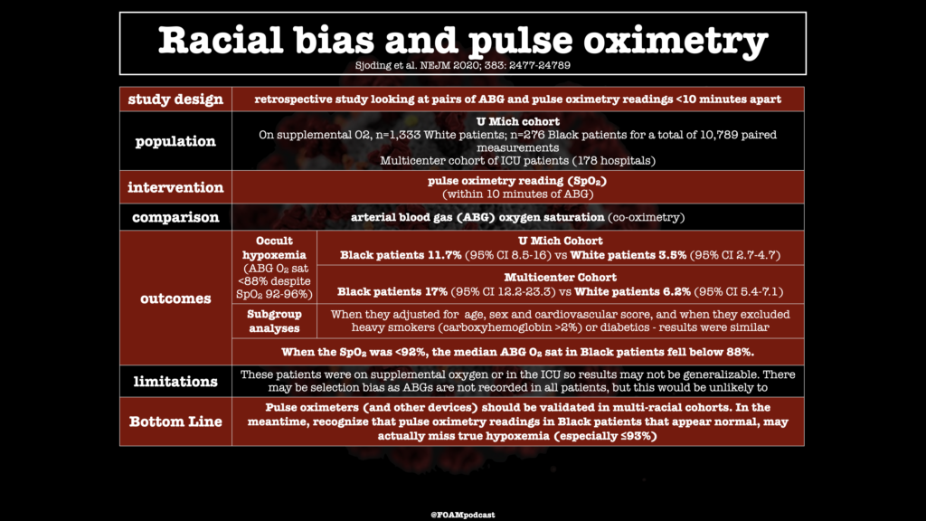
Pulse oximetry is one of the key vital signs that we use to triage patients and make treatment and disposition decisions. Especially during the COVID-19 pandemic, when hospitals are full, many hospitals are using pulse oximetry thresholds to determine outpatient versus inpatient management. Despite the ubiquity of pulse oximetry, prior studies have found that the devices might not be as accurate in patients with different skin pigmentation. In this podcast we review a new article in NEJM.

Sjoding et al. NEJM 2020

Episode 59 – Syncope (and the PESIT study)

ITUNES OR LISTEN HERE

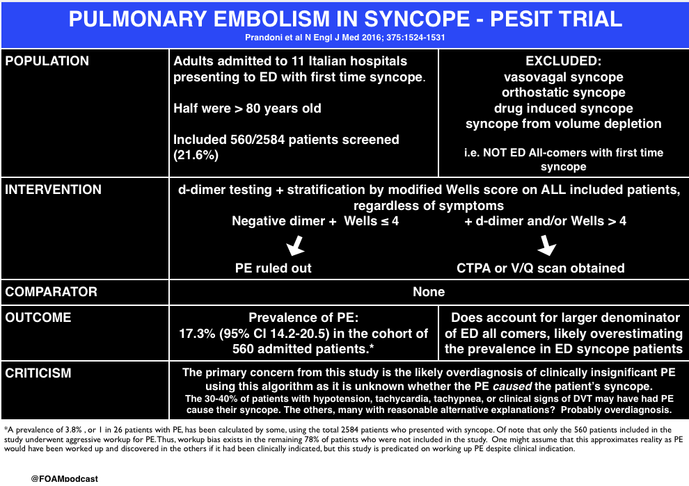
The PESIT study in the New England Journal of Medicine stirred up controversy in the FOAM world earlier in October 2016.  In this episode we cover the following posts on this article on pulmonary embolism in syncope:

screen-shot-2016-10-25-at-7-52-02-pm

Core Content

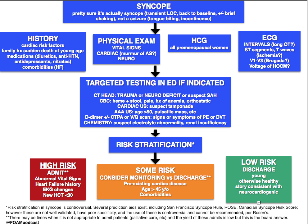
We delve into core content on syncope usingRosen’s Emergency Medicine (8th edition) and Tintinalli’s Emergency Medicine (8th edition) Chapter 52

screen-shot-2016-10-31-at-4-41-19-pm

 Rosh Review Emergency Board Review Questions

An 83-year-old is being evaluated in the emergency department after an episode of syncope. The woman was preparing dinner when she felt her heart start to race. The next thing she remembers is waking up on the floor. She experienced a similar episode about three weeks ago. She has never had anything like this before. Her past medical history is remarkable for hypertension, hyperlipidemia and hypothyroidism. Her medications include lisinopril, atorvastatin and levothyroxine. On physical exam her blood pressure is 142/83, heart rate 76/min, and respiration rate 13/min. Cardiac auscultation reveals no murmur. The remainder of her physical exam is normal. Electrocardiogram reveals normal sinus rhythm with left axis deviation. No cardiac rhythm abnormalities are detected. What is the most likely etiology of this patient’s syncope?

A. Aortic stenosis

B. Cardiac dysrhythmia

C. Orthostatic hypotension

D. Vasovagal

[accordion]
[toggle title=”Answer” state=”closed”]

B. Cardiac dysrhythmia is the most likely cause of this woman’s syncope. Cardiac dysrhythmias are a common cause of syncope in the elderly population. It is characterized by a brief or absent prodrome and palpitations immediately preceding the event. Several episodes over a short period of time in someone with no history of syncope suggest a dysrhythmia. Given this patient’s short prodrome, palpitations and history of a previous similar event makes a cardiac dysrhythmia the most likely etiology.

Aortic stenosis (A) is unlikely the cause of her syncope. Aortic stenosis is associated with a crescendo-decrescendo systolic ejection murmur. Syncope related to aortic stenosis typically occurs during exertion and is associated with very severe disease. This patient’s syncopal episode occurred while stationary. Additionally, she has no systemic symptoms of aortic stenosis.Vasovagal (D) is the most common cause of syncope in the general population. It is usually triggered by provoking factors such a blood draw or an intense emotion. Prodromal symptoms include feeling warm, sweating, nausea, and pallor. This woman does not report any of these symptoms. Orthostatic hypotension (C) causes syncope upon assuming an upright position from supine or sitting. It is often caused by hypovolemia, medications or autonomic nervous system disorders. This woman was standing while preparing dinner making orthostatic hypotension unlikely.


[/toggle]
[/accordion]

An 18-year-old woman presents after having a syncopal episode. She is complaining of a 2-day history of lower abdominal pain and vaginal spotting. Her BP is 86/42, HR is 128, RR is 18 breaths, and oxygen saturation is 99% on room air. She is drowsy, but answers questions appropriately. What is the most appropriate next step in management?


A. Establish large-bore IV access and administer an IV fluid bolus

B. Initiate rapid sequence induction and orotracheal intubation

C. Perform a bedside urine pregnancy testing

D. Perform an ultrasound of the abdomen to assess for free fluid

[accordion]
[toggle title=”Answer” state=”closed”]

A. The patient is hypotensive and tachycardic. She is suffering from hypovolemic shock secondary to a ruptured ectopic pregnancy. Therefore she requires immediate intravenous access and volume resuscitation with Lactated Ringer’s or normal saline.  Emergency Department management of unstable patients includes rapid assessment of the ABC’s (Airway, Breathing, Circulation). This patient is phonating, has a respiratory rate of 18 breaths per minute and an oxygen saturation of 99% on room air.  There is no concern that her airway or breathing is in immediate jeopardy, therefore she would not require immediate rapid sequence induction and orotracheal intubation (B). Although a bedside pregnancy test (C) and abdominal ultrasound (D) would help make a diagnosis of ruptured ectopic pregnancy, the next step would be to resuscitate the patient.

[/toggle]
[/accordion]

References

  1. De Lorenzo RA. “Syncope.” Chapter 15. Rosen’s Emergency Medicine (8 ed). pp 131-145
  2. Chapter 52. Tintinalli’s Emergency Medicine: A Comprehensive Review (8 ed).
  3. Serrano LA, Hess EP, Bellolio MF et al. Accuracy and Quality of Clinical Decision Rules for Syncope in the Emergency Department: A Systematic Review and Meta-analysis. Annals of Emergency Medicine. 56(4):362-373.e1. 2010.

Episode 36 – Pneumothorax

(ITUNES OR LISTEN HERE)

The Free Open Access Medical Education (FOAM)

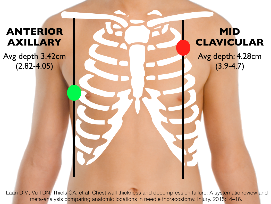
HEFT EMcast has produced an excellent podcast summary of the literature demonstrating that needle decompression at the 2nd intercostal space at the midclavicular line (2ICS MCL) is likely to fail.  They review a systematic review and meta-analysis by Laan et al in 2015 that suggests the fifth intercostal space at the anterior axillary line (5ICS AAL) is less likely to fail [1].

Problems with the 2ICS MCL (For more detail on this, see this post) 

  • Chest Wall Thickness – to achieve a success rate >90% of the time with needle decompression at the 2ICS MCL, a [1,2]
  • The 2ICS MCL is Difficult to Find– one study of emergency physicians found that providers could find the 2ICS MCL 60% of the time. Similarly, a blinded cadaveric study in which naval corpsmen who had just had a refresher course in needle decompress were asked to needle decompress cadavers at the 2ICS MCL and 5ICS AAL.  They found 80% correct placement in the 5ICS AAL group compared with a mere 30% in the 2ICS MCL group [3,4].
  • Important vascular structures nearby – the heart and great arteries lie nearby the 2ICS MCL and given the frequent misplacement of these catheters, these structures may be injured. Of note, placement aiming for the 5ICS AAL, if too inferiorly placed, could result in liver or splenic injury.

Needle Thoracostomy: 5th ICS Anterior Axillary Line = Less Likely to Fail

Core Content

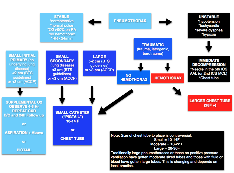
We delve into core content on pneumothorax and empyema using Rosen’s Medicine (8e),  Chapters 77 and Tintinalli’s Emergency Medicine: A Comprehensive Study Guide  (8e) Chapters 68

Pneumothorax

Screen Shot 2016-03-17 at 11.31.24 AM

Smaller bore percutaneous tubes (seldinger technique) are becoming increasingly common, even in traumatic pneumothorax.  These tubes have the advantage of smaller size, less pain, and reduced infection.  However, they may be more prone to kinking and may be less desireable for some fluid collections [6-19].  See this post for more on percutaneous tubes.

Open pneumothorax (“sucking chest wound”): communication between the surrounding environment and the pleural space, often due to penetrating trauma but may occur in its absence.

  • Treatment:  3 sided dressing (occlusive dressing could create tension) followed by chest tube (not inserted into wound).

Empyema  

Infection between the visceral pleura which covers the lung and the parietal pleura which covers the thoracic wall.

Causes: 

  • Parapneumonic effusions (i.e. effusions that came as a result of an underlying pneumonia)- most common causes of pneumonia (s. pneumo or s. aureus. or H flu if not vaccinated)
  • Trauma (iatrogenic or otherwise, i.e. retained hemothorax):  gram negative bacilli.

Clinical symptoms:

  • Fever
  • Cough
  • Pleuritic chest pain
  • Decreased breath sounds.

Phases:

  • Exudative (early) – drainage, antibiotics
  • Fibropurluent (middle) – fibrinolytics +/- video assisted thorascopic surgery (VATS)
  • Organizational (late) – VATS and/or surgery

Generously Donated Rosh Review Questions

Question 1. A 22-year-old woman with a history of asthma presents with chest pain. Over the last several days, the patient has been coughing due to an asthma exacerbation. Today she developed sharp chest pain in the middle of her chest.  A chest X-ray is shown below.[polldaddy poll=9349650]

[expand title=”Answer”] D. Pneumomediastinum is free air contained within the mediastinum of the chest, most commonly due to air originating in the alveolar space. Air escapes from ruptured alveoli along the peribronchial vascular sheaths to the hilum and into the mediastinum. However, other causes like Boorhaeve’s syndrome and traumatic chest injury can lead to pneumomediastinum. The patient’s outcome is typically related to the underlying cause. In most cases, patients are hemodynamically stable and only require supportive care and observation. The condition rarely progresses to become tension pneumomediastinum and therefore surgical intereventions are not necessary. Approximately 14% of patients with pneumomediastinum have an associated pneumothorax. CT scan of the chest (A) may identify other pathology causing pneumomediastinum. However, in this patient with a history of asthma and recent coughing, a ruptured alveolus is the likely culprit. Additional investigation with CT scan is not necessary. High flow oxygen therapy (B) is often used for nitrogen washout in the treatment of a pneumothorax. In patients with pneumomediastinum, this is not necessary. A pig tail catheter (C) has been reported in some case reports however, surgical intervention with a catheter is rarely needed in the condition because of its stability. This differs from isolated peumopericardium because pneumocardium should not rise above the level of the pericardial reflection which it does in this case. In addition, the history of chest pain after coughing due to asthma is also consistent with pneumomediastinum. [/expand]

Question 2. You are caring for an intubated patient diagnosed with sepsis that is awaiting a bed in the intensive care unit. The patient has peripheral intravenous access, however you decide to place a right-sided subclavian central venous catheter in order to measure central venous pressure, assess volume status and determine if vasopressors are indicated.  Shortly after successful line placement, the patient becomes hypotensive and develops high peak airway pressures. [polldaddy poll=9349659]

[expand title=”Answer”] C. The patient likely has a tension pneumothorax that resulted from subclavian central line placement combined with the fact that the patient is on positive pressure ventilation (PPV). The patient requires emergent needle thoracostomy in the right second intercostal space midclavicular line to alleviate the tension. Pneumothorax is a common complication associated with subclavian venipuncture and occurs in up to 6% of attempts. Patients on positive pressure ventilation are more likely to develop a tension pneumothorax due to increased intrapleural pressures.Tension pneumothorax is a clinical diagnosis not a radiographic diagnosis. Clinical signs and symptoms include hypotension, absent breath sounds on the affected side, trachea deviation away from the affected side and sudden resistance to ventilation for patients on PPV. This is life threatening and requires immediate attention. Waiting for radiology to come and perform a chest X-ray (A) can result in patient death. Patients may develop hypotension as a result of intubation and positive pressure ventilation. This generally occurs immediately following initiation of PPV and is secondary to decreased venous return. This is treated with an IV fluid bolus (B) however would not cause elevated peak airway pressures as in this scenario. Emergent pericardiocentesis (D) is performed for treatment of cardiac tamponade, which manifests as hypotension, muffled heart tones and jugular venous distention. This is unlikely to be the problem in this scenario. [/expand]

References:

  1.  Laan D V., Vu TDN, Thiels CA, et al. Chest wall thickness and decompression failure: A systematic review and meta-analysis comparing anatomic locations in needle thoracostomy. Injury. 2015:14–16. doi:10.1016/j.injury.2015.11.045.
  2. Hecker M, Hegenscheid K, Völzke H, et al. Needle decompression of tension pneumothorax. J Trauma Acute Care Surg. 2016;80(1):119–124. doi:10.1097/TA.0000000000000878.
  3. Aho JM, Thiels CA, El Khatib MM, et al. Needle thoracostomy. J Trauma Acute Care Surg. 2016;80(2):272–277. doi:10.1097/TA.0000000000000889.
  4. Ferrie EP, Collum N, McGovern S. The right place in the right space? Awareness of site for needle thoracocentesis. Emerg Med J. 2005;22(11):788–789. doi:10.1136/emj.2004.015107.
  5. Inaba K, Karamanos E, Skiada D, et al. Cadaveric comparison of the optimal site for needle decompression of tension pneumothorax by prehospital care providers. doi:10.1097/TA.0000000000000849.
  6.  Laws D et al. BTS guidelines for the insertion of a chest drain. Thorax. 2003 May;58 Suppl 2:ii53-9.
  7.  Benton IJ, Benfield GF. Comparison of a large and small-calibre tube drain for managing spontaneous pneumothoraces. Respir Med. 2009 Oct;103(10):1436-40.
  8. Dull KE, Fleisher GR. Pigtail catheters versus large-bore chest tubes for pneumothoraces in children treated in the emergency department. Pediatr Emerg Care. 2002 Aug;18(4):265-7.
  9. Gammie JS et al. The pigtail catheters for pleural drainages: a less invasive alternative to tube thoracostomy. JSLS. 1999 Jan-Mar;3(1):57–61.
  10.  Kuo HC, et al. Small-bore pigtail catheters for the treatment of primary spontaneous pneumothorax in young adolescents. Emerg Med J. 2013 Mar;30(3):e17.
  11. Repanshek ZD, Ufberg JW, Vilke GM, Chan TC, Harrigan RA. Alternative Treatments of Pneumothorax. J Emerg Med. 2013 Feb;44(2):457-466.
  12. Hassani B, Foote J, Borgundvaag B. Outpatient management of primary spontaneous pneumothorax in the emergency department of a community hospital using a small-bore catheter and a Heimlich valve. Acad Emerg Med. 2009 Jun;16(6):513-8.
  13. Kulvatunyou N, Vijayasekaran A, Hansen A, et al. Two-year experience of using pigtail catheters to treat traumatic pneumothorax: a changing trend. J Trauma. 2011 Nov;71(5):1104-7.
  14. Rivera L, O’Reilly EB, Sise MJ, et al. Small catheter tube thoracostomy: effective in managing chest trauma in stable patients. J Trauma. 2009 Feb;66(2):393–9
  15. Kulvatunyou N, et al. A prospective randomized study of 14-French pigtail catheters vs 28F chest tubes in patients with traumatic pneumothorax: impact on tube-site pain and failure rate. EAST Annual Surgical Assembly, Oral paper 12, Jan 17, 2013.
  16. Kulvatunyou N, Joseph B, Friese RS, et al. 14 French pigtail catheters placed by surgeons to drain blood on trauma patients. J Trauma Acute Care Surg. 2012;73(6):1423–1427.
  17. Russo RM, Zakaluzny SA, Neff LP, et al. A pilot study of chest tube versus pigtail catheter drainage of acute hemothorax in swine. J Trauma Acute Care Surg. 2015;79(6):1038–1043.
  18.  Liu YH, et al. Ultrasound-guided pigtail catheters for drainage of various pleural diseases. Am J Emerg Med. 2010 Oct;28(8):915-21
  19.  Inaba K, Lustenberger T, Recinos G. Does size matter? A prospective analysis of 28-32 versus 36-40 French chest tube size in trauma. The journal of trauma and acute care surgery. 72(2):422-7. 2012.


Episode 36 – Rib and Sternal Fractures

(ITUNES OR Listen Here)

The Free Open Access Medical Education (FOAM)

This week we cover a joint piece between the Ultrasound Podcast and SonoIn5 on diagnosis of rib and sternal fractures with ultrasound.

Technique:  Linear probe, in line with the long axis of the bone (vertical for sternum, horizontal-ish for ribs).

Diagnosis: Cortical disruption (step off). Excellent sensitivity for sternal fractures [1-3]

Sternal Fracture
Sternal Fracture

  • Caution with sternal fractures as the sternomanubrial joint can mimic fracture, but looks more “bumpy” (see below)

Core Content – Rib and Sternal Fractures

Tintinalli (7e) Chapters 258, 259; Rosen (8e) Chapter 45 

Rib Fractures

Diagnosis:

  • Chest x-ray initial test of choice – may miss 50% of fractures, unclear if this is clinically significant [6]
  • Ultrasound has found to have excellent sensitivity [7]
  • Rib films are NOT recommended [4-6].

Complications: Traumatic rib fractures may be associated with other traumatic injuries such as pneumothorax, hemothorax, or in the case of lower rib fractures, intra-abdominal injury. However, rib fractures themselves have been associated with mortality, most often as sequelae of pulmonary embarrassment including pneumonia, intubation, and death. Mortality in elderly patients with rib fractures is significantly higher than the younger counterparts at 22% and 10% respectively [8,9].

  • Mortality is between 3-13%
  • Risk stratification (see this post): Battle and colleagues developed a prognostic scoring system, not externally validated and unclear if it would change practice, that highlights common sense predictors of poorer outcomes:
    • Age (>65)
    • Higher number of rib fractures
    • Chronic lung disease
    • Hypoxia (<90%)
    • Pre-injury anticoagulant use [11]

Treatment

  • Analgesia:
    • Often includes NSAIDS (ibuprofen), acetaminophen, and narcotics +/- gabapentin (ibuprofen and gabapentin depending on renal function)
    • Epidural analgesia – highly recommended in the EAST guidelines [14].
    • Paracostal analgesia (ex: ON-Q pump) – not sufficient evidence for EAST recommendation (2005) [14]
  • Pulmonary Hygiene (formerly pulmonary toilet):  involved incentive spirometry, coughing, mobilization (up, out of bed), and possibly chest physical therapy
  • ORIF, “rib fixation” or “rib plating,” is increasingly common in the US and studies have found improvements in ICU LOS and ventilator days [15]

Disposition

  • Many rib fracture patients will need to be admitted to the hospital for pain control, observation, and pulmonary hygiene.
    • Some rib fracture patients may benefit from care at trauma centers.  Lee et al  wrote that 3+ rib fractures exists as an indication for transfer to a level 1 trauma center and many places ascribe to this, it depends on the hospital and physicians.
    • While patients in the ED may look good, patients may benefit from high intensity floors (ie stepdown units) and many patients get observed in ICUs, again, depending on local practice patterns. Some protocols risk stratify patients (i.e. to the ICU vs floor) by incentive spirometry.
  • Patients with adequate pain control who are low risk (younger, <3 rib fractures, good effort on incentive spirometry) may be discharged from the ED with analgesia and education on importance of pulmonary hygiene

Sternal Fractures – more common with ubiquity of airbags and seatbelts.

Diagnosis:  Classically the “gold standard” has been lateral x-ray. However, CT technology has improved since those studies. Ample literature suggests that ultrasound has excellent sensitivity [1-3].

Complications: Historically, sternal fractures were associated with injuries of the great vessels, high mortality, and blunt cardiac injury (BCI) [16-18].  The most recent iteration of the EAST guidelines states, “the presence of a sternal fracture alone does not predict the presence of BCI and thus should not prompt monitoring in the setting of normal ECG result and troponin I level” (Level 2) [18].

Treatment: Analgesia. Most patients with isolated sternal fractures (no pneumothorax, hemothorax, BCI, or hemodynamic instability) that have adequate pain control can be discharged from the ED [1-2].

Blunt Cardiac Injury

A broad category including a range of injuries from clinically silent dysrhythmias to cardiac wall rupture or vasospasm. BCI often results from high impact injury and should be considered in patients with significant thoracic trauma including rib fractures, sternal fracture, pneumothorax, hemothorax, and pulmonary contusion.

Diagnosis: There is no gold standard test.  One can rule out BCI with a normal ECG and a single normal troponin I [18].

Management: If an ECG or troponin is abnormal, admit to telemetry for monitoring and echo.

Generously Donated Rosh Review Questions 

Question 1.  A 23-year-old man presents with chest pain after a motor vehicle collision. The patient’s chest struck the steering wheel. He has no other complaints or injuries. Chest X-ray is unremarkable. ECG shows sinus tachycardia with anterior ST depressions. A troponin is sent and is positive at 3.50 mg/dl. [polldaddy poll=9134639]

Question 2A 20-year-old man presents with left rib pain after falling while playing soccer and striking his chest. Vital signs are normal. On physical examination, the patient has tenderness to palpation over the 4th rib in the midaxillary line. [polldaddy poll=9134640]

Question 3.  A 32-year-old woman was the restrained driver involved in a head-on motor vehicle collision (MVC) 2 days prior to presentation. She is complaining of chest pain and bruising to her chest. Her blood pressure is 118/78 mm Hg, pulse is 88 beats/minute, respirations are 18 breaths/minute and oxygen saturation is 96% on room air. You note bony tenderness and ecchymosis to her sternum. You order a chest X-ray and diagnose a non-displaced sternal fracture. [polldaddy poll=9134643]

Answers

  1. This patient presents with a myocardial contusion and should have an echocardiogram performed to look for any cardiac damage. Myocardial contusion describes a nebulous condition. It can occur through several mechanisms including a direct blow to the chest and compressive force over a prolonged period of time. Histologically, the disorder has similar findings to those seen after acute myocardial infarction. The majority of contusions heal spontaneously but small pericardial effusions may develop. Delayed rupture after resorption of hematoma is feared but rare complication. Patients with myocardial contusion will present after trauma with external signs of trauma and typically have other concomitant thoracic lesions (pulmonary contusion, pneumothorax, hemothorax). Patients will typically have tachycardia (up to 70%). ECG may show dysrrhythmia or ST changes but may also be normal. Although it is not effective to admit all patients for workup for myocardial contusion and the disease has a very low rate of cardiac complications, in the presence of ECG changes and elevated biomarkers, observation and echocardiography are a reasonable approach. Echocardiogram can be used to diagnose pericardial effusion, thrombi formation and valvular disruption.Cardiac catheterization (A) is not necessary after a myocardial contusion as coronary artery obstruction is not part of the pathophsyiology. The patient should not be discharged home (B)without an echocardiogram. Pericardiocentesis (D) is only necessary in the presence of a large pericardial effusion or one causing cardiac tamponade.
  2. This patient presents with signs and symptoms consistent with a rib facture. A chest X-ray should be performed to rule out any other pathology including pneumothorax and pulmonary contusion. Rib fractures are a common injury after thoracic trauma and the incidence increases with increasing age. They may be associated with a number of potential complications including pulmonary contusions, hemothorax, penumothorax and post-traumatic pneumonia. Fractures are most common at the posterior angle, which represents the weakest area. The ribs most commonly fractured are the 4th – 9th ribs. The 9th – 11th ribs are mobile, which reduces the risk of fracture. However, fractures of these ribs are more likely to be associated with intraabdominal injuries. Rib fractures should be suspected based on history and clinical evaluation. Patients will present with chest pain and tenderness over the area. Imaging should be obtained to rule out the more serious associated complications of pneumothorax, hemothorax and pulmonary contusion. Chest X-ray is the appropriate modality for this but often will not demonstrate the presence of a single rib fracture when it is in fact present. This is particularly true of non-displaced fractures. Rib belts (B) are discouraged as they may decrease the depth of respiration and lead to atelectasis and pneumonia. CT scan of the chest (D) is not routinely required for management of a simple rib fracture. Analgesia and discharge home (A) is likley to occur once more serious pathology is ruled out with a chest X-ray. Patients with rib fractures should also receive an incentive spirometer to help reduce the complication of pneumonia.
  3. Isolated, non-displaced sternal fractures are associated with low overall mortality rates. Fractures and dislocations of the sternum are caused primarily by anterior blunt chest wall trauma during a head-on MVC. Isolated fractures of the sternum most commonly occur when the chest wall is thrust against a diagonal seatbelt strap during rapid deceleration in a frontal impact MVC. They are more common in older individuals and women. Most fractures are transverse and non-displaced and can be diagnosed on a lateral chest radiograph. Although a fracture of the sternum can be seen following major thoracic trauma, its presence alone does not indicate severe underlying thoracic injury. However, if other significant underlying thoracic injuries are suspected, a CT-scan of the thorax should be performed

References:

  1. You JS, Chung YE, Kim D, Park S, Chung SP. Role of sonography in the emergency room to diagnose sternal fractures. Journal of clinical ultrasound : JCU. 38(3):135-7. 2010. [pubmed]
  2. Engin G, Yekeler E, Güloğlu R, Acunaş B, Acunaş G. US versus conventional radiography in the diagnosis of sternal fractures. Acta radiologica (Stockholm, Sweden : 1987). 41(3):296-9. 2000. [pubmed]
  3. Jin W, Yang DM, Kim HC, Ryu KN. Diagnostic values of sonography for assessment of sternal fractures compared with conventional radiography and bone scans. J Ultrasound Med. 2006 Oct. 25(10):1263-8; quiz 1269-70.
  4. ”Pulmonary Trauma” Tintinalli’s Emergency Medicine: A Comprehensive Study Guide.  7th ed. Ch 258.
  5.  “Thoracic Trauma” Rosen’s Emergency Medicine. 8th ed. Chapter 45.
  6. Henry TS, Kirsch J. ACR Appropriateness Criteria® rib fractures. Journal of thoracic imaging. 29(6):364-6. 2014. [pubmed]
  7. Chan SS. Emergency bedside ultrasound for the diagnosis of rib fractures. The American journal of emergency medicine. 27(5):617-20. 2009. [pubmed]
  8.  Ziegler DW, Agarwal NN. The morbidity and mortality of rib fractures. J. Trauma. 1994;37(6):975–9.
  9. Bulger EM, Arneson M a, Mock CN, Jurkovich GJ. Rib fractures in the elderly. J. Trauma. 2000;48(6):1040–6
  10. Flagel BT, Luchette F a, Reed RL, et al. Half-a-dozen ribs: the breakpoint for mortality. Surgery. 2005;138(4):717–23; discussion 723–5.
  11. Battle CE, Hutchings H, Evans P. Risk factors that predict mortality in patients with blunt chest wall trauma: a systematic review and meta-analysis. Injury. 2012;43(1):8–17.
  12. Livingston DH, Shogan B, John P, Lavery RF. CT diagnosis of Rib fractures and the prediction of acute respiratory failure. The Journal of trauma. 64(4):905-11. 2008. [pubmed]
  13. Battle CE, Hutchings H, Lovett S.  Predicting outcomes after blunt chest wall trauma: development and external validation of a new prognostic model Critical Care 2014, 18:R98
  14. Pain Management in Blunt Thoracic Trauma (BTT)J Trauma. 59(5):1256-1267, November 2005.
  15. Doben AR, Eriksson EA, Denlinger CE. Surgical rib fixation for flail chest deformity improves liberation from mechanical ventilation. Journal of critical care. 29(1):139-43. 2014. [pubmed]
  16. Screening for Blunt Cardiac Injury. J Trauma. 73(5):S301-S306, November 2012
  17. Karangelis D, Koufakis T, Spiliopoulos K, Tsilimingas N, Bouliaris K, Desimonas N. Management of isolated sternal fractures using a practical algorithm. J Emerg Trauma Shock. 7(3):170-. 2014. [article]
  18. Dua A, McMaster J, Desai PJ et al. The Association between Blunt Cardiac Injury and Isolated Sternal Fracture. Cardiology Research and Practice. 2014:1-3. 2014. [article]

Episode 35 – The Trachea

(ITUNES OR Listen Here)

The Free Open Access Medical Education (FOAM)

Jane Brody wrote an article, “What Comes After the Heimlich Maneuver” that ran in the NY Times and stirred up a ruckus on Twitter. This is a reasonable article on choking and details the limitations of the Heimlich maneuver.  Unfortunately, the article ends  instructing the layperson do to a cricothyrotomy (cric) with a sharp knife and “something like a straw or casing of a ballpoint pen (first remove the ink cartridge).  “

Dr. Seth Trueger (@MDaware) wrote a post, Bad Idea Jeans, discouraging this practice saying that deciding which patient needs a cric is one of the more difficult but more important parts of this procedure.

On another note, our friend Dr. Andy Neill has found that medical students are able to perform crics with Papermate pens on cadavers [1]. However, it appears that most pens may not be suitable [2]. Further, while medical students are nearly lay people, we do not think this the cric should be within the domain of lay people (especially without patients already declared dead and preserved).

Cricothyrotomies – In reality, this is a bloody procedures that should only be done by those with proper training when the airway cannot be otherwise secured. The actual procedure has been detailed by those far smarter and with more experience than the FOAMcast crew. We recommend checking out Dr. Scott Weingart’s compilation of resources here.

The anatomy of a cricothyrotomy by Dr. Andy Neill

Core Content – Tracheostomy Emergencies and Neck Infections

Tintinalli (7e) Chapters 242, 119

Tracheostomy (trach) (we refer to tracheostomy and tracheotomy interchangeably although there are some technical differences)

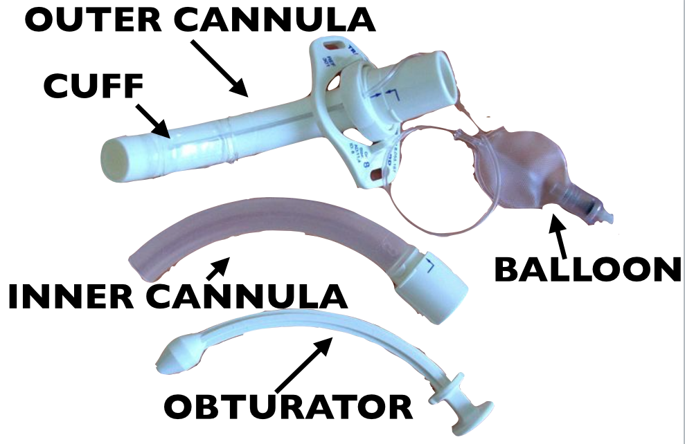
Anatomy of the trach tube (may vary)
Screen Shot 2015-10-07 at 8.40.17 AM

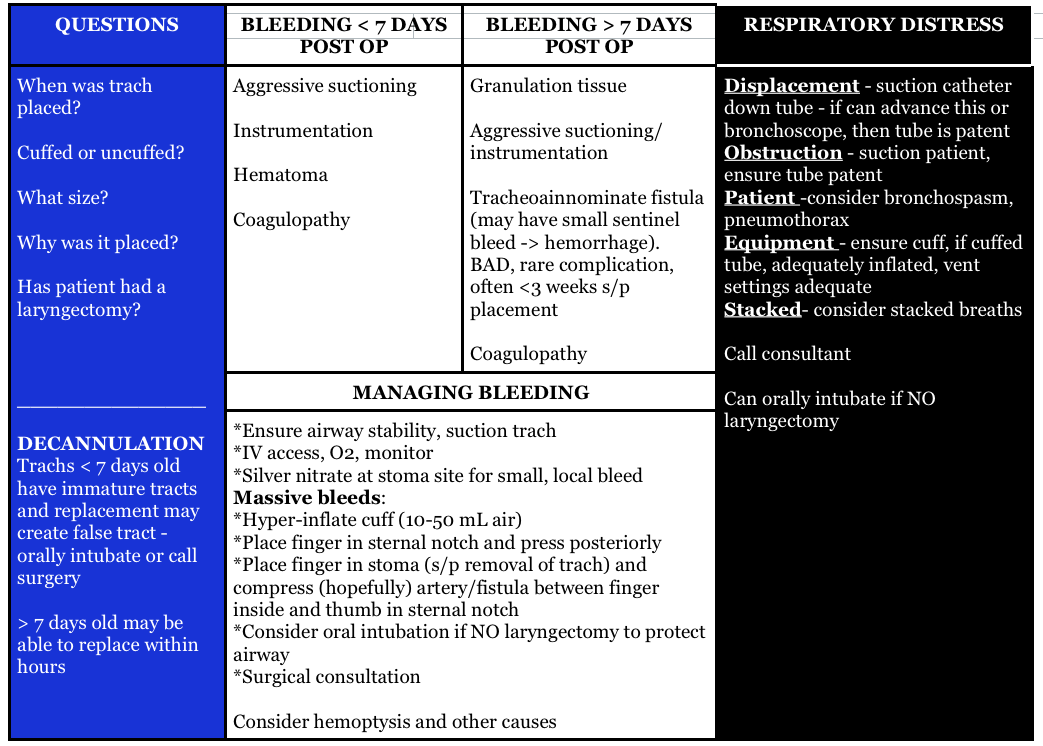
Things we have to know about trach emergencies
Screen Shot 2015-10-07 at 7.43.30 AM

Pediatric Trachea (Stridor) Pearls

Screen Shot 2015-10-07 at 8.20.11 AM

Generously Donated Rosh Review Questions 

Question 1. A 72-year-old man who is 1 week out from an ischemic stroke presents with respiratory distress. He had a tracheostomy placed 6 days ago for sudden respiratory failure. The patient is hypoxic and tachypneic on presentation with minimal breath sounds bilaterally. There is no subcutaneous air around the stoma.[polldaddy poll=9110296]

Question 2A 3-year-old boy presents in severe respiratory distress. His mother informs you that he has been ill for the last 5 days, initially with a low-grade fever and “barky cough.”  He was seen at an urgent care facility 4 days ago and given a  “breathing treatment” and discharged on steroids. He has become progressively worse despite compliance with the steroid regimen, which prompted his mother to call an ambulance this morning. He is otherwise healthy and up-to-date on his immunizations. On examination, the child is toxic in appearance and febrile. His oropharynx is clear. You hear both inspiratory and expiratory stridor.  [polldaddy poll=9110301]

Answers

1.The patient’s presentation is concerning for airway obstruction and the first step in management is suctioning of the tracheostomy tube. Tracheostomy tubes are placed for long-term mechanical ventilation in patients with anticipated prolonged or permanent respiratory failure. The two most common complications are obstruction and dislodgement. Sudden onset of respiratory failure often indicates mucous plugging or equipment failure. Suctioning of the tracheostomy is a simple procedure that may quickly relieve the patient’s symptoms. 2 to 3 ml of normal saline should be instilled into the tube followed by suctioning. Patients with slower decline in respiratory status may have a worsening of their underlying pulmonary pathology or may have developed a pulmonary infection.  A cricothyrotomy (A) will not lead to effective oxygenation or ventilation as the cricothyroid membrane is above the tracheostomy site. The tracheostomy tube should not be removed and replaced with either an endotracheal tube (C) or a new tracheostomy tube (B) at this time because the tracheostomy tract has not matured at 6 days (this usually occurs at 15-30 days). If equipment failure in the form of a tracheostomy tube malfunction is suspected, the tube should be replaced with fiberoptic visualization to ensure that a false lumen isn’t created.

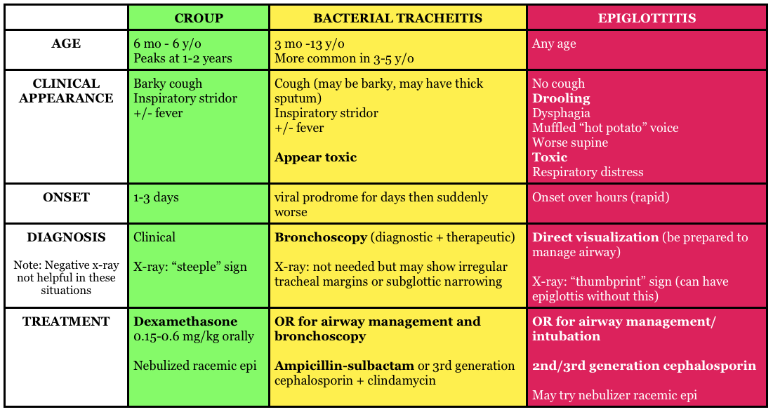
2.The patient is suffering from acute bacterial tracheitis. Bacterial tracheitis is the result of severe inflammation of the epithelial lining of the trachea leading to thick mucopurulent secretion production. This clinically manifests as viral prodrome with fever, URI symptoms, barky cough and stridor that intensifies and progresses to include a toxic appearing child with signs of airway obstruction, inspiratory and expiratory stridor, cyanosis, and severe respiratory distress. Another clue is that the child has been treated with medications (aerosolized epinephrine and steroids) for croup and has not improved clinically. Bacterial tracheitis is most common in children between the ages of 3 to 5 years. Most patients require orotracheal intubation for respiratory distress and ICU admission. The patient should be started on broad-spectrum intravenous antibiotics. Croup (B) is the most common cause of upper airway distress and obstruction in children between 6 months to 6 years of age with peak incidence at 2 years of age. Croup begins as a prodrome of low-grade fever and URI symptoms and is characterized by a barky cough, inspiratory stridor, and hoarse voice. Children are less toxic in appearance and rarely develop respiratory failure. The mainstays of treatment are steroids and aerosolized epinephrine. Epiglottitis (C) is characterized by abrupt onset of fever and sore throat and children classically present with difficulty in breathing, anxiety, stridor and drooling. This is less common in vaccinated children, such as the patient above and typically occurs in slightly older children. There is generally not a prodrome associated with epiglottitis. Peritonsillar abscess (D) occurs more commonly during adolescence and presents with trismus, unilateral sore throat, fever, tonsillar asymmetry, and uvula deviation away from the affected tonsil. The age of this patient and normal oropharynx examination make this diagnosis very unlikely.

References:

  1. Neill A, Anderson P. Observational cadaveric study of emergency bystander cricothyroidotomy with a ballpoint pen by untrained junior doctors and medical students. Emergency medicine journal : EMJ. 30(4):308-11. 2013. [pubmed]
  2. Owens D, Greenwood B, Galley A, Tomkinson A, Woolley S. Airflow efficacy of ballpoint pen tubes: a consideration for use in bystander cricothyrotomy. Emergency medicine journal : EMJ. 27(4):317-20. 2010. [pubmed]
Episode 33 – Hemoptysis

(ITUNES OR Listen Here)

The Free Open Access Medical Education (FOAM)

Dr. Ryan Radecki of Emergency Medicine Literature of Note reviews Gestational Age D-Dimers covering an article by Murphy and colleagues in BJOG.  

The paper: The authors took blood samples from 760 healthy pregnant patients at one point during their pregnancy. They propose a continuous increase for a normal d-dimer cut off throughout gestation.

  • 1-12 weeks:    n=33, 81% with normal d-dimer
  • 19-21 weeks:  n=53, 32% with normal d-dimer
  • 28-36 weeks: n=8, 6% with normal d-dimer
  • 39-40 weeks: 0, 0% normal d-dimer
  • Postpartum day 2: n=12, 8% with normal d-dimer

Dr. Radecki’s “Take Home:

  • Dr. Kline has advocated for the following d-dimer cut offs in pregnancy: 1st trimester 750 ng/mL, 2nd trimester 1000 ng/mL, and 3rd trimester 1250 ng/mL(based on a standard cut-off of 500 ng/mL) and this may be reasonable but is not rooted in robust evidence.

Interestingly, this post was followed by another post covering an article by Hutchinson et al from Am J Roentgenol showing that of 174 CTPAs initially read as positive, 45 were read as negative by chest radiologist upon blinded retrospective review.  That means 25.9% of this cohort diagnosed with PE apparently had negative CT scans.

Core Content – Hemoptysis

Tintinalli (7e) Chapter 66;  Rosen’s Emergency Medicine (8e) Chapter 24

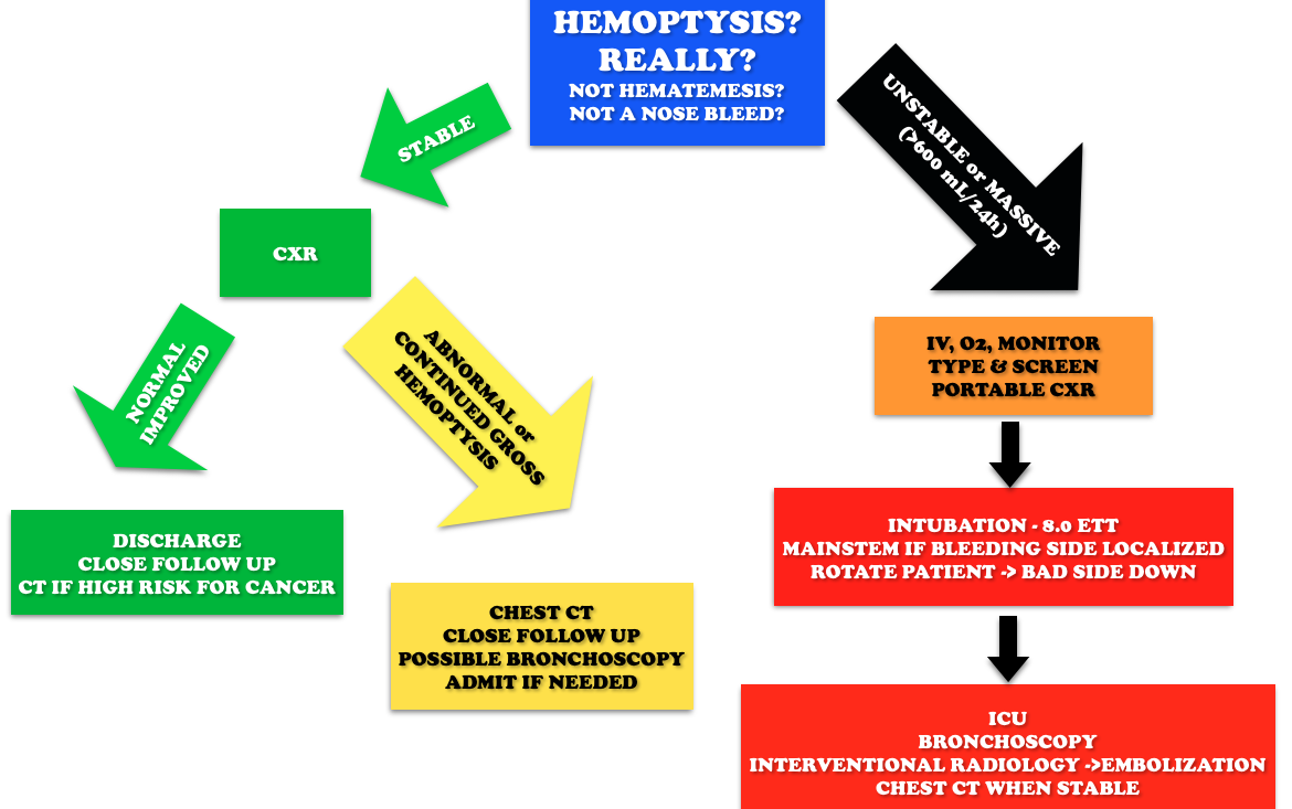
Etiology: Most common causes are bronchitis (often blood tinged sputum), infection (abscess, pneumonia, tuberculosis), neoplasm (lung cancer).  Other causes include iatrogenic causes (bronchoscopy, biopsy, aspirated foreign body), anticoagulation, and autoimmune diseases such as granulomatous polyangiitis (Wegener’s), lupus, and Goodpasture’s.

Workup:

Hemoptysis Workup

Generously Donated Rosh Review Questions 

Question 1. A 50-year-old man, nonsmoker, presents to the ED with a 2-day history of cough now associated with frank hemoptysis. He denies any constitutional symptoms. Vital signs are BP 125/70, HR 80, RR 16, and pulse oximetry is 98% on room air. On exam, his lung fields are clear; the remainder of the exam is unremarkable. A chest radiograph is performed, which is normal. [polldaddy poll=9039260]

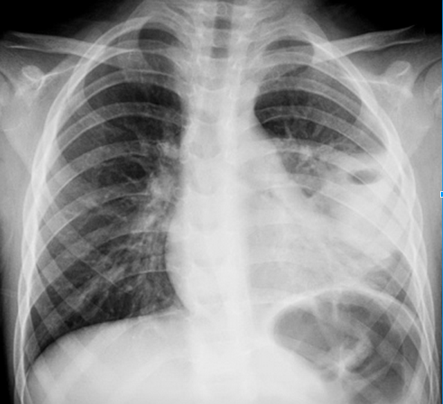
Question 2. A 55-year-old man, smoker, presents to the ED with hemoptysis and dyspnea for 4 weeks. His VS are T 37°C, BP 146/76 mm Hg, HR 85 bpm, RR 20 per minute, and oxygen saturation 96% on RA. His lung exam reveals distant breath sounds on the left side. His chest X-ray is shown below. [polldaddy poll=9039262]

Rosh Review
Rosh Review

Answers

1.C. The patient is hemodynamically stable with a normal chest radiograph, so he does not require ICU admission (A). Patients with massive hemoptysis require ICU admission. The decision to perform a bronchoscopy (B) in this patient will be left up to the pulmonologist. Given the overall clinical picture, urgent bronchoscopy is not required in this case. With massive hemoptysis, an emergent bronchoscopy is indicated. Bronchitis (D) typically presents with the abrupt onset of cough with blood-streaked purulent sputum. The patient in the clinical scenario has persistent frank hemoptysis, which mandates further investigation. In a patient who does not smoke, is under the age of 40, and has a normal chest radiograph and scant hemoptysis, treatment for bronchitis can be initiated with outpatient follow-up.

2. B. Although bronchitis (A) is the most common cause of hemoptysis (responsible for 15%-30% of cases), patients present with cough as the dominant symptom and have abnormal lung exams and normal chest x-rays. The cough may be productive of sputum. The diagnosis of pneumonia (C) requires focal findings on physical exam or infiltrates on radiographic imaging and is typically accompanied by a fever. Patients with lung cancer are at increased risk for pulmonary embolism (D). This patient’s Wells score is 2 (one point each for hemoptysis and malignancy), which makes the likelihood of PE 16% in an ED population. Given the lung mass seen on chest x-ray, lung cancer is more likely than PE.

References: