Episode 11 – Ebola and Transmission Precaution Pearls

Episode 11 (iTunes or listen here)

The Free Open Access Medical education (FOAM)

We review Mount Sinai Emergency Medicine Residency’s blog post on Ebola. The Pearls:

  • Signs and symptoms of ebola: Fever (>101.5F, 41C), severe HA, myalgias, vomiting, abdominal pain, unexplained hemorrhage, hypotension plus an epidemiologic risk factor in the past 3 weeks.
  • Risk factors:  contact with blood or other body fluids of a patient known or suspected to have ebola, residence or travel to endemic areas, and direct handling of bats, rodents, or primates from disease endemic areas.
  • CDC recommends screening in those with :
    • percutaneous/mucous membrane exposure or direct skin contact with body fluids of a person with a confirmed or suspected case of ebola without appropriate personal protective equipment
    • laboratory processing of body fluids of suspected or confirmed ebola cases without appropriate PPE or standard biosafety precautions
    • participation in funeral rites or other direct exposure to human remains in the geographic area where the outbreak is occurring without appropriate personal protective equipment.
  • Personal protective equipment is key in prevention of ebola spread.  Ebola is not airborne but due to the case mortality rate, fear, and questionable history of aerosol transmission in the past, we treat it like it is.  Recommended protection in the United States:  fluid impermeable gown, N95 respirator, eye shield and in situations with large amounts of fluids -double gloving, disposable shoe covers, and leg coverings (CDC recommendations).

Check out the CDC website on Ebola

EMDocs post on Ebola

EMCRIT Ebola algorithm


The Bread and Butter

We summarize some key topics from the following readings, Tintinalli (7e) Chapters 157, 148  but, the point isn’t to just take our word for it.  Go enrich your fundamental understanding yourself! Airborne Precautions – used for patients known to be or suspected of being infected with organisms transmitted by airborne droplets and small particle residue (<5 micrometers) of evaporated droplets containing microorganisms that can be spread by air currents.

  • Require special, negative pressure rooms with special ventilation and filtration and the N95 respirators.
  • Limited movement of patient within the health care setting and in the ED they need to be in a room with the door closed.
  • Recommended for: Measles, Varicella, Tuberculosis

Droplet Precautions – used for patients known to have or suspected of having serious illnesses transmitted by large particle droplets (>5 micrometers) produced by the patient during talking, sneezing, or coughing or during procedures.

  • Use a mask and wash your hands.  N95 respirator recommended for procedures like bronchoscopy, suctioning, etc.
  • Non Sterile gown if one anticipates substantial contact with the patient or if the patient is incontinent or has wound drainage not contained by dressings.
  • Limit transportation and movement of the patient – they should wear a mask when transported throughout the hospital.
  • Recommended for: Serious infections, Pertussis, Parvovirus B19, Mumps, Rubella

Varicella – Herpes virus that causes chickenpox (primary infection) and a secondary reactivation (herpes zoster/shingles) as the virus may lie latent in dorsal root ganglia.

  • Use Airborne precautions as it’s spread via respiratory secretions but may also spread (although less infectious) from the fluid of the non-crusted vesicles.
  • Symptoms of chickenpox: fever, malaise, headache and a vesicular rash that appears in crops with lesions at varying stages, including papules, vesicles, and crusted lesions, predominantly on the torso and face.
  • Most infections are minor and self-limited but increased sequelae exist in the immunocompromised and elderly.  These subgroups may benefit from antivirals
  • Immunizations now prevalent although individuals can still get mild chickenpox after immunization.
  • Varicella-zoster immune globulin exists but use as postexposure prophylaxis is essentially limited to non-immune pregnant women and the severely immunosuppressed. Healthy non-immunue individuals can be vaccinated after exposure and, if they are high risk and develop symptoms, they can get antivirals.

Generously Donated Rosh Review Questions

Question 1. [polldaddy poll=8241932]

Question 2.  A 3-year-old boy presents to the ED with 3 days of fever, cough, and runny nose. On exam, you note conjunctival injection and an erythematous, nonblanching, nonvesicular, maculopapular rash behind his ears and on his hairline, with a few spots on his chest. [polldaddy poll=8241939]

References:

Safe Management of Patients with Ebola Virus Disease (EVD) in U.S. Hospitals.”  CDC.  August 6, 2014

Cline DM.  “Chapter 157. Occupational Exposures, Infection Control, and Standard Precautions”  Tintinalli’s Emergency Medicine:  A Comprehensive Study Guide (2011).

Answers

1. Chickenpox is a highly contagious but generally benign and self-limited viral disease caused by the varicella-zoster virus (also known as human herpesvirus 3). The disease is characterized by the sudden onset of fever, malaise, and a pustular maculopapular rash that can occur anywhere on the skin or mucus membranes. The lesions then become vesiculated followed by scabbing over the course of 3-4 days before resolving. Skin lesions appear in crops with multiple lesions of various stages appearing on the skin at the same time. Uncomplicated infection is generally treated with supportive measures, including antipyretic, antipruritic, and pain control medications. Antivirals such as acyclovir, valacyclovir, and foscarnet may also be initiated in severe disease or immunosuppressed individuals. Parents should be cautioned to avoid giving their children aspirin or aspirin containing medications due to the risk of developing Reye’s syndrome.

The lesions of chickenpox appear suddenly rather than gradually (A). Smallpox lesions may appear similar to chickenpox lesions, however they are found in the same stage (B) of development. Rubella (German measles) is associated with the sudden onset of a maculopapular rash that first appears on the face then rapidly spreads inferiorly to the neck, trunk, and extremities and fades by the 3rd day (C).

2. Rubeola, or measles, is associated with fever and rash with cough, conjunctivitis, coryza, and Koplik spots. The characteristic rash is erythematous, nonblanching, and maculopapular. It begins on the head, usually behind the ears and around the hairline, with subsequent spread down the face, to the trunk, and extremities (centrifugal spread). The rash may coalesce into salmon-colored patches and typically disappears within 1 week. Koplik spots or pinpoint-sized white lesions on a red background that appear on the buccal mucosa opposite the molars are pathognomonic.

Roseola (A) is a viral infection with the onset of a rash that occurs upon resolution of a high fever. It is common in ages 6–18 months. Rubella (B) is often referred to as “three-day measles.” It is a mild illness, except for congenital infection, which can cause major birth defects. It is associated with fever, rash, and prominent lymphadenopathy, with tender posterior auricular, cervical, and occipital nodes. Varicella (D) (chicken pox) is associated with a flu-like illness and the formation of macules that progress to fluid-filled vesicles in an erythematous base (“dew drops on a rose petal”). Crops of lesions typically appear at the same time with vesicles in various stages of healing.
Episode 10 – Pediatric GI Emergencies

Episode 10 (iTunes or listen here)

The Free Open Access Medical education (FOAM)

We review Dr. Natalie May’s brilliant post on the St. Emlyn’s blog, “When Sick Means Sick: Emesemantics and Vomiting in Kids”  in which she dissects emesis descriptors such as bilious, projectile, and coffee-ground.

The Pearls:

  • Ask for color descriptors or look at the emesis yourself rather rely on typical descriptors of emesis.
  • Bilious vomiting, medical speaking, means emesis that appears like cooked, green spinach – not the yellow color that parents often mean. While sometimes normal in older children with gastroenteritis, in neonates or anyone sick appearing, this represents a surgical emergency such as volvulus, malrotation, necrotizing enterocolitis etc.
Photo: Laurent Nguyen, Wikimedia Commons
  • Projectile vomiting – Most vomit is projected at least a short distance, so parents may say even reflux is projectile.  Observe a test feed to gauge whether a baby is vomiting or has true projectile vomiting which may represent idiopathic hypertrophic pyloric stenosis.
  • Coffee ground emesis – this is a blackish-brown gritty emesis but parents may mean any brown-ish vomit.  This is typically indicative of upper GI bleed, which is pretty rare in pediatric patients.

The Bread and Butter

We summarize some key topics from the following readings, Tintinalli (7e) Chapters 111,124; Rosen’s 8(e) Chapter 172 but, the point isn’t to just take our word for it.  Go enrich your fundamental understanding yourself!

Neonatal Jaundice

Physiologic Jaundice – Jaundice in healthy, full-term newborns typically develops during the 2nd – 3rd day of life and resolves by the 5th or 6th day.  This occurs a little later in Asian and premature infants.

  • Mean peak total serum bilirubin is 6 mg/dL
  • Use the nomogram in infants >35 weeks gestational age to determine need for phototherapy

Non-physiologic Jaundice – bad.

  • Jaundice in the first 24 hours
  • Bilirubin rising faster than 5 mg/dL in 24 hours •Clinical jaundice >1 week
  • Direct bilirubin >2 mg/dL
  • In healthy term infants total serum bilirubin concentration >15 mg/dL (so remember: average of 6 but more than 15 is bad. But check out the nomogram in non-preemies).

Indirect Neonatal Jaundice – 3 main causes, listed below. Treatment is with phototherapy and mitigation of underlying causes.

  • Increased lysis – this can be due to lysis of red blood cells or sequestration of blood – ABO incompatibility, splenic sequestration, spherocytosis.
  • Decreased hepatic uptake/decreased conjugation – Immature transfer enzymes (babies grow out of this), breastmilk jaundice (lack certain enzymes, idiopathic hypertrophic pyloric stenosis (unclear why), as well as Gilberts, Crigler Najjar Syndrome
  • Increased enterohepatic uptake (i.e. too much reclaimed from the gut) – obstruction or breastfeeding jaundice (dehydrated babies who are breast feeding).

Direct Bilirubinemia

  • Etiology – biliary tree obstruction or biliary atresia, enzyme deficiencies (cystic fibrosis, alpha-1 antitrypsin deficiency, glycogen storage diseases)
  • Conjugated bilirubin is non-toxic so treat the underlying cause

Emergency Department Diagnostics:

  • Total and Fractionated Bilirubin,  Blood Type with Rh factor, Coomb’s test, blood count, Reticulocyte count, consider sepsis work-up .

More FOAM

Intussusception

Presentation: Abdominal pain, vomiting, bloody or guaiac positive stool.  The classic triad is not useful and present in 15-20%.  While intussusception is most common at approximately 1 year of age and, moreover 2 months -6 years, it can present at any time, including the elderly.

Etiology:  The bowel telescopes on itself and

Diagnosis:  Clinical, ultrasound (target sign), or diagnostic and therapeutic air contrast enema. It’s also reasonable to get plain films, if desired.

Treatment:  Air contrast enema in radiology results in approximately 60% success so most recommend a surgery consult in the event there’s a complication or failure in radiology.  Also, give these patients 20cc/kg fluid bolus and treat their pain.

More FOAM:

Generously donated Rosh Review questions (scroll for answers)

Question 1. A 10-month-old previously healthy boy presents with 1 day of bilious vomiting and fever. The patient is ill-appearing. Physical examination reveals a distended and diffusely tender abdomen with guarding and rebound. [polldaddy poll=8224476]

Question 2. A 2-year-old ex-33 week premature girl presents with vomiting, diarrhea and poor feeding. The patient has episodes of fussiness and inconsolable crying followed by periods of lethargy and sleeping. During periods of fussiness, the patient draws her legs up to her chest. [polldaddy poll=8225869]

Also, check out “Ketamine, The Album” – A musical written by and for emergency physicians as a tribute to ketamine


Answers.

1.Correct Answer ( A ) This patient presents with signs and symptoms concerning for an obstruction secondary to a volvulus and requires emergent surgical evaluation. Malrotation is a relatively common occurrence (1 in 500 live births) and about 75% of patients with malrotation will develop volvulus. During embryonic development, rotation of the gut arrests. This allows for the small bowel to twist around the superior mesenteric artery causing an acute obstruction. Patients will present with sudden onset of abdominal distension and bilious emesis. These infants will be ill-appearing and possibly toxic on presentation. Although a number of diagnostic modalities can be employed for definitive diagnosis, the priority in an ill-appearing infant with bilious emesis is emergent surgical consultation. All other interventions risk delaying definitive management. While waiting for the surgical consultation, the patient should have an IV placed, fluid resuscitation begun and a nasogastric tube placed for decompression of the stomach. Additionally, broad spectrum antibiotics should be administered. After consultation, an upper GI series may be obtained for definitive diagnosis.

Stool cultures (B) are useful when there is a suspicion for infectious process such as a parasitic or bacterial infection. Laboratory studies (C) will provide limited data and should not delay definitive management by a surgeon. The patient should receive intravenous hydration, not oral rehydration (D) as there is a high likelihood that this patient will be taken to the operating room. As such, the patient should be kept NPO.

2. D.  This patient presents with symptoms concerning for intussusception and should have an emergent ultrasound performed to make the diagnosis. Intussusception is defined as the telescoping of one segment of the intestine into another. It is the most common cause of obstruction in children younger than 2 years of age. The classic triadof intussusception is abdominal pain, vomiting and bloody stools but all three features are only present in about 33% of patients. Bowel movements may be loose with mucous and blood and appear like “currant jelly.” Often patients will have cycles of severe abdominal pain lasting 10 to 15 minutes during which they are inconsolable. These episodes are followed by periods of painlessness during which the child may be lethargic. Palpation of the abdomen may reveal a sausage-like mass in the right upper quadrant representing the actual intussusception. The lead point for the telescoping may be due to Henoch-Schonlein purpura vasculitis, Meckel’s divericulum, lymphoma or polyps in children over 5 years of age. In younger children, enlarge Peyer’s patches may be the culprit. These occur after viral infections. Ultrasound of the abdomen is the best initial modality for identifying the intussusception. It may reveal the classic findings of a target sign or “pseudokidney” sign. Sensitivity and specificity of ultrasound approach 100%.  Abdominal X-ray (A) may show intussusception but may be negative in up to 20% of patients. CT(B) and MRI (C) of the abdomen and pelvis  are also unreliable in the diagnosis.

Episode 8 – Acid-Base and Hyponatremia

Episode 8 (iTunes or Listen Here)

The Free Open Access Medical education (FOAM)

This week we review Dr. David Story’s talk from SMACC GOLD, “Is Chloride a Poison?”  Dr. Story discusses the Stewart ion approach to acid-base, driven by the independent variable, the Strong Ion Difference (SID), which is the difference between the sums of concentrations of the strong cations and strong ions (typically Sodium and Chloride). He also reviews literature that suggests that there may be morbidity and even mortality associated with large volume infusions of 0.9% NaCl (NS), although more research is required in this arena to determine the patient oriented sequelae. Perhaps we should be using more balanced solutions such as lactated ringers (LR).

Also, SMACC  is awesome, listen to the talks from SMACC GOLD and come meet us in Chicago next June!

Literature on the topic:

Other FOAM Acid-Base Resources:

The Bread and Butter

We summarize some key topics from the following readings, Tintinalli (7e) Chapters 19,21; Rosen’s (3e) Chapters 124, 125 …but, the point isn’t to just take our word for it.  Go enrich your fundamental understanding yourself!

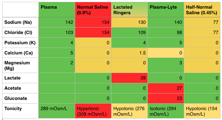
IV Fluids – Know the Composition (PV card from ALiEM)

Red - Too Much, Yellow - Too Little, Green - Just Right
Red – Too Much, Yellow – Too Little, Green – Just Right

Costs – NS (0.9%) is the cheapest, coming in at just over a US dollar per Liter and LR is slightly more expensive (most estimates are approximately $0.50 more per liter).  Plasma-lyte is more expensive, costing several dollars more per liter.

Downsides of Normal Saline (NS, 0.9%)

  • Hypertonic, hypernatremic, hyperchloremic – it has a little too much of everything and is acidic, with a pH of 5.0 [1]
  • The SID (Strong Ion Difference) of NS is 0, far less than the physiologic or normal SID of 38.  This is where the non-anion gap acidosis comes into play [1].

Caution with Lactated Ringers (LR)

  • LR contains calcium and some labs studies have shown that this may cause clotting; thus, major societies say LR is incompatible with blood products.  There are some studies to show that this may not be as big of a deal as previously thought: Albert et alCull et alLorenzo et al.
  • May interfere with Lactate clearance -A study of healthy individuals demonstrated that LR did not affect serum lactate levels [2].  However, often we are doing these large volume resuscitations in the critically ill who may have hepatic insufficiency and, thereby, reduced lactate clearance.  There is worry that LR may increase the serum lactate, making it difficult to gauge resuscitation by lactate clearance.  Paul Marino’s ICU Book states that significant skewing of the lactate is unlikely unless the patient’s ability to hepatically clear lactate is nil and the patient has gotten several liters of LR [1].

FOAM Resources:

Hyponatremia  – Na+ <135 mEq/L

Symptoms: often asymptomatic but may see vague symptoms such as nausea, vomiting, myalgias, lethargy.  Values <120 mEq  more associated with symptoms and <113 mEq, may see seizures/coma.

Causes:

  • Hypovolemic – Extra-renal: dehydration (vomiting, diarrhea, small bowel obstruction, burns), infusion of hypotonic fluids, Renal: thiazide diuretics, Renal Tubular Acidosis, osmotic diuresis, aldosterone/mineralocorticoid deficiency
  • Euvolemic – SIADH (syndrome of inappropriate secretion of antidiuretic hormone), water intoxication, drugs (NSAIDs, APAP, TCAs, sulfonylureas, morphine, carbamazepine, etc), beer potomania
  • Hypervolemic – think organ failure and people who are third spacing fluids. Congestive Heart Failure, Liver Failure, Renal Failure

Other FOAM Hyponatremia Resources:

Generously donated Rosh Review questions (scroll for answers)

Question 1.[polldaddy poll=8191357]

Question 2. A 23-year-old woman presents with seizures. The patient received 2 mg of lorazepam by EMS but continues to seize. Serum lab tests show the following: sodium 118, potassium 3.6, chloride 90, bicarbonate 21, BUN/Cr 10/1.0, glucose 89. [polldaddy poll=8191360]

Additional References:

1.Tizard, H.  Chapter 12: Colloid and Crystalloid Resuscitation.  Marino’s The ICU Book, ed 4.Lippincott Williams & Wilkins, 2007.

2.Didwania A, Miller J, Kassel D, Jet al. Effect of intravenous lactated Ringer’s solution infusion on the circulating lactate concentration: Part 3. Results of a prospective, randomized, double-blind, placebo-controlled trial.Crit Care Med. 1997 Nov;25(11):1851-4.

Answers.

1.C -The syndrome of inappropriate secretion of ADH (SIADH) is defined by the secretion of ADH in the absence of an appropriate physiologic stimulus. Its hallmark is an inappropriately concentrated urine, despite the presence of a low serum osmolality and a normal circulating blood volume. Causes of SIADH include central nervous system disorders, pulmonary disease, drugs, stress, pain, and surgery. Therefore, the above patient, with a known history of lung cancer and hyponatremia, most likely has SIADH and exhibits the following lab findings: serum osmolarity low, urine osmolarity high, urine sodium high. Psychogenic polydipsia (D) is a rare cause of euvolemic hyponatremia and is seen in psychiatric patients who consume large amounts of free water (in excess of 1 L/hr). This large consumption overwhelms the kidney’s ability to excrete free water. Patients will exhibit serum osmolarity low, urine osmolarity low, urine sodium low. Diabetes insipidus (B) results in the loss of large amounts of dilute urine from the loss of concentrating ability in the distal nephron. This may be due to a central cause—such as the lack of ADH secretion from the pituitary—or a nephrogenic cause—such as the lack of responsiveness to circulating ADH. Laboratory workup that invariably shows serum osmolarity high, urine osmolarity high, urine sodium low (A) rarely occurs.

2.A-This patient presents with prolonged seizure activity and hyponatremia and should emergently be treated withhypertonic saline. Hyponatremia is defined as a serum sodium level <135 mEq/L and is the second most common electrolyte abnormality after hypokalemia. The symptoms and signs of hyponatremia depend on the patient’s volume status, the cause and the rapidity of the change in serum sodium. Typically, patients with acute changes will have more severe symptoms including nausea, vomiting, confusion, stupor and seizures. Chronic hyponatremia will typically present with mild neurologic symptoms as well as lower serum sodium levels than acute hyponatremia. In patients without neurologic symptoms, volume status should be assessed and additional labs should be sent off to determine the cause of hyponatremia (urine sodium, osmolarity etc.). Patients with neurologic symptoms should be aggressively treated with 3% hypertonic saline. When correcting serum sodium, it is important to increase the serum sodium by no more than 0.5 mEq/L/hour and by no more than 10 – 12 mEq/day. More rapid changes can lead to central pontine myelinolysis, a crippling neurologic disease.

Episode 4 – Transfusions and Ingested Foreign Bodies

Episode 4 – Transfusion Emergencies (iTunes or Listen Here

The Free Open Access Medical Education (FOAM) –from Dr. Ryan Radecki’s erudite blog, Emergency Medicine Literature of Note..

Infections & Transfusions” – a JAMA meta-analysis found that higher hemoglobin targets were associated with an increased incidence of infection with a number needed to harm of 20-38.  The  group with a target level of 7-9 g/dL had an infection rate of 11.8% (95% CI, 7.0%-16.7%)  compared with an infectious complication rate of 16.9% (95% CI, 8.9%-25.4%) in the “liberally” transfused group.

Grilling Injuries on Memorial Day” – Grilling isn’t risk free.  Dr. Radecki reviewed a case series of six individuals who presented in one year to a hospital after ingesting meat cooked on a grill.  Three patients had neck pain with wire grill bristles removed via laryngoscopy and three had abdominal pain necessitating removal – 2 by colonoscopy and 1 with urgent surgery secondary to intestinal perforation.

The Bread and Butter

We summarize some key topics from the following readings,  Tintinalli (7e) Chapters 233; Rosen’s (8e) Chapter 7…but, the point isn’t to just take our word for it.  Go enrich your fundamental understanding yourself!

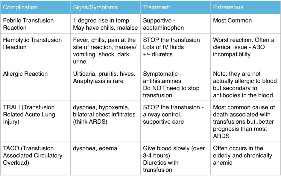
Transfusion Reactions

Immediate Reactions – great FOAM summary from Life in the Fast Lane: Transfusion Risks, Transfusion Reactions 

Screen Shot 2014-06-08 at 9.39.12 PM


The Ones Our Patients Care About (Infectious – statistics are US based)

  • Bacterial contamination is rare: 1/500,000 – 1/1,000,000
    • Most common pathogen: Yersinia Entercolitica
  • More common in platelets: 1/1000-1/2000 per Rosen, CDC, and the AABB  (Tintinalli cites 1 in 6 million)
  • Most Common virus: Parvovirus B19 (1 in 10,000).  The others are very very rare: HIV and Hepatitis C > 1 in 1 million, Hepatitis B 1 in 100,000-200,000

Ingested Foreign Bodies

  • Beware the button battery. These can cause necrosis within hours in the esophagus and must be removed ASAP.
  • Objects that are irregular, very sharp, or have dimensions greater than 2.5cm in width or 6 cm in length that are still in the stomach or duodenum – call GI to have these removed via endoscopy.

Generously donated Rosh Review questions (scroll for answers)

Question 1 A 55-year-old woman is receiving a blood transfusion due to persistent vaginal bleeding and a hemoglobin of 5 mg/dL. While receiving the transfusion, she develops fever, chills, back pain, pain at the site of transfusion, and tachycardia. [polldaddy poll=8109900]

Question 2 A 28-year-old man presents with a 1-day history of rectal bleeding. In the ED, he is hypotensive, thrombocytopenic, and is found to be passing melena. He receives a transfusion of platelets and packed red blood cells as part of his resuscitation. Twenty minutes after the start of his platelet transfusion, his BP is 90 mm Hg systolic, he becomes dyspneic, and his oxygen saturation drops from 99% on room air to 91% on 2L of oxygen supplementation. On exam, you note rales at the lung apices and that he is using accessory muscles to breathe. His chest radiograph shows diffuse interstitial infiltrates. [polldaddy poll=8109907]

Question 3 [polldaddy poll=8109908]


References:

Emery M.  Blood and Blood Products.  Rosen’s Emergency Medicine. 2014: 8th ed. p 75-80.e2

Coil CJ, Santen SA.  Transfusion Therapy  Tintinalli’s Emergency Medicine: A Comprehensive Review. 7th ed.

Hillyer CD, Josephson CD, Blajchman MA, et al. Bacterial contamination of blood components: risks, strategies, and regulation: joint ASH and AABB educational session in transfusion medicine. Hematology Am Soc Hematol Educ Program. 2003:575-89.

1. C – Up to 20% of all transfusions may lead to some type of adverse reaction. Although most of these reactions are minor, some are life-threatening. The patient is having an acute intravascular hemolytic reaction. This occurs when the recipient’s antibodies recognize and induce hemolysis of the donor’s red blood cells and may result in activation of the coagulation system and disseminated intravascular coagulation. This type of reaction typically presents with back pain, pain at the site of transfusion,headache, fever, hypotension, dyspnea, tachycardia, chills, bronchospasm, pulmonary edema, bleeding, and development of renal failure. First, stop the transfusion. Then initiate intravenous hydration to maintain diuresis.

2.D- This patient is most likely suffering from transfusion-related acute lung injury (TRALI), one of the leading causes of transfusion-related mortality. It is most closely associated with platelet and fresh frozen plasma transfusions, though cases have been reported with packed red blood cells since there is some residual plasma in the packed cells. Symptoms begin abruptly during transfusion or within 6 hours and resemble adult respiratory distress syndrome with noncardiogenic pulmonary edema, dyspnea, hypoxemia, and bilateral infiltrates on chest radiograph.

3. A- The patient is experiencing an allergic reaction without serious signs or symptoms. The transfusion does not need to be stopped for such a reaction; an antihistamine will help to relieve symptoms.

Episode 3 – Ear Emergencies

Episode 3 – Ear Emergencies (iTunes

The Free Open Access Medical Education (FOAM) – SMART EM Pseudoaxioms 2 and Literature Update

The podcast reviews:

  • Bullous Myringitis, a painful infection of the tympanic membrane, is typically caused by viruses, strep. pneumoniae and moraxella and, less commonly, mycoplasma.  The boards have caught up with this. Check out this review by Mellick.
  • PHANTOM-S trial: Use of the STEMO (Stroke Emergency Mobile – an ambulance with neurologist and a CT scanner for suspected strokes) reduced mean alarm-to-treatment time by 25 minutes (95% CI, 20-29; P < .001) without an appreciable neurologic benefit for this intensive intervention.
  • An article by Gregg et al in the NEJM discussed incredible improvements in diabetes outcomes as a result of tight glycemic control. Dr. Newman asserts that much of this is secondary to dilution, as the definition of diabetes changed in 1997.  This resulted in more people with less severe disease being diagnosed with diabetes.

The Bread and Butter

We summarize some key topics from the following readings,  Tintinalli (7e) Chapters 237; Rosen’s (8e) Chapter 92.  A good read on common ear emergencies from EBMedicine…but, the point isn’t to just take our word for it.  Go enrich your fundamental understanding yourself!

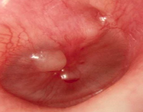
Bullous Myringitis

This is a painful infection characterized by bullae on the tympanic membrane (TM), which has a richly innervated outer epithelium (hence the severe otalgia). Patients may collect fluid behind their TM or have a concomitant otitis media.

Etiology: viruses, typical otitis media pathogens.  Mycoplasma and chlamydia have been associated with bullous myringitis but the association is unclear and these are not the most common causes.

Treatment: pain control, pain control, pain control.  Antibiotics are optional in most cases. But then again, antibiotics are not necessary in most cases of otitis media.  Both Rosen and Tintinalli are on board with this.

Perforated Tympanic Membrane

Photo:Didier Descouens (Wikimedia Commons)

Etiology: infection, trauma (q-tips, instrumentation), changes in pressure (diving, flying)

Treatment:  Keep the ear canal dry, follow up with ENT.  Most of these patients can go home.  If the injury is in the posteriorsuperior aspect of the TM or secondary to penetrating trauma, they should see ENT within 24 hours because they may have damage to the bones of the middle ear.

Auricular Hematoma

Etiology: Blunt trauma (often associated with boxing, fights, or termed “rugby ear“)

Treatment:  Incision, drainage, and compression dressing/splint.  Photo guide to repair.

Ototoxic Agents (great table in Tintialli 8 e, Ch 237, p1551)

Risk of hearing loss typically increases with exposure to medication (dose and length of use) and issues with clearance such as renal insufficiency may cause medications to hang around longer than anticipated or at higher levels.

Loop Diuretics: furosemide, bumetanide, ethacrynic acid

Salicylates (aspirin and quinine), NSAIDs

Antibiotics that end in -mycin or -micin: aminoglycosides (gentamicin), vancomycin, erythromycin

Chemotherapeutic agents: vincristine, vinblastine, cisplatin, carboplatin

Topical agents: ethanol, polymixin B, neomycin

Sudden Hearing Loss Differential Diagnosis 

Occurs over the span of three days.

Differential Diagnosis categories for any ailment can be remembered by the mnemonic VINDICATE

  • Vascular – sickle cell, polycythemia
  • Infectious/Inflammatory – viruses (zoster oticus or herpes, EBV, CMV, mumps), syphilis, labyrinthitis, temporal arteritis
  • Neoplasms – leukemia, masses,  acoustic neuroma
  • Drugs –  loop diuretics, antibiotics that end in -mycin or -micin (aminoglycosides like gentamicin, vancomycin, erythromycin), salicylates and NSAIDs, chemotherapeutic agents (cisplatin, carboplatin, vinblastine, vincristine, and topical agents (ethanol, polymyxin B, neomycin)
  • Iatrogenic/idiopathic – perforated TM, idiopathic endolymphatic hydrops (Meniere’s disease) – vertigo, hearing loss, tinnitus
  • Autoimmune -granulomatosis with polyangitis (Wegener’s)
  • Trauma – ruptured TM
  • Endocrine – diabetes, high cholesterol

Necrotizing Otitis Externa (Malignant Otitis Externa) – an infection that can turn into osteomyelitis of the skull

Presentation: otalgia, headache, and swelling and tenderness around the ear particularly in the setting of a prolonged course of otitis externa.  Diagnosis often requires CT scan to gauge involvement.

Epidemiology: Diabetics, immunocompromised

Etiology: Pseudomonas (90%)
Treatment:   Pediatrics- imipenem or an aminoglycoside and an antispeudomonal penicillin.  Adults – cephalosporin or quinolone

Dispo: Mild cases with good follow up can get oral quinolones as outpatients. More severe cases – admission, IV antibiotics, and perhaps surgical debridement.

Generously donated Rosh Review questions (scroll for answers)

Question 1.[polldaddy poll=8093105]Screen Shot 2014-06-01 at 9.28.25 PM

Question 2.

A 36-year-old woman presents to the ED complaining of decreased hearing and increased fullness to the right ear. Over the last week, she has used cotton-tipped applicators to attempt to remove cerumen from her right ear. On exam, you notice a cerumen-impacted external canal on the right. You irrigate the right ear with warm saline using an 18-gauge IV catheter and a plastic curette to remove the cerumen. During the procedure, the patient has sudden and complete hearing loss to the right ear. [polldaddy poll=8093098]


Question 3. A 16-year-old girl presents complaining of pain behind her left ear. She thought the pain was due to an ear infection and took three of her boyfriend’s leftover antibiotic tablets without seeing her primary care physician. Her ear pain improved for a couple of days, but now she is complaining of fever and discharge from the external auditory canal. Her vitals are T 38.4°C, BP 120/80, HR 108, and RR 18. On physical examination, she has postauricular tenderness, swelling, and erythema. You note purulent otorrhea through a perforated tympanic membrane. [polldaddy poll=8096505]

Answers:

1. D-Bullous myringitis was previously linked to Mycoplasma pneumoniae but it appears, based on middle ear aspirate culture results, that typical acute otitis media pathogens are the true cause. Among these, Streptococcus pneumoniae is most common.
2. C – The patient does not require admission (A) to the hospital. ENT care can be arranged for as an outpatient. Traumatic tympanic membrane perforations do not require otic antibiotics (B) unless the ear was contaminated such as from diving in seawater or the rupture is secondary to infection. The patient should receive more than ac otton ball (D) in her ear. Her management should include analgesia and ENT follow-up because complications of tympanic membrane rupture include facial nerve palsy, vertigo, and hearing loss.

3. C – This patient has necrotizing otitis externa.

References:

Silverberg M, Lucchesi M.  Common Disorders of the External, Middle, and Inner Ear.  Tintinalli’s Emergency Medicine, A Comprehensive Study Guide, ed 7. New York, McGraw-Hill, 2011, (Ch) 237:p 1556-1557.