Episode 16 – Headaches

(ITUNES OR LISTEN HERE)

The Free Open Access Medical Education (FOAM)

This week we review a post from Dr. Rob Orman’s ERCast, Is it really a sinus headache?

POUND- 4 criteria is very indicative of migraine (+LR 24), 3 criteria also likely (+LR 3), although most of this comes from the outpatient literature [1].

  • Pounding headache
  • hOurs: headache lasts 4-72 h without medication
  • Unilateral headaches
  • Nausea
  • Disabling: disrupts daily activities

The Bread and Butter

We summarize some key topics from the following readings, Tintinalli (7e) Chapter 159 ; Rosen’s 8(e) Chapter 20, 103 – but, the point isn’t to just take our word for it.  Go enrich your fundamental understanding yourself!

In Emergency Medicine, our job is to investigate and think about the life and limb threatening causes, even to mundane problems.   Things such as intracranial bleeds, meningitis, masses – these are huge deals and are covered well and hammered into our heads.  For FOAM core content on this, check out the St. Emlyn’s podcast.  On this episode, we’re running a mini-ophthalmology headache special and focusing on headaches that treatment may render “sight saving.”

Temporal Arteritis – often in patients older than 50 years of age and more common in those with a history of polymyalgia rheumatica. May be accompanied by visual changes including the “classic” amaurosis fugax or “curtain” of unilateral vision loss.  If not treated, these patient can lose vision permanently.

  • Unilateral or localized headache, often in the temporal or retro-orbital area
  • Jaw claudication (pain with chewing) – most specific sign
  • Decreased pulse in temporal artery or tenderness
  • Sedimentation Rate (ESR) >50

Treatment

  • Prednisone 40-60 mg if thinking about diagnosis
  • Temporal artery biopsy within 48 hrs

Acute Angle Closure Glaucoma – Classically, these patients present with unilateral mid-dilated pupils and severe nausea, vomiting, and headaches.  The history can, naturally, be less classic and more vague.  Also, if not treated, this can lead to vision loss.

  • Elevated intraocular pressure (>20 mmHg)
  • Decreased visual acuity
  • Fixed irregular semidilated (midposition) pupil
  • Slit lamp — shallow AC (closed angle), injected conjunctiva; corneal microcystic edema (cloudy)

Treatment –

  • Ophthalmology consult stat
    • They may want topical b-blocker, cholinergic, alpha-2 agonist, eye drops or administration of acetazolamide

Idiopathic Intracranial Hypertension (Pseudotumor Cerebri) – Common in young, overweight women or those on oral contraceptives.  Untreated, they can suffer vision loss.

  • Elevated opening pressure (>20-25 cm H20) on lumbar puncture

Treatment

  • Neuro follow up
  • Acetazolamide +/- furosemide
  • Therapeutic lumbar punctures

Cerebral Venous Sinus Thrombosis – may present as atypical headache with stroke like symptoms in patients without known vascular risk factors.  The neurological findings may be transient.  Often associated with post-partum patients, patients with hypercoaguable states (Factor V mutations, protein C or S deficiency, antithrombin III deficiency, etc), patients on OCPs.

Diagnosis – CTV or MRV (magnetic resonance venography) after CT scan, which may be normal.

Treatment – Anticoagulation, although this is somewhat controversial

Generously Donated Rosh Review Questions (Scroll for Answers)
Question 1. A 73-year-old woman with a history of hypertension presents with a unilateral headache for 3 weeks. She states that she has a throbbing pain at her right temple and has pain in her jaw with opening and closing. The vision in her right eye has worsened over the previous day. Her blood pressure is 173/100.
 [polldaddy poll=8340282]
Question 2. A 71-year-old woman presents to the ED with daily headaches for 2 months. She describes the headache as a dull pain that is most intense in the morning and resolves by the afternoon. On exam you note 4/5 motor weakness of the left upper and lower extremity.
[polldaddy poll=8340292]
References:
1. Detsky ME,McDonald DR, Baerlocher MO, Tomlinson GA,McCrory DC, Booth CM. Does this patient with headache have a migraine or need neuroimaging? JAMA. 2006 Sep 13;296(10):1274-83.

2. Chapter 20, 103.  Rosen’s Emergency Medicine, 8e.

3.Chapter 159.  Tintinalli’s Emergency Medicine: A Comprehensive Study Guide, 7e. New York, NY: McGraw-Hill; 2011

Answers
1. D. This patient presents with a unilateral, subacute headache with associated jaw claudication and vision change; symptoms consistent with temporal arteritis. Temporal arteritis or giant cell arteritis is a systemic inflammatory process of small and medium-size arteries. The most commonly involved vessels are the ophthalmic vessels and the extracranial branches of the aortic arch. The disease typically affects patients over 70 years of age and is more common in women than in men. Patients present with a subacute headache that is throbbing in nature and may be present for weeks to months. Often, patients will have symptoms for more than 2 months. Patients may also report jaw claudication secondary to vascular insufficiency of the masseter and temporalis muscles. Physical examination may reveal tenderness over the temporal artery. Systemic symptoms may also be present including fever, joint pains, and weight loss. Diagnostic testing in the Emergency Department generally begins with an erythrocyte sedimentation rate (ESR) with a cutoff of 50 mm/hour although the level may be >100 mm/hour. However, the ESR will be normal in 10-25% of patients. The gold standard diagnostic test is a temporal artery biopsy. In patients with a high-clinical likelihood of temporal arteritis, treatment should be initiated regardless of initial diagnostic testing as delay can lead to permanent visual loss. Prednisone should be started at 60 – 120 mg/day.

Carbamazepine (A) is the treatment of choice for trigeminal neuralgia, not temporal arteritis. The patient does not present with symptoms consistent with hypertensive emergency requiring emergent antihypertensive treatment withlabetalol (B). A non-contrast head CT scan (C) is not helpful in temporal arteritis as the disease does not involve the intracranial contents.

2.  B  More than half of patients diagnosed with a brain tumor complain of headache. However, the headache associated with brain tumor is highly variable. Patients may describe it as continuous or intermittent, unilateral or bilateral, sharp or dull. It is associated with neurologic deficits less than 10% of the time. However, in the setting of aneurologic deficit and chronic headache (as in this scenario with motor weakness), a mass lesion should be strongly considered as the cause. Patients may also complain of nausea, vomiting, visual change, and gait disturbance. Headaches due to brain tumors are classically associated with pain that is worse in the morning (as in this case). However, this is rare.

Central venous thrombosis (A) results from hypercoagulable states and is associated with acute to subacute headaches with vomiting and sometimes seizures. Risk factors include the use of oral contraceptives, postpartum or postoperative states, and any hypercoagulable state such as factor V Leiden mutation, antithrombin III deficiency, protein S or C deficiency, or polycythemia. The diagnosis is usually made by MRI venogram. Migraine headache (C) is classified as a primary headache and can be quite variable in presentation. These headaches can be associated with nausea, vomiting, photophobia, and phonophobia. The headache may also be preceded or accompanied by an aura that develops gradually over minutes, usually lasts 60 minutes, and is reversible. Auras may include neurologic symptom but commonly include scintillating scotomas (dark spots) or flashing lights. Temporal arteritis (D) occurs almost exclusively in patients older than 50 years and is much more common in women. Headache is the most common symptom of temporal arteritis and usually occurs over the frontotemporal region. It is strongly associated with a history of polymyalgia rheumatic. It is not associated with focal neurologic deficits, but it can lead to vision loss due to ischemic optic neuritis.
Episode 15 – Atrial Fibrillation/Flutter

(iTunes or listen here)

The Free Open Access Medical Education (FOAM)

This week we review a post from Academic Life in Emergency Medicine, written by Brent Reed on selecting rate control agents in the management of atrial fibrillation.  This is a follow up post to Bryan Hayes’ summary of emergency department (ED) management of acute atrial fibrillation.  Both are high yield.

Acute Management:

  • No clear cut winner in the beta-blocker vs. calcium channel blocker battle

Long Term Atrial Fibrillation Management in General:

Avoid beta-blockers in:

  • Obstructive lung disease (asthma/COPD)
  • Peripheral vascular disease
  • Diabetics
  • Severe congestive heart failure (CHF)
  • Erectile dysfunction

Avoid calcium-channel blockers in:

  • Severe CHF and acute decompensated heart failure (ADHF)

Of note, in patients

The Maryland Critical Care Project has a great post with many of Dr. Amal Mattu’s key FOAM talks embedded on Tachydysrhythmias You Gotta Know.

The Bread and Butter

We summarize some key topics from the following readings, Tintinalli (7e) Chapter 280, 295 ; Rosen’s 8(e) Chapter  50 – a well written chapter, but, the point isn’t to just take our word for it.  Go enrich your fundamental understanding yourself!

Atrial Fibrillation

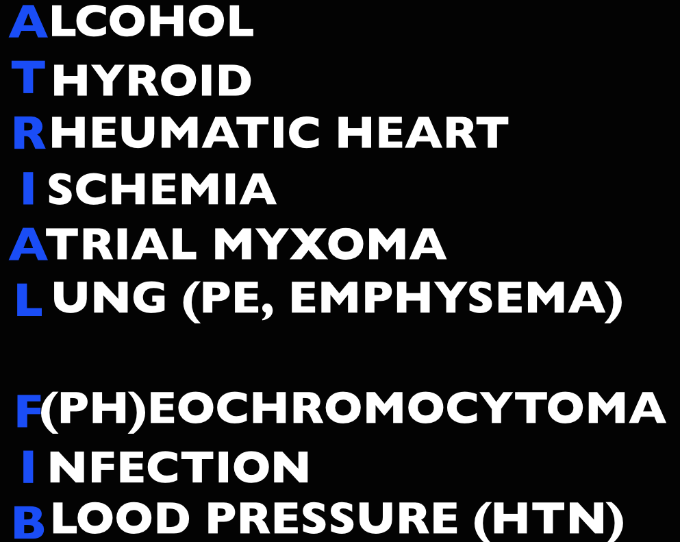
Etiology

Screen Shot 2014-09-16 at 3.57.07 PM

Management

Unstable (hypotension, pulmonary edema, altered mental status, chest pain) – cardioversion.

  • Pads in either an anterior-lateral (AL) or anterior-posterior (AP) position followed by synchronized cardioversion at 100-200 J biphasic.  Current literature shows no significant difference in pad placement [1]
  • If cardioversion fails, try amiodarone load or diltiazem. Check out EMCrit on Crashing A Fib

Stable

  • Rate control. A target of <120 beats per minute is acceptable in the ED [2-3].  First line agents are nodal blocking agents such as diltiazem and metoprolol
    • Diltiazem 0.25 mg/kg IV over 2 minutes with a peak effect in 2-7 minutes. Can repeat at 0.35 mg/kg IV over 2 minutes.
    • Metoprolol 5-10 mg IV.
  • Rhythm control with cardioversion.  While there’s no proven benefit to rhythm control, many patients would prefer to be in sinus rhythm and ED cardioversion of stable new-onset atrial fibrillation is appropriate in a select population, notably, when the onset is <48 hours (or <72 hours per Rosen).  The pooled literature suggests a thromboembolism rate <0.8% [4].
    • Note: A recent article in JAMA by Nuotio et al found a higher rate of embolic events in patients who were electively cardioverted after >12 hours in atrial fibrillation.The 30 day risk of thromboembolism when cardioverted between 12-48 hours was 1.1%, compared to the ~2% risk if cardioverted after 48 hours. While the risk is still small, it is higher than the ~0.3% risk of thromboembolism with anticoagulation on board.
  • Treat the underlying cause (ex: sepsis, pulmonary embolism, hyperthyroidism, etc)
  • May also consider Amiodarone, Digoxin (mean >11 hours to rate control) [3]

In atrial fibrillation with pre-excitation (WPW), an often wide and irregular rhythm with different/changing morphologies to the QRS do NOT treat with an AV Nodal blocking agent as this may result in death (Adenosine, Beta-blocker, Calcium-channel blocker, etc). Treat with procainamide or shock

Disposition – Admit patients that present unstable, with underlying co-morbidities, or those that are not rate controlled.  Depending on the patient’s follow up and local practice patterns, the

Atrial Flutter

How to Avoid Misdiagnosing Atrial Flutter – Dr. Amal Mattu

Management – same as atrial fibrillation

  • More sensitive to electrical cardioversion, less sensitive to chemical cardioversion

Multifocal Atrial Tachycardia

Irregular narrow complex tachycardia with p waves of at least 3 morphologies (this can be difficult to see, so look in multiple leads, particularly V2)

 Etiology -often seen in advanced pulmonary disease

Management  – Treat the underlying cause, do NOT cardiovert MAT

 Learn from the master ECG educator, Dr. Amal Mattu

Generously Donated Rosh Review Questions (Scroll for Answers)

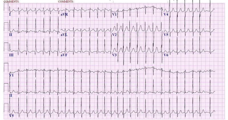
Question 1.  A 72-year-old man with a history of hypertension, diabetes, and congestive heart failure presents to the ED with heart palpitations for the past 4 days. He denies any chest pain, shortness of breath, abdominal pain, or history of similar palpitations. In the ED, his vital signs are BP 135/75, HR 138, RR 14, and oxygen saturation 98% on room air. His ECG is seen below. Which of the following is the most appropriate next step in management?

Rosh Review
Rosh Review

A. Chemical cardioversion

B. Rate Control

C. Synchronized cardioversion

D. Warfarin

Question 2.  When do you worry about giving calcium channel blockers, beta-blockers, or digoxin in a patient with atrial fibrillation?

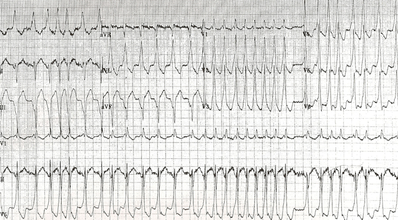
Question 3. An 18-year-old woman presents with palpitations and near syncope. Her vitals are T 98.7F, HR 199, BP 113/66, RR 32, and oxygen saturation 94%. Her ECG is shown below. What treatment is indicated?

Rosh Review
Rosh Review

A. Administer adenosine 6 mg IV

B. Administer diltiazem 10 mg IV

C. Administer lopressor 10 mg IV

D. Administer procainamide 100mg IV

References

1.  Kirkland S, Stiell I, AlShawabkeh T, Campbell S, Dickinson G, Rowe BH. The Efficacy of Pad Placement for Electrical Cardioversion of Atrial Fibrillation/Flutter: A Systematic Review. Acad Emerg Med. 2014;21(7):717–726.

2. Chapter.  Rosen’s Emergency Medicine, 8e.

3.Chapter.  Tintinalli’s Emergency Medicine: A Comprehensive Study Guide, 7e. New York, NY: McGraw-Hill; 2011

4. Cohn BG, Keim SM, Yealy DM. Is Emergency Department Cardioversion of Recent-onset Atrial Fibrillation Safe and Effective? J Emerg Med. 2013;45(1):117–27.

Answers

1.  B. Atrial fibrillation is caused by chaotic, disorderly firing from a second focus within the atria, resulting in uncoordinated atrial contractions. Patients with atrial fibrillation may present with palpitations, chest pain, shortness of breath, or they may be asymptomatic. Atrial fibrillation can be classified as chronic or paroxysmal, with paroxysms lasting minutes to days. On ECG, there are irregularly irregular narrow QRS complexes. In addition, no discernible p-waves are noted, rather fibrillatory waves are seen. Unless the patient is hemodynamically unstable, the mainstay of therapy is rate control. This is achieved through medications that act on the AV node such as calcium channel blockers (eg diltiazem or verapamil), beta-blockers, or digoxin. Due to digoxin’s slow onset of action and side effects, it is considered a second line medication.

If atrial fibrillation has been present for >48 hours, there is an increased risk of atrial thrombus formation. An echocardiogram should be obtained in these patients to exclude thrombus formation prior to rhythm control. Patients with chronic atrial fibrillation usually are placed on warfarin (D) or a similar anticoagulant to prevent thromboembolism.Chemical cardioversion (A) (amiorodone, procainamide or flecainide) can be attempted in patients with paroxysmal atrial fibrillation for less than 48 hours. Synchronized cardioversion (C) is used in patients who are hemodynamically unstable. This can be achieved by administering 50 – 100 J of electricity in synchronization mode.

2.  If a patient has an accessory pathway, such as Wolff-Parkinson-White Syndrome.

3. D. This patient presents with near syncope in the setting of atrial fibrillation with abberant conduction most likely secondary to Wolff-Parkinson-White (WPW) syndrome and should be chemically or electrically cardioverted. WPW syndrome refers to the presence of an accessory pathway between the right atrium and right ventricle. This accessory pathway has a shortened refractory period and can bypass normal conduction down the AV node. Because of the shortened refractory time, the accessory pathway in WPW can conduct atrial impulses much faster than the AV node can allowing for a ventricular rate between 150 and 300 beats per minute. Any tachycardia greater than 200 beats per minute in an adult should raise suspicion for an accessory pathway.

Patients with WPW can be asymptomatic or may present with severe tachydysrhythmias. The most common presenting dysrhythmia is reentrant tachycardia (70-80%) and second is atrial fibrillation (10-30%). In these tachydysrhythmias, the patient can conduct orthodromically (down the AV node and back up the accessory pathway), antidromically (down the accessory pathway and up the AV node) or in both directions. Patients who have any antidromic conduction will present with wide complex tachycardias. In patients with irregularly irregular wide-complex tachycardias, atrial fibrillation with WPW is the most common diagnosis. If the patient is unstable, electrical cardioversion should be pursued immediately as these patients run the risk of degrading into ventricular tachycardia and ventricular fibrillation. If the patient is stable, procainamide can be administered for chemical cardioversion. Procainamide is a class Ia anitdysrhythmic agent. The dose of procainamide (D) is 18-20 mg/kg administered at a rate of 20-30 mg/min.

In patients with WPW, antidysrhythmic agents that block the AV node are contraindicated. Blocking the AV node causes unopposed electrical conduction down the accessory pathway. This can lead to ventricular dysrhythmias. Additionally, the accessory pathway in WPW responds paradoxically to AV nodal blocking agents by further decreasing its refractory time. Adenosine (A), beta-blockers (C), calcium-channel blockers (B) and digoxin all block the AV node.

Episode 4 – Transfusions and Ingested Foreign Bodies

Episode 4 – Transfusion Emergencies (iTunes or Listen Here

The Free Open Access Medical Education (FOAM) –from Dr. Ryan Radecki’s erudite blog, Emergency Medicine Literature of Note..

Infections & Transfusions” – a JAMA meta-analysis found that higher hemoglobin targets were associated with an increased incidence of infection with a number needed to harm of 20-38.  The  group with a target level of 7-9 g/dL had an infection rate of 11.8% (95% CI, 7.0%-16.7%)  compared with an infectious complication rate of 16.9% (95% CI, 8.9%-25.4%) in the “liberally” transfused group.

Grilling Injuries on Memorial Day” – Grilling isn’t risk free.  Dr. Radecki reviewed a case series of six individuals who presented in one year to a hospital after ingesting meat cooked on a grill.  Three patients had neck pain with wire grill bristles removed via laryngoscopy and three had abdominal pain necessitating removal – 2 by colonoscopy and 1 with urgent surgery secondary to intestinal perforation.

The Bread and Butter

We summarize some key topics from the following readings,  Tintinalli (7e) Chapters 233; Rosen’s (8e) Chapter 7…but, the point isn’t to just take our word for it.  Go enrich your fundamental understanding yourself!

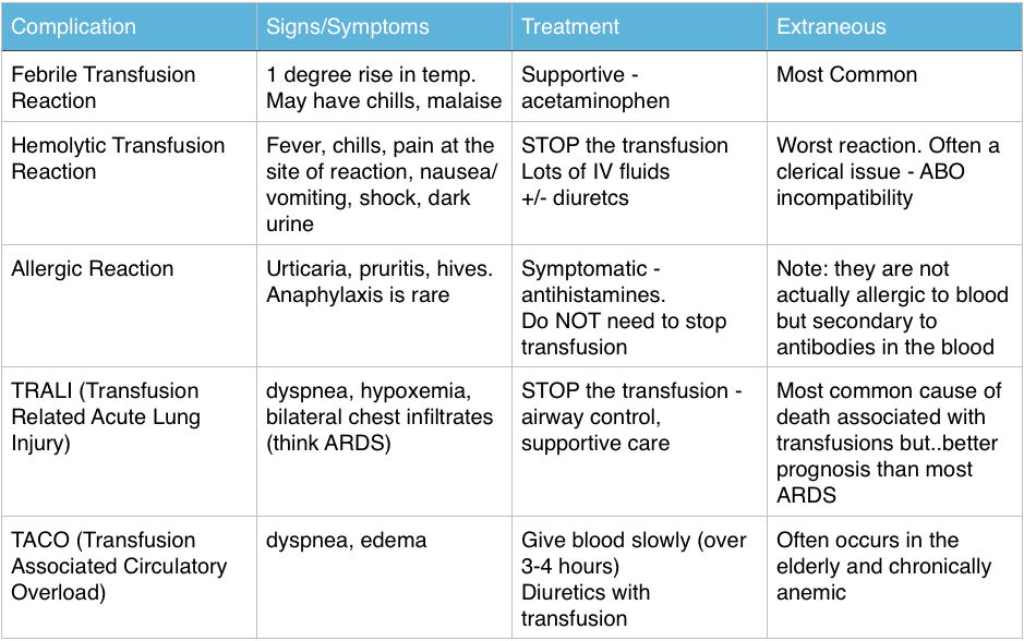
Transfusion Reactions

Immediate Reactions – great FOAM summary from Life in the Fast Lane: Transfusion Risks, Transfusion Reactions 

Screen Shot 2014-06-08 at 9.39.12 PM


The Ones Our Patients Care About (Infectious – statistics are US based)

  • Bacterial contamination is rare: 1/500,000 – 1/1,000,000
    • Most common pathogen: Yersinia Entercolitica
  • More common in platelets: 1/1000-1/2000 per Rosen, CDC, and the AABB  (Tintinalli cites 1 in 6 million)
  • Most Common virus: Parvovirus B19 (1 in 10,000).  The others are very very rare: HIV and Hepatitis C > 1 in 1 million, Hepatitis B 1 in 100,000-200,000

Ingested Foreign Bodies

  • Beware the button battery. These can cause necrosis within hours in the esophagus and must be removed ASAP.
  • Objects that are irregular, very sharp, or have dimensions greater than 2.5cm in width or 6 cm in length that are still in the stomach or duodenum – call GI to have these removed via endoscopy.

Generously donated Rosh Review questions (scroll for answers)

Question 1 A 55-year-old woman is receiving a blood transfusion due to persistent vaginal bleeding and a hemoglobin of 5 mg/dL. While receiving the transfusion, she develops fever, chills, back pain, pain at the site of transfusion, and tachycardia. [polldaddy poll=8109900]

Question 2 A 28-year-old man presents with a 1-day history of rectal bleeding. In the ED, he is hypotensive, thrombocytopenic, and is found to be passing melena. He receives a transfusion of platelets and packed red blood cells as part of his resuscitation. Twenty minutes after the start of his platelet transfusion, his BP is 90 mm Hg systolic, he becomes dyspneic, and his oxygen saturation drops from 99% on room air to 91% on 2L of oxygen supplementation. On exam, you note rales at the lung apices and that he is using accessory muscles to breathe. His chest radiograph shows diffuse interstitial infiltrates. [polldaddy poll=8109907]

Question 3 [polldaddy poll=8109908]


References:

Emery M.  Blood and Blood Products.  Rosen’s Emergency Medicine. 2014: 8th ed. p 75-80.e2

Coil CJ, Santen SA.  Transfusion Therapy  Tintinalli’s Emergency Medicine: A Comprehensive Review. 7th ed.

Hillyer CD, Josephson CD, Blajchman MA, et al. Bacterial contamination of blood components: risks, strategies, and regulation: joint ASH and AABB educational session in transfusion medicine. Hematology Am Soc Hematol Educ Program. 2003:575-89.

1. C – Up to 20% of all transfusions may lead to some type of adverse reaction. Although most of these reactions are minor, some are life-threatening. The patient is having an acute intravascular hemolytic reaction. This occurs when the recipient’s antibodies recognize and induce hemolysis of the donor’s red blood cells and may result in activation of the coagulation system and disseminated intravascular coagulation. This type of reaction typically presents with back pain, pain at the site of transfusion,headache, fever, hypotension, dyspnea, tachycardia, chills, bronchospasm, pulmonary edema, bleeding, and development of renal failure. First, stop the transfusion. Then initiate intravenous hydration to maintain diuresis.

2.D- This patient is most likely suffering from transfusion-related acute lung injury (TRALI), one of the leading causes of transfusion-related mortality. It is most closely associated with platelet and fresh frozen plasma transfusions, though cases have been reported with packed red blood cells since there is some residual plasma in the packed cells. Symptoms begin abruptly during transfusion or within 6 hours and resemble adult respiratory distress syndrome with noncardiogenic pulmonary edema, dyspnea, hypoxemia, and bilateral infiltrates on chest radiograph.

3. A- The patient is experiencing an allergic reaction without serious signs or symptoms. The transfusion does not need to be stopped for such a reaction; an antihistamine will help to relieve symptoms.