Foamcastini – Do We Make Saves?

ITUNES OR LISTEN HERE

We are at SMACC in Dublin – thanks to the Rosh Review, an excellent board review question bank.

Do We Make Saves?

Dr. Mervyn Singer “Is Survival Predetermined in the Critically  Ill?”

  • Many critical care studies are negative, but in some cases this may be because critically ill patients don’t have a uniform prognosis.
  • Dr. Singer argues that some people may be “destined to die” and some may be “destined to live.”  Interventions may be harmful or futile in one group but beneficial in the other.  For example, Dr. Singer references the CORTICUS trial of steroids in septic shock [1]. This was a negative trial.  Dr. Singer asserts that some evidence (of not great quality), purports that the sickest patients could benefit from steroids, while this same intervention could be deleterious in the healthier ones.
  • Problem: many of the studies that go back and re-analyze these groups looking at the sickest or least sick patients? They perform subgroup analyses, a form of data dredging that must be taken with a huge grain of salt.

Favorite Pearls

Dr. Suzanne Mason – “Acute Care of the Elderly”

  • Hospital admission may not benefit geriatric patients – interdisciplinary interventions involving nurses, consultants, pharmacists, physical therapy may be best.
  • If there is a single, free intervention that providers can do it’s assessing for polypharmacy. Polypharmacy in the elderly is a huge problem.  Check the patient’s medication list and beware adding new medications that may not be absolutely necessary.

Dr. Victoria Brazil – “So You Think You’re a Resuscitationist?”

  • The Dunning-Kruger effect is real in medicine and this is why we MUST have and provide feedback.  A review of the Dunning-Kruger effect can be found here.  Essentially, people tend to overestimate what they know (i.e. overly confident in their knowledge). The exception?  Masters tend to underestimate their knowledge.

Screen Shot 2014-03-15 at 9.36.53 AM

  • Our perception of reality is very skewed so, again, feedback is crucial. For example, Cemalovic and colleagues found that intubators underestimated the time they took to intubate: they estimated 23.5 seconds on average vs the 45.5 seconds intubation actually took. Additionally, they thought 13% of their patients desaturated during intubation but 23% actually desaturated [2].

Also, there was an excellent tribute to the late Dr. John Hinds, a reminder that by living profoundly, you can impact people across the world.

References

  1. Sprung CL, Annane D, Keh D et al. Hydrocortisone Therapy for Patients with Septic Shock. N Engl J Med. 358(2):111-124. 2008. [article]
  2. Cemalovic N, Scoccimarro A, Arslan A, Fraser R, Kanter M, Caputo N. Human factors in the emergency department: Is physician perception of time to intubation and desaturation rate accurate? Emergency medicine Australasia : EMA. 28(3):295-9. 2016. [pubmed]

Episode 51 – Ocular Trauma

(ITUNES OR LISTEN HERE)

The Free Open Access Medical Education (FOAM)

We cover this short video from EMRAP on lateral canthotomies.  This is one of those rare procedures that is vision saving; hence, it is worthy of frequent review.

Lateral Canthotomy and Cantholysis

Indication:  Suspect orbital compartment syndrome -afferent pupillary defect (APD), “tense eye” with taught lids, and high intraocular pressure (IOP) ≥ 40 mmHg. The APD is really a critical feature, as it is your indicator that there is optic nerve compromise from the intraorbital pressure

Clinical presentation:  History of orbital trauma, often in the setting of orbital fractures with decreased visual acuity, proptosis, chemosis

Procedure: Call ophthalmology.

  • Analgesia – inject lidocaine with epinephrine into the area of the lateral canthus
  • Devascularize – use straight kelly clamps to crush the lateral canthus for 1-2 minutes
  • Incise – Use scissors to cut the lateral canthus 1-2 cm. Then,  find the inferior crus of the lateral canthus ligament (looks kind of like a wishbone from a turkey but you rarely are going to be able to see the tendon so you’re “strumming” it with the closed tips of your scissors inferomedially toward the nose) – cut the inferior crus of the ligament to release pressure (this is the key step)

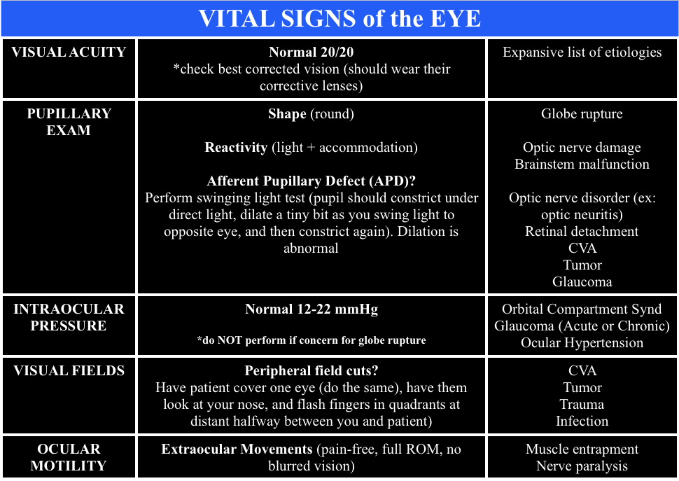
When calling ophthalmology, ensure you have the “Vital Signs of the Eye”

  • Vital Signs of the Eye - @FOAMpodcast
    Vital Signs of the Eye – @FOAMpodcast

Core Content

We delve into core content on eye trauma using Rosen’s (8th edition) Chapter 71 and Chapter 241 in Tintinalli (8th edition)

Orbital Fractures

Orbital Fractures

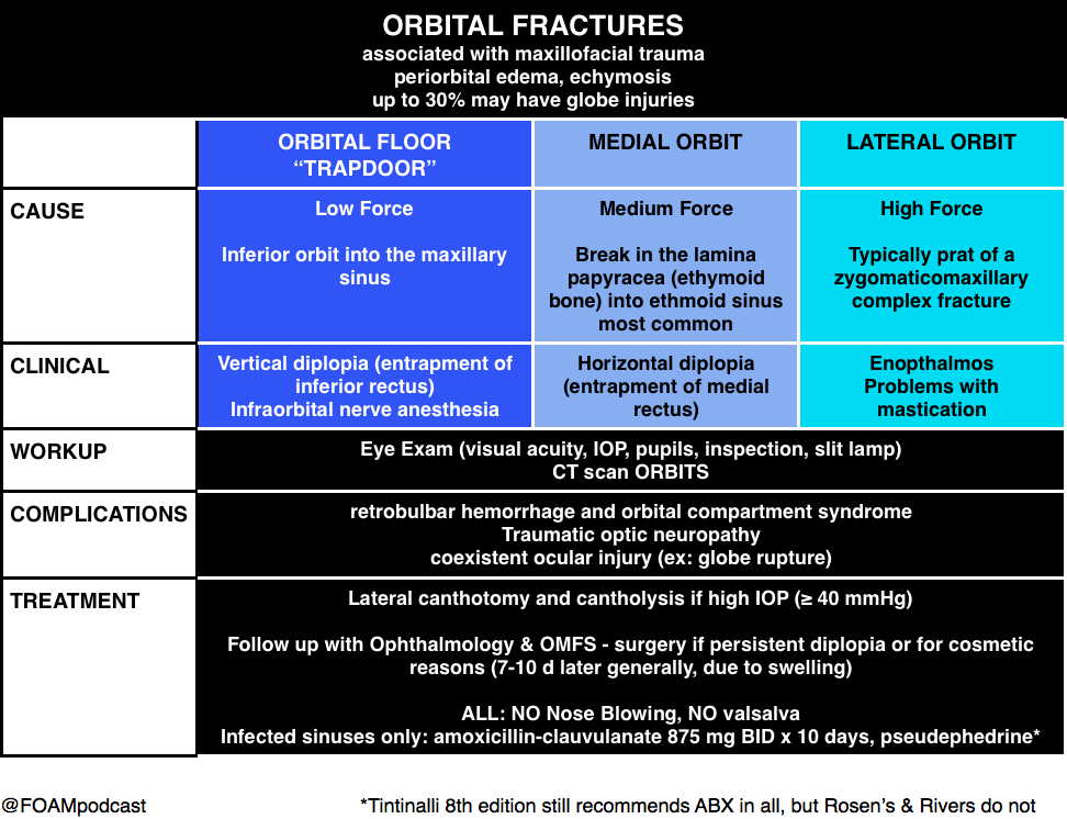
Note: entrapment is a clinical diagnosis.  A CT cannot comment on function, only structure.  Extraocular movements are a critical part of the fracture exam.

Pearl: young people are prone to “greenstick” fractures where the floor can actually pinch the muscle and trigger the oculocardiac reflex, which can be deadly.  They often have a “white eye” without too much impressive on exam but refuse to look in a certain direction due to nausea.
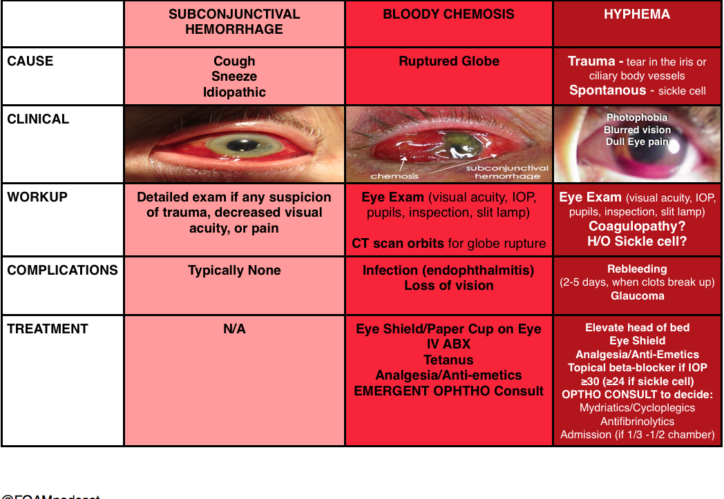
Hyphema

(and things confused with hyphema)

Hyphema

Burns

Ocular Burns

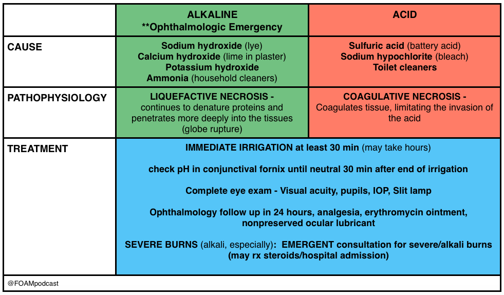
Pearl: An important thing to keep in mind is that a “white” eye after such an exposure is actually an ominous sign, as it implies ischemia of the limbal blood vessels, which portends a pretty bad prognosis.  People usually think that the more red an eye, the worse when, in truth, it’s often the other way around.

Generously Donated Rosh Review Questions

A 43-year-old construction worker presents with right eye pain. He states he was using a nail gun when he felt something hit his eye. Visual examination reveals a small nail penetrating the globe. What management should be pursued?

A. CT scan of the orbit and ophthalmology consultation

B. Measure intraocular pressure and consult ophthalmology

C. Perform lateral canthotomy and consult ophthalmology

D. Remove the foreign body, start topical antibiotics and send to ophthalmology for follow up

[accordion]
[toggle title=”Answer” state=”closed”]

This patient presents with a globe injury and should have a protective shield placed, intravenous antibiotics started, CT scan of the orbit performed and ophthalmology consulted emergently. Foreign body penetration of the globe is often associated with hammering, drilling, mechanical grinding or sanding. Any patient who presents with a foreign body sensation after one of these activities should increase suspicion for a penetrating injury of the globe. CT scan, MRI and ultrasound can all be used for diagnosis but MRI should be avoided if the suspected foreign body is metal containing. Many of these patients will require operative management and should be kept NPO. A protective shield should be placed to protect the eye but patching should be avoided as it may increase pressure on the eye. Emergent ophthalmologic consultation should be obtained

[/toggle]
[/accordion]

A 23-year-old man presents after a fight. His eye is seen below. Physical examination reveals intact extraocular movements, normal fluorescein staining, normal intraocular pressure, and normal visual acuity.

content_image-_medial_lower_lid_laceration

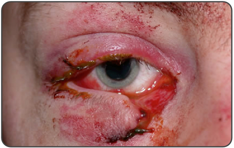
What management is indicated?

A. Delayed closure

B.  Laceration repair by the Emergency Physician

C. Ophthalmology consultation for repair

D. Tissue adhesive for repair

[accordion]
[toggle title=”Answer” state=”closed”]

C. Ophthalmology consultation for repair. This patient presents with a complex eyelid laceration possibly involving the canalicular system and should have a consultation with either ophthalmology or plastic surgery regarding repair. It is important to search for a penetrating globe injury in any patient with an eyelid laceration because of the proximity of structures. Simple horizontal and partial thickness lid lacerations can be repaired primarily by an Emergency Physician. However, more complicated lacerations should be considered for specialist repair because of the high likelihood of cosmetic or functional complications, or both. In general, lacerations through the orbital septum, lacerations with tissue loss, lacerations involving the lid margins, lacerations involving the levator or canthal tendons and those involving the canalicular system should be repaired by a skilled ophthalmologist or plastic surgeon. Injury to the canalicular system should be suspected in any laceration involving the medial lower eyelid. Tissue adhesive (D) is contraindicated this close to the eye and should not be used in an injury that requires precision alignment of tissue. Delayed closure (A) will likely lead to worse cosmetic outcomes. Primary repair by the Emergency Physician (B) is not recommended if the canalicular system may be involved.

[/toggle]
[/accordion]


Thanks to our peer reviewer, Michael Westafer, MD – Ophthalmologist and Glaucoma Fellow at Mayo Clinic

References

  1. “Eye Emergencies.”  Chapter 241.  Tintinalli’s Emergency Medicine: A Comprehensive Review. 8th edition.  
  2. Sharma R and Brunette D.  “Ophthalmology.”  Chapter 71.  Rosen’s Emergency Medicine.  8th edition, 909-930.

Episode 50 – Seizures

(ITUNES OR LISTEN HERE)

The Free Open Access Medical Education (FOAM)

We cover Special Seizures from EMin5.com by Dr. Anna Pickens. This is a short video summarizing important diagnoses to consider when a seizure doesn’t stop after the first or second round of benzodiazepines.

[expand title=”Female“]

Consider eclampsia, which typically occurs >20 weeks gestation to 6 weeks post-partum. Magnesium 4-6 grams IV, followed by infusion. Watch for apnea, check reflexes. [/expand]


[expand title=”Possible history of alcohol abuse“]

Consider alcohol withdrawal seizure. These patients respond to benzodiazepines and will typically NOT respond to antiepileptics such as phenytoin. They just need larger doses of benzodiazepines, so ramp up the dosages of those (10-100 mg of diazepam) and may require phenobarbital or propofol. [/expand]


[expand title=”Possible isoniazid exposure“]

Isoniazid toxicity can cause seizures, coma, metabolic acidosis, often within only 30 minutes of ingestion. The treatment here is pyridoxine (vitamin B6). These patients will need large doses and many recommend empirically giving 5 GRAMS intravenously (note: this is takes many vials as the typical dose of this medication is 50-100 MILLIGRAMS). This is a great review by First10inEM [/expand]


[expand title=”Consider hyponatremia“]

Treat with 100 mL of 3% hypertonic saline over 10-15 minutes. Can repeat x 1. Beware of rapidly correcting sodium in these patients due to central pontine myelinolysis/osmotic demyelination syndrome. [/expand]

Core Content

We delve into core content on seizures using Rosen’s (8th edition) Chapter 18 and Chapter 171 in Tintinalli (8th edition)

Screen Shot 2016-05-24 at 10.16.02 PM

Febrile Seizures AAP Guidelines

Febrile Seizures

Generously Donated Rosh Review Questions

A fifteen-year-old girl presents for evaluation and clearance for sports play. She has played team sports in the past, and would like to join the swim team this year. She was recently diagnosed with a seizure disorder. Her seizures are usually in the mornings, are generalized tonic-clonic seizures, and last for 1-2 minutes. They occur once per week. She is currently taking topiramate for seizure control. Her physical exam and vital signs are reassuring. Which of the following is the best recommendation for this patient?A. Allow participation in swimming as this is not a contact sport
B. Deny participation in all sports until seizures are better controlled
C. Permit patient to join the swim team as long as she has rectal diazepam with her at all times
D. Refuse participation in swimming as seizures are poorly controlled

[expand title=”Answer”]

D. Refuse participation in swimming as seizures are poorly controlled. Swimming (A) is a danger to children with poorly controlled seizures, as there is a risk that the child will have a seizure during the exercise and could suffer near-drowning or death. Some sporting events are safe for children with epilepsy (B), such as running. While the child participating in non-contact and non-aquatic sports may still have a seizure, the risk of morbidity and mortality to the child and other participants is low. Rectal diazepam (C) is a pharmacologic therapy that can stop seizures once they begin; however, the risk for morbidity and mortality remains high for children with poorly controlled seizures.

[/expand]

A full-term 3-week-old girl is brought in by her parents who report that she has been “acting funny” for 2 hours. They noticed that she has been moving her lips nonstop. She was a full-term, normal, spontaneous vaginal delivery and has been feeding well with adequate wet diapers since hospital discharge. She is afebrile and vital signs are normal. The anterior fontanelle is flat, and red reflexes are present. Heart, lung, and abdominal exams are normal. Her neurologic exam is positive for root, suck, and Moro reflexes, upgoing Babinski reflexes, and rhythmic lip-smacking movements. What is the most appropriate next step to take with this baby?
A. Administer a benzodiazepine
B. Initiate EEG monitoring
C. Perform a CT scan of the brain
D. Provide reassurance that this is normal behavior

[expand title=”Answer”]

This baby is having neonatal seizures, which are often subtle and more likely to be focal than tonic clonic. The most common manifestations are lip smacking, eye deviation, staring, rhythmic blinking, and bicycling movements. The patient should receive a benzodiazepine, such as lorazepam, midazolam, or diazepam, to stop the seizure. This should be followed by a workup that is directed at finding the underlying cause because neonatal seizures are less likely to be idiopathic than seizures in older children. A full septic workup is mandated for neonates with seizures, whether or not they are febrile. This includes CBC, blood culture, chemistry, UA, urine culture, chest x-ray, and CSF analysis. The infant should receive antibiotics and acyclovir and should be admitted to the hospital for further evaluation. Trauma can also cause neonatal seizures. It is important to look for signs of trauma such as bruising, bulging fontanelles, and retinal hemorrhages; the history may also include poor feeding, lethargy, or vomiting. Any infant with suspected head trauma should have a CT scan (C) and undergo a full child-abuse workup. An EEG (B) can be considered after more life-threatening causes of seizures, such as infection or trauma, are ruled out. A newborn (< 1 month old) with abnormal behavior should never be sent home with parental reassurance (D) only. Many new parents mistake normal behaviors for abnormal ones, but any truly abnormal behavior needs further investigation.[/expand]


References:

Kornegay JG. Chapter 171: Seizures Tintinalli’s Emergency Medicine: A Comprehensive Review (8e).

McMullan JT, Davitt AM, Pollack CV. “Seizures.” Rosen’s Emergency Medicine, Chapter 18, 156-161.e1


Episode 49 – The AAP BRUE (formerly ALTE) Guidelines

(ITUNES OR LISTEN HERE)

The Free Open Access Medical Education (FOAM)

We review the American Academy of Pediatrics guideline on Brief Resolved Unexplained Events (Formerly Apparent Life-Threatening Events) and Evaluation of Lower-Risk Infants.

Screen Shot 2016-05-04 at 9.33.37 AM

Note: Some have voiced concerns that these guidelines potentially downplay the event. The concern is that the yield of these workups and admissions may be low, but possibly worthwhile.  At FOAMcast, we are not qualified to critique these guidelines but there are helpful tables and charts in them to realize that these recommendations really are only for specific events and children AFTER thorough history and physical.

Generously Donated Rosh Review Question

A 6-week-old boy is brought to the emergency room because of cyanosis. He was sleeping comfortably in a supine position right after a feeding when he suddenly choked, became limp and lips turned blue. The mother witnessed the event and blew to the face of the boy. The whole episode lasted for about two minutes. EMS was called and upon arrival at the house, the boy was back to his usual self. At the ER, he boy has normal vital signs with normal physical examination findings.

A. Admit for cardiorespiratory monitoring

B. Discharge after reassuring the parents

C. Observe for four hours in the ER

D. Request for complete blood count

[accordion]
[toggle title=”Answer” state=”closed”]

A. Admit for cardiorespiratory monitoring. The boy in the vignette had an apparent life-threatening event (ALTE) which is not a specific diagnosis but a description of an acute, unexpected episode that is frightening to the caretaker. ALTE includes one or more of the following features: apnea, color change (may be cyanotic, pallid, erythematous or plethoric), marked change in muscle tone (limpness or rigidity) and choking or gagging. A specific cause for ALTE can be identified in over one-half of patients after a careful history, physical examination, and appropriate laboratory evaluation. The remaining cases are considered idiopathic if no cause can be identified after a thorough assessment. Common etiologies for ALTE include gastroesophageal reflux, neurologic problems (such as seizures), and respiratory infection. The history of an ALTE must be taken seriously, even if the infant appears entirely well by the time he or she is evaluated. In-hospital observation with cardiorespiratory monitoring is indicated for infants whose initial evaluation suggests physiologic compromise. Hospital admission may provide important clinical information where additional episodes may be witnessed by medical personnel during the observation period. In addition, serious underlying medical conditions may become apparent.  Discharging the patient after reassuring the parents (B) and observing the patient for four hours in the ER (C) are not appropriate management strategies for the infant in the vignette who needs admission for cardiorespiratory monitoring. Requesting for complete blood count (D) is not routinely done in the evaluation of an ALTE and would not aid in the management for the infant in the vignette.

[/toggle]
[/accordion]

References:

Tieder JS, Bonkowsky JL, Etzel RA, Franklin WH, Gremse DA, Herman B et al. Subcommittee on Apparent Life Threatening Events. Brief Resolved Unexplained Events (Formerly Apparent Life-Threatening Events) and Evaluation of Lower-Risk Infants: Executive Summary.  Pediatrics. In Press

Episode 48 – Urine Drug Screen, Cocaine, and PCP

(ITUNES OR LISTEN HERE)

The Free Open Access Medical Education (FOAM)

We review a post by Dr. Seth Trueger (@MDaware) on false positives of common medications in the urine drug screen.  We delve into posts by Dr. Bryan Hayes (@PharmERToxGuy) on false negatives for benzodiazepines and opioids in the urine drug screen.

Screen Shot 2016-04-25 at 8.29.32 PM

Core Content

We delve into core content on cocaine and phencyclidine (PCP) using Rosen’s (8th edition), Chapter 154 and Tintinalli (8th edition)

Cocaine

Cocaine
Cocaine

Note: Beta-blockers are contra-indicated in cocaine induced hypertension and chest pain.  Much of the ischemia induced by cocaine is thought to be due to vasospasm, predominantly from alpha-1 receptor effects.  Beta-blockers block the relaxation provided by beta-2 stimulus on muscles, leaving alpha-1 constricting vessels, “unopposed.”  This is largely theoretical/based on canine literature as there are only two human studies on this, (a) 10 humans given propranolol + cocaine with vasospasm and (b) 9 humans given labetalol + cocaine without vasospasm [4,5]. However, recent papers attempting to dispel this teaching don’t quite prove the point. One retrospective chart review looked at cocaine positive urine screens in patients with chest pain and found no worsened troponins. Cocaine stays positive in the urine for 3 days so it is not clear that these were patients presenting with cocaine associated chest pain [6].

Generously Donated Rosh Review Questions

  1. An 18-year-old man is brought to the ED by the police after being found running around a parking lot, screaming at bystanders. He reportedly smoked phencyclidine (PCP) earlier that day. His vital signs are notable for a heart rate of 130 beats per minute and a blood pressure of 150/86 mm Hg. On physical exam, he is diaphoretic, with vertical nystagmus and equal pupils and appears acutely agitated.

A. Administer haloperidol for acute agitation

B. Administer IV fluids for presumed rhabdomyolysis and benzodiazepines for agitation

C. Apply physical restraints; this is adequate treatment for most cases

D. Monitor for traumatic injuries; they are the most common cause of morbidity and mortality with PCP use

E. Urinary acidification to increase PCP elimination

[accordion]
[toggle title=”Answer” state=”closed”]

The most common cause of morbidity and mortality in patients with phencyclidine (PCP) intoxication is rhabdomyolysis. Fluid hydration with normal saline is the initial therapy for rhabdomyolysis. Creatinine kinase (CK) should be obtained in patients with PCP intoxication. If abnormal, serial levels should be obtained until a downward trend is noted. Serum creatinine should also be monitored for evidence of renal insufficiency. PCP is a dissociative anesthetic drug that works on the NMDA glutamate receptor. People who abuse PCP can have sensations of euphoria initially, but this can be followed by an emergence reaction that is characterized by dysphoria and psychosis. If initial calming measures such as placing the patient in a calm environment fail and chemical restraints are needed, benzodiazepines are the treatment of choice. Benzodiazepines are also the preferred medication for acute agitation. Haloperidol (A) may cause dysrhythmias and hypotension. It also lowers the seizure threshold and may precipitate PCP-induced movement disorders. PCP is a weak base and its excretion is increased in acidic urine. Physical restraints (C) may be required for patients with PCP intoxication, but they should be used in conjunction with chemical restraints (preferably benzodiazepines). An agitated patient fighting against physical restraints is at risk for worsening rhabdomyolysis, which, if unrecognized, can precipitate cardiac arrest. Traumatic injuries (D)frequently occur in PCP-intoxicated patients; however, rhabdomyolysis with subsequent renal insufficiency is the most common cause of associated morbidity and mortality. Patients with PCP intoxication should be evaluated for suspected trauma, and any injuries should be stabilized and treated. Acidification of the urine (E) can cause a metabolic acidosis, which is potentially harmful and has not demonstrated improved outcomes. For these reasons, urinary acidification is not routinely recommended.

[/toggle]
[/accordion]

2. A 39-year-old woman presents with chest pain and difficulty breathing that began shortly after smoking crack cocaine. Vital signs are BP 190/85 mm Hg, HR 105, RR 18, T 99.1℉. The ECG demonstrates ST segment depression and T wave inversions in the lateral leads.

A. Diazepam

B. Metoprolol

C. Morphine

D. Phenylephrine

[accordion]
[toggle title=”Answer” state=”closed”]

Diazepam, a benzodiazepine, should be administered to this patient who is hyperadrenergic from cocaine intoxication. Signs and symptoms of cocaine intoxication include, dilated pupils, diaphoresis, tachycardia, hypertension and hyperthermia. Many patients experience euphoria, though some will develop acute psychosis. Benzodiazepines decrease the cocaine-induced hyperadrenergic state. Reduction of sympathetic tone induces coronary and peripheral vasodilation. Coronary artery dilation directly improves myocardial blood flow. Peripheral vasodilation reduces preload and afterload. Reductions in preload and afterload decrease blood pressure and improve myocardial oxygen demand. Several factors, including, excess sympathetic stimulation, dehydration, hyperthermia, and cocaine-induced cardiac sodium channel blockade, may cause patients with cocaine intoxication to develop dysrhythmias. These contributing factors should be treated with benzodiazepines, IV fluid resuscitation and temperature management. In some patients, cocaine-induced cardiac sodium channel blockade may cause wide complex tachycardia that should be treated with sodium bicarbonate. Hyperthermia should be managed aggressively with a target temperature of less than or equal to 102.0℉. Severe agitation, aggression or psychosis should be initially managed with benzodiazepines. Most antipsychotic agents have pronounced anticholinergic side effects. This may worsen dysrhythmias.and decrease sweating, further complicating temperature management

[/toggle]
[/accordion]

  1. Rao R, Hoffman RS.  Cocaine and other Sympathomimetics. Rosen’s Emergency Medicine (8e). Chapter 154, 1999-2006.e2
  2. “Cocaine and Amphetamines.” Tintinalli’s Emergency Medicine: A Comprehensive Study Guide (8e). Chapter 187
  3. “Prison Medicine.” Tintinalli’s Emergency Medicine: A Comprehensive Study Guide (8e). Chapter 301
  4. Lange RA, Cigarroa RG, Flores ED, et al. Potentiation of cocaine, induced coronary vasoconstriction by beta adrenergic blockade. Ann Intern Med. 1990;112:897–90
  5. Boehrer JD, Moliterno DJ, Willard JE, Hillis LD, Lange RA. Influence of labetalol on cocaine-induced coronary vasoconstriction in humans. Am J Med. 1993;94(6):608–610
  6. Ibrahim M, Maselli DJ, Hasan R, Hamilton A. Safety of β-blockers in the acute management of cocaine-associated chest pain. The American Journal of Emergency Medicine. 31(3):613-616.


Episode 47 – Left Bundles and Implantable Cardiac Devices

(ITUNES OR LISTEN HERE)

The Free Open Access Medical Education (FOAM)

We cover a post from Dr. Smith’s ECG blog investigating ways to read ischemia on a ventricular paced ECG.  In A Patient with Ischemic symptoms and a Biventricular Pacemaker, Dr. Smith asserts that the modified Sgarbossa criteria may work in ventricular paced rhythms as well as Left Bundle Branch Blocks (LBBB).

@FOAMpodcast
@FOAMpodcast

Core Content

We delve into core content on implantable cardiac devices using Rosen’s (8th edition), Chapter 80 and Tintinalli (8th edition)

Screen Shot 2016-04-09 at 10.06.12 AM

Generously Donated Rosh Review Questions

Question 1.  A 44-year-old man with an automatic implantable cardioverter-defibrillator (AICD) in place presents with weakness and palpitations. Vitals are HR 180, BP 83/53, RR 28. His ECG is shown below.  [polldaddy poll=9378494]

rosh review[expand title=”Answer”]Electrical cardioversion.  This patient presents with unstable ventricular tachycardia and should immediately be electrically cardioverted. AICDs are placed for a number of indications but the goal is the treatment of ventricular dysrhythmias, particularly ventricular tachycardia (VT) and ventricular fibrillation (VFib). All AICDs have a right ventricular lead that is used for sensing. During insertion, the cardiologist determines the parameters for the device to deliver a shock. Despite the presence of these devices, patients may still present in ventricular dysrhythmias that have not been shocked if the device is malfunctioning. Patients may also experience inappropriate shocks. Regardless of the presence of the device, if a patient presents in an unstable dysrhythmia,electrical cardioversion or defibrillation (depending on the circumstance) should still be performed. This patient presents with ventricular tachycardia and hypotension and device failure. It is recommended that if a shock is to be delivered in a patient with an AICD that the pads not be placed over the device site. Otherwise, the presence of a device is not a contraindication to external electrical cardioversion/defibrillation.
Amiodarone (A) and procainamide (D) are antidysrhythmic agents that can be used in the treatment of stable ventricular tachycardia. Placing a magnet over the AICD site (C) may be beneficial in treating patients who are receiving inappropriate shocks” [/expand]

Question 2.  A 76-year-old woman presents to the Emergency Department with generalized weakness and fatigue. She had a pacemaker placed one month ago. [polldaddy poll=9378495]

rosh review

[expand title=”Answer”] A. Failure to capture can range from complete absence of pacemaker activity to pacemaker spikes being seen but not resulting in depolarization of the myocardium. Complete absence of activity can be due to battery depletion, fracture of the pacemaker lead (which is uncommon with today’s technology) or disconnection of the lead from the generator. Intermittent failure to capture is commonly due to lead displacement and is most likely to happen within the first month of placement. Failure to pace can also be due to impaired endocardium, which despite an intact and normally functioning pacemaker, will not depolarize properly. Causes include ischemia or infarction, hyperkalemia or use of class III antiarrhythmic drugs. Overpacing (B) can occur when atrial flutter develops during dual chamber pacing. The pacemaker may sense the atrial flutter waves resulting in a rapidly paced ventricular rate. A “runaway pacemaker”, a pacemaker that causes extreme increases in pacing rates due to malfunction, is very unlikely with current pacemaker technology. In both of these cases, placing a magnet over the pacemaker will switch it from demand to fixed mode and may terminate the tachycardia. Oversensing (C) occurs when the pacemaker senses electrical activity that is not of cardiac origin and erroneously inhibits the generator. This may result in bradycardia. Undersensing (D) occurs when the pacemaker can not adequately sense the intrinsic electrical activity of the heart. If the pacemaker is in an inhibit mode, this can result in the pacemaker firing inappropriately. [/expand]

References:

1. Sgarbossa EB, Pinski SL, Barbagelata et al. Electrocardiographic diagnosis of evolving acute myocardial infarction in the presence of left bundle-branch block. N Engl J Med. 1996 Feb 22;334(8):481

2.  Smith SW, Dodd KW, Henry TD et al. Diagnosis of ST-elevation myocardial infarction in the presence of left bundle branch block with the ST-elevation to S-wave ratio in a modified Sgarbossa rule. Ann Emerg Med. 2012 Dec;60(6):766-76.

3.Cai Q, Mehta N, Sgarbossa EB, Pinski SL, Wagner GS, Califf RM, Barbagelata A. The left bundle-branch block puzzle in the 2013 ST-elevation myocardial infarction guideline: from falsely declaring emergency to denying reperfusion in a high-risk population. Are the Sgarbossa Criteria ready for prime time? Am Heart J. 2013 Sep;166(3):409-13. doi: 10.1016/j.ahj.2013.03.032.

Episode 47 – Left Bundles and Implantable Cardiac Devices

(ITUNES OR LISTEN HERE)

The Free Open Access Medical Education (FOAM)

We cover a post from Dr. Smith’s ECG blog investigating ways to read ischemia on a ventricular paced ECG.  In A Patient with Ischemic symptoms and a Biventricular Pacemaker, Dr. Smith asserts that the modified Sgarbossa criteria may work in ventricular paced rhythms as well as Left Bundle Branch Blocks (LBBB).

@FOAMpodcast
@FOAMpodcast

Core Content

We delve into core content on implantable cardiac devices using Rosen’s (8th edition), Chapter 80 and Tintinalli (8th edition)

Screen Shot 2016-04-09 at 10.06.12 AM

Generously Donated Rosh Review Questions

Question 1.  A 44-year-old man with an automatic implantable cardioverter-defibrillator (AICD) in place presents with weakness and palpitations. Vitals are HR 180, BP 83/53, RR 28. His ECG is shown below.  [polldaddy poll=9378494]

rosh review

Question 2.  A 76-year-old woman presents to the Emergency Department with generalized weakness and fatigue. She had a pacemaker placed one month ago. [polldaddy poll=9378495]

rosh review

Answers

  1. Electrical cardioversion.  This patient presents with unstable ventricular tachycardia and should immediately be electrically cardioverted. AICDs are placed for a number of indications but the goal is the treatment of ventricular dysrhythmias, particularly ventricular tachycardia (VT) and ventricular fibrillation (VFib). All AICDs have a right ventricular lead that is used for sensing. During insertion, the cardiologist determines the parameters for the device to deliver a shock. Despite the presence of these devices, patients may still present in ventricular dysrhythmias that have not been shocked if the device is malfunctioning. Patients may also experience inappropriate shocks. Regardless of the presence of the device, if a patient presents in an unstable dysrhythmia,electrical cardioversion or defibrillation (depending on the circumstance) should still be performed. This patient presents with ventricular tachycardia and hypotension and device failure. It is recommended that if a shock is to be delivered in a patient with an AICD that the pads not be placed over the device site. Otherwise, the presence of a device is not a contraindication to external electrical cardioversion/defibrillation.
    Amiodarone (A) and procainamide (D) are antidysrhythmic agents that can be used in the treatment of stable ventricular tachycardia. Placing a magnet over the AICD site (C) may be beneficial in treating patients who are receiving inappropriate shocks.
  2. A. Failure to capture can range from complete absence of pacemaker activity to pacemaker spikes being seen but not resulting in depolarization of the myocardium. Complete absence of activity can be due to battery depletion, fracture of the pacemaker lead (which is uncommon with today’s technology) or disconnection of the lead from the generator. Intermittent failure to capture is commonly due to lead displacement and is most likely to happen within the first month of placement. Failure to pace can also be due to impaired endocardium, which despite an intact and normally functioning pacemaker, will not depolarize properly. Causes include ischemia or infarction, hyperkalemia or use of class III antiarrhythmic drugs. Overpacing (B) can occur when atrial flutter develops during dual chamber pacing. The pacemaker may sense the atrial flutter waves resulting in a rapidly paced ventricular rate. A “runaway pacemaker”, a pacemaker that causes extreme increases in pacing rates due to malfunction, is very unlikely with current pacemaker technology. In both of these cases, placing a magnet over the pacemaker will switch it from demand to fixed mode and may terminate the tachycardia. Oversensing (C) occurs when the pacemaker senses electrical activity that is not of cardiac origin and erroneously inhibits the generator. This may result in bradycardia. Undersensing (D) occurs when the pacemaker can not adequately sense the intrinsic electrical activity of the heart. If the pacemaker is in an inhibit mode, this can result in the pacemaker firing inappropriately.

References:

1. Sgarbossa EB, Pinski SL, Barbagelata et al. Electrocardiographic diagnosis of evolving acute myocardial infarction in the presence of left bundle-branch block. N Engl J Med. 1996 Feb 22;334(8):481

2.  Smith SW, Dodd KW, Henry TD et al. Diagnosis of ST-elevation myocardial infarction in the presence of left bundle branch block with the ST-elevation to S-wave ratio in a modified Sgarbossa rule. Ann Emerg Med. 2012 Dec;60(6):766-76.

3.Cai Q, Mehta N, Sgarbossa EB, Pinski SL, Wagner GS, Califf RM, Barbagelata A. The left bundle-branch block puzzle in the 2013 ST-elevation myocardial infarction guideline: from falsely declaring emergency to denying reperfusion in a high-risk population. Are the Sgarbossa Criteria ready for prime time? Am Heart J. 2013 Sep;166(3):409-13. doi: 10.1016/j.ahj.2013.03.032.

Episode 36 – Pneumothorax

(ITUNES OR LISTEN HERE)

The Free Open Access Medical Education (FOAM)

HEFT EMcast has produced an excellent podcast summary of the literature demonstrating that needle decompression at the 2nd intercostal space at the midclavicular line (2ICS MCL) is likely to fail.  They review a systematic review and meta-analysis by Laan et al in 2015 that suggests the fifth intercostal space at the anterior axillary line (5ICS AAL) is less likely to fail [1].

Problems with the 2ICS MCL (For more detail on this, see this post) 

  • Chest Wall Thickness – to achieve a success rate >90% of the time with needle decompression at the 2ICS MCL, a [1,2]
  • The 2ICS MCL is Difficult to Find– one study of emergency physicians found that providers could find the 2ICS MCL 60% of the time. Similarly, a blinded cadaveric study in which naval corpsmen who had just had a refresher course in needle decompress were asked to needle decompress cadavers at the 2ICS MCL and 5ICS AAL.  They found 80% correct placement in the 5ICS AAL group compared with a mere 30% in the 2ICS MCL group [3,4].
  • Important vascular structures nearby – the heart and great arteries lie nearby the 2ICS MCL and given the frequent misplacement of these catheters, these structures may be injured. Of note, placement aiming for the 5ICS AAL, if too inferiorly placed, could result in liver or splenic injury.
Needle Thoracostomy: 5th ICS Anterior Axillary Line = Less Likely to Fail

Core Content

We delve into core content on pneumothorax and empyema using Rosen’s Medicine (8e),  Chapters 77 and Tintinalli’s Emergency Medicine: A Comprehensive Study Guide  (8e) Chapters 68

Pneumothorax

Screen Shot 2016-03-17 at 11.31.24 AM

Smaller bore percutaneous tubes (seldinger technique) are becoming increasingly common, even in traumatic pneumothorax.  These tubes have the advantage of smaller size, less pain, and reduced infection.  However, they may be more prone to kinking and may be less desireable for some fluid collections [6-19].  See this post for more on percutaneous tubes.

Open pneumothorax (“sucking chest wound”): communication between the surrounding environment and the pleural space, often due to penetrating trauma but may occur in its absence.

  • Treatment:  3 sided dressing (occlusive dressing could create tension) followed by chest tube (not inserted into wound).

Empyema  

Infection between the visceral pleura which covers the lung and the parietal pleura which covers the thoracic wall.

Causes: 

  • Parapneumonic effusions (i.e. effusions that came as a result of an underlying pneumonia)- most common causes of pneumonia (s. pneumo or s. aureus. or H flu if not vaccinated)
  • Trauma (iatrogenic or otherwise, i.e. retained hemothorax):  gram negative bacilli.

Clinical symptoms:

  • Fever
  • Cough
  • Pleuritic chest pain
  • Decreased breath sounds.

Phases:

  • Exudative (early) – drainage, antibiotics
  • Fibropurluent (middle) – fibrinolytics +/- video assisted thorascopic surgery (VATS)
  • Organizational (late) – VATS and/or surgery

Generously Donated Rosh Review Questions

Question 1. A 22-year-old woman with a history of asthma presents with chest pain. Over the last several days, the patient has been coughing due to an asthma exacerbation. Today she developed sharp chest pain in the middle of her chest.  A chest X-ray is shown below.[polldaddy poll=9349650]

[expand title=”Answer”] D. Pneumomediastinum is free air contained within the mediastinum of the chest, most commonly due to air originating in the alveolar space. Air escapes from ruptured alveoli along the peribronchial vascular sheaths to the hilum and into the mediastinum. However, other causes like Boorhaeve’s syndrome and traumatic chest injury can lead to pneumomediastinum. The patient’s outcome is typically related to the underlying cause. In most cases, patients are hemodynamically stable and only require supportive care and observation. The condition rarely progresses to become tension pneumomediastinum and therefore surgical intereventions are not necessary. Approximately 14% of patients with pneumomediastinum have an associated pneumothorax. CT scan of the chest (A) may identify other pathology causing pneumomediastinum. However, in this patient with a history of asthma and recent coughing, a ruptured alveolus is the likely culprit. Additional investigation with CT scan is not necessary. High flow oxygen therapy (B) is often used for nitrogen washout in the treatment of a pneumothorax. In patients with pneumomediastinum, this is not necessary. A pig tail catheter (C) has been reported in some case reports however, surgical intervention with a catheter is rarely needed in the condition because of its stability. This differs from isolated peumopericardium because pneumocardium should not rise above the level of the pericardial reflection which it does in this case. In addition, the history of chest pain after coughing due to asthma is also consistent with pneumomediastinum. [/expand]

Question 2. You are caring for an intubated patient diagnosed with sepsis that is awaiting a bed in the intensive care unit. The patient has peripheral intravenous access, however you decide to place a right-sided subclavian central venous catheter in order to measure central venous pressure, assess volume status and determine if vasopressors are indicated.  Shortly after successful line placement, the patient becomes hypotensive and develops high peak airway pressures. [polldaddy poll=9349659]

[expand title=”Answer”] C. The patient likely has a tension pneumothorax that resulted from subclavian central line placement combined with the fact that the patient is on positive pressure ventilation (PPV). The patient requires emergent needle thoracostomy in the right second intercostal space midclavicular line to alleviate the tension. Pneumothorax is a common complication associated with subclavian venipuncture and occurs in up to 6% of attempts. Patients on positive pressure ventilation are more likely to develop a tension pneumothorax due to increased intrapleural pressures.Tension pneumothorax is a clinical diagnosis not a radiographic diagnosis. Clinical signs and symptoms include hypotension, absent breath sounds on the affected side, trachea deviation away from the affected side and sudden resistance to ventilation for patients on PPV. This is life threatening and requires immediate attention. Waiting for radiology to come and perform a chest X-ray (A) can result in patient death. Patients may develop hypotension as a result of intubation and positive pressure ventilation. This generally occurs immediately following initiation of PPV and is secondary to decreased venous return. This is treated with an IV fluid bolus (B) however would not cause elevated peak airway pressures as in this scenario. Emergent pericardiocentesis (D) is performed for treatment of cardiac tamponade, which manifests as hypotension, muffled heart tones and jugular venous distention. This is unlikely to be the problem in this scenario. [/expand]

References:

  1.  Laan D V., Vu TDN, Thiels CA, et al. Chest wall thickness and decompression failure: A systematic review and meta-analysis comparing anatomic locations in needle thoracostomy. Injury. 2015:14–16. doi:10.1016/j.injury.2015.11.045.
  2. Hecker M, Hegenscheid K, Völzke H, et al. Needle decompression of tension pneumothorax. J Trauma Acute Care Surg. 2016;80(1):119–124. doi:10.1097/TA.0000000000000878.
  3. Aho JM, Thiels CA, El Khatib MM, et al. Needle thoracostomy. J Trauma Acute Care Surg. 2016;80(2):272–277. doi:10.1097/TA.0000000000000889.
  4. Ferrie EP, Collum N, McGovern S. The right place in the right space? Awareness of site for needle thoracocentesis. Emerg Med J. 2005;22(11):788–789. doi:10.1136/emj.2004.015107.
  5. Inaba K, Karamanos E, Skiada D, et al. Cadaveric comparison of the optimal site for needle decompression of tension pneumothorax by prehospital care providers. doi:10.1097/TA.0000000000000849.
  6.  Laws D et al. BTS guidelines for the insertion of a chest drain. Thorax. 2003 May;58 Suppl 2:ii53-9.
  7.  Benton IJ, Benfield GF. Comparison of a large and small-calibre tube drain for managing spontaneous pneumothoraces. Respir Med. 2009 Oct;103(10):1436-40.
  8. Dull KE, Fleisher GR. Pigtail catheters versus large-bore chest tubes for pneumothoraces in children treated in the emergency department. Pediatr Emerg Care. 2002 Aug;18(4):265-7.
  9. Gammie JS et al. The pigtail catheters for pleural drainages: a less invasive alternative to tube thoracostomy. JSLS. 1999 Jan-Mar;3(1):57–61.
  10.  Kuo HC, et al. Small-bore pigtail catheters for the treatment of primary spontaneous pneumothorax in young adolescents. Emerg Med J. 2013 Mar;30(3):e17.
  11. Repanshek ZD, Ufberg JW, Vilke GM, Chan TC, Harrigan RA. Alternative Treatments of Pneumothorax. J Emerg Med. 2013 Feb;44(2):457-466.
  12. Hassani B, Foote J, Borgundvaag B. Outpatient management of primary spontaneous pneumothorax in the emergency department of a community hospital using a small-bore catheter and a Heimlich valve. Acad Emerg Med. 2009 Jun;16(6):513-8.
  13. Kulvatunyou N, Vijayasekaran A, Hansen A, et al. Two-year experience of using pigtail catheters to treat traumatic pneumothorax: a changing trend. J Trauma. 2011 Nov;71(5):1104-7.
  14. Rivera L, O’Reilly EB, Sise MJ, et al. Small catheter tube thoracostomy: effective in managing chest trauma in stable patients. J Trauma. 2009 Feb;66(2):393–9
  15. Kulvatunyou N, et al. A prospective randomized study of 14-French pigtail catheters vs 28F chest tubes in patients with traumatic pneumothorax: impact on tube-site pain and failure rate. EAST Annual Surgical Assembly, Oral paper 12, Jan 17, 2013.
  16. Kulvatunyou N, Joseph B, Friese RS, et al. 14 French pigtail catheters placed by surgeons to drain blood on trauma patients. J Trauma Acute Care Surg. 2012;73(6):1423–1427.
  17. Russo RM, Zakaluzny SA, Neff LP, et al. A pilot study of chest tube versus pigtail catheter drainage of acute hemothorax in swine. J Trauma Acute Care Surg. 2015;79(6):1038–1043.
  18.  Liu YH, et al. Ultrasound-guided pigtail catheters for drainage of various pleural diseases. Am J Emerg Med. 2010 Oct;28(8):915-21
  19.  Inaba K, Lustenberger T, Recinos G. Does size matter? A prospective analysis of 28-32 versus 36-40 French chest tube size in trauma. The journal of trauma and acute care surgery. 72(2):422-7. 2012.


Episode 45 – Diverticulitis

(ITUNES OR LISTEN HERE)

The Free Open Access Medical Education (FOAM)

Life in the Fast Lane Research and Reviews (LITFL R&R) #121  featured a section on the new American Gastroenterology Association (AGA) guidelines on diverticulitis. The game changer?  Antibiotics aren’t a requirement in select patients with uncomplicated acute diverticulitis [1].

The guidelines based this recommendation on two studies, previously covered by Dr. Ryan Radecki on Emergency Medicine literature of note over the past 3 years. This post details a prospective observational study on antibiotics for acute diverticulitis [2].  In another post, Dr. Radecki discusses an RCT of antibiotics (ABX) vs IV fluids only.

  • 623 patients with an episode with a short history and with clinical signs of diverticulitis, with fever (>38 Celsius) and inflammatory parameters, verified by computed tomography (CT), and without any sign of complications (fistula, perforation, abscess) or signs of sepsis
  • Randomized to IVF only or IVF + antibiotics
  • Primary Outcome – 6 patients (1.9%) developed complications in the no ABX arm vs 3 patients (1.0%) in the ABX arm (not statistically significant). Overall study complication rate was 1.4% [3].

Of note, since 2012, the Cochrane Review suggests that antibiotics may not be necessary in uncomplicated appendicitis [4].

A note on LITFL R&R – every week this blog post features 5-10 high yield articles, culled from contributors across the globe from all kinds of literature – pediatrics, critical care, emergency medicine, etc. It is difficult to keep up with the literature and some have estimated that the number needed to read (NNR) to of 20-200, depending on the journal [5].  Those looking for high yield articles may find their time well spent focused on this cherry picked selection of articles.

Core Content

We delve into core content on diverticula and clostridium difficile using Rosen’s Medicine (8e),  Chapters 31, 173 and Tintinalli’s Emergency Medicine: A Comprehensive Study Guide  (7e) Chapters 76, 85.

Diverticulosis

Diverticula are small herniations through the wall of the colon (small outpouchings). Often this is asymptomatic, identified incidentally on imaging or colonoscopy. Most common cause of lower gastrointestinal bleeding (LGIB) in adults in the U.S.

Diverticulitis

Diverticulitis Algorithm

Clostridium Difficile (c. diff)

C. Difficile


Note on testing – asymptomatic carriage rates of c.diff vary based on the population but may be between 3-50%.  Textbooks quote a 3% carriage rate in newborns and rates of 20%-50% in hospitals and long term care facilities, respectively [10,11].

C. diff historically has a unique odor, refrains of “it smells like c. diff” echo in the halls.  Yet this does not perform very well, essentially a coin flip based on a 2013 study by  Rao and colleagues.  They  had 18 nurses smell 10 stool samples (5 c. diff positive and 5 c. diff neg) and found the median percent correct identification of c. diff positive vs negative was 45% [6]. 

Rosh Review Questions

Question 1. [polldaddy poll=9330955]

Question 2.A 75-year-old woman presents with several days of voluminous watery stools. She was discharged from the hospital one week ago following treatment for pneumonia. Stool studies reveal C. difficile toxin. [polldaddy poll=9333580]

Answers:

  1. C. Patients who present with uncomplicated diverticulitis should be treated with oral antibiotics for 7-10 days. Diverticulitis is an inflammation of the diverticulum in the large intestine. In uncomplicated cases of diverticulitis, patients present with abdominal pain typically in the left lower quadrant with tenderness to palpation in the same area. Patients should not have peritoneal signs or masses on examination. Complicated diverticulitis is defined as the presence of either extensive inflammation or complications such as abscess, peritonitis or obstruction. Patients with uncomplicated diverticulitis can be empirically treated with antibiotics (typically as an outpatient) for 7-10 days. Patients with uncomplicated diverticulitis typically do not require CT imaging (A). Patients with complicated diverticulitis should be treated with intravenous antibiotics (B) and admitted to the hospital. Ultrasound (D) has shown promise in diagnosing diverticulitis but CT is the imaging modality of choice.
  2. C.C. difficile infection is caused by a spore-forming obligate anaerobic bacillus that causes a spectrum of disease ranging from diarrhea to pseudomembranous colitis. C. difficile is the most common cause of infectious diarrhea in hospitalized patients in the United States. Risk factors for infection include broad-spectrum antibiotic use, particularly clindamycin, though other antibiotics have also been implicated. Additional risk factors include prolonged hospitalization, advanced age, and underlying comorbidities. The spectrum of clinical manifestations includes frequent watery stools to a more toxic clinical presentation with profuse stools (up to 20-30 per day), crampy abdominal pain, fever, leukocytosis, and hypovolemia. C. difficile colitis should be suspected in patients who develop diarrhea while taking or after recent cessation of antibiotics, or among recently discharged patients who develop diarrhea. Diagnosis is confirmed by identification of C.difficile toxin in the stool. Colonoscopy, while not usually necessary for diagnosis, reveals characteristic yellowish plaques in the intestinal lumen, confirming pseudomembranous colitis. Treatment for C. difficile infection depends on disease severity. Previously healthy patients with very mild symptoms may be managed by cessation of the offending antibiotic and close clinical monitoring. Oral metronidazole, 500 mg po every 6 hours for 10-14 days is the treatment for moderately severe colitis. Severely ill patients should be hospitalized and treated with oral vancomycin, 125 mg po every 6 hours for 10-14 days.

References:

  1. Stollman N, Smalley W, Hirano I, AGA Institute Clinical Guidelines Committee. American Gastroenterological Association Institute Guideline on the Management of Acute Diverticulitis. Gastroenterology. 2015;149(7):1944–9. doi:10.1053/j.gastro.2015.10.003.
  2. Isacson D, Thorisson A, Andreasson K, Nikberg M, Smedh K, Chabok A. Outpatient, non-antibiotic management in acute uncomplicated diverticulitis: a prospective study. Int J Colorectal Dis. 2015;30(9):1229–1234. doi:10.1007/s00384-015-2258-y.
  3. Chabok A, Phlman L, Hjern F, Haapaniemi S, Smedh K. Randomized clinical trial of antibiotics in acute uncomplicated diverticulitis. Br J Surg. 2012;99(4):532–539. doi:10.1002/bjs.8688.
  4. Shabanzadeh DM1, Wille-Jørgensen P.Antibiotics for uncomplicated diverticulitis.  Cochrane Database Syst Rev. 2012 Nov 14;11:CD009092. doi: 10.1002/14651858.CD009092.pub2. 
  5. McKibbon KA, Wilczynski NL, Haynes RB. What do evidence-based secondary journals tell us about the publication of clinically important articles in primary care journals? BMC Med. 2004;2:33.
  6. Rao K, Berland D, Young C, Walk ST, Newton DW. The Nose Knows Not: Poor Predictive Value of Stool Sample Odor for Detection of Clostridium difficile. Clinical Infectious Diseases. 56(4):615-616. 2012.
  7. “Chapter 85: Diverticulitis.” Tintinalli’s Emergency Medicine: A Comprehensive Study Guide, 7e.New York, NY: McGraw-Hill; 2011. p 578-581.
  8. “Disorders of the Large Intestine.” Rosen’s Emergency Medicine, 8th e. p 1261-1275.
  9. “Gastrointestinal Bleeding.”  Rosen’s Emergency Medicine, 8th e. p 248-253.
  10.  “Infectious Diarrheal Disease and Dehydration.” Rosen’s Emergency Medicine, 8th ep 2188-2204.
  11. “Chapter 76: Disorders Presenting Primarily with Diarrhea.” Tintinalli’s Emergency Medicine: A Comprehensive Study Guide, 7e.New York, NY: McGraw-Hill; 2011. p 534-535

Sepsis: Redefined

(ITUNES OR LISTEN HERE)

The Society of Critical Care Medicine (SCCM)  and the European Society of Intensive Care Medicine (ESICM) redefined sepsis with the Third International Consensus Definitions for Sepsis and Septic Shock (Sepsis-3). Sepsis is life-threatening organ dysfunction due to dysregulated host responses to infection.  Septic shock is a subset of sepsis where underlying circulatory and cellular/metabolic abnormalities are profound enough to substantially increase mortality [2]. 

2003 vs 2016 Definitions of Sepsis [1-4]

Sepsis 3.0

Systemic Inflammatory Response Syndrome (SIRS) is out.  

  • Includes normal responses to infection (eg. fever and tachycardia is not dysregulated infection; it’s just infection)
  • SIRS even MISSES up to ⅛ very septic ICU pts (NEJM 2015) [5]. 

qSOFA is in. a qSOFA score of 2 or 3 or a rise in the SOFA score of 2.  The SOFA score requires a ton of lab values so the authors wanted something that could be assessed at triage, hence the quick SOFA score (qSOFA). The authors derived and retrospectively validated this score and compared it to SIRS in a cohort of 148,907 patients [3]

qSOFA scoring
qSOFA scoring   —@FOAMpodcast

Issues:

  • Unclear how to interpret studies (EGDT through ProCESS, PROMISE, ARISE) with new definitions.
  • CMS is not going to adapt.
  • Not endorsed by ACEP or SAEM as emergency providers were not included.
  • qSOFA has not been prospectively validated. It’s unclear how it will perform in this fashion

Notes: Sepsis rates have increased over the past 10 years and it appears that mortality has decreased.  However, less sick patients are included  in this. It appears that the Sepsis 3 authors were hoping for a more specific definition.

References:

  1. Levy MM, Fink MP, Marshall JC. 2001 SCCM/ESICM/ACCP/ATS/SIS International Sepsis Definitions Conference. Critical care medicine. 31(4):1250-6. 2003.
  2.  Singer M, Deutschman CS, Seymour CW, et al: The Sepsis Definitions Task Force The Third International Consensus Definitions for Sepsis and Septic Shock (Sepsis-3).
    AMA. 2016;315(8):801-810. doi:10.1001/jama.2016.0287.
  3.  Seymour CW, Liu V, Iwashyna TJ, et al. Assessment of clinical criteria for sepsis: For the Third International Consensus Definitions for Sepsis and Septic Shock (Sepsis-3).JAMA. 2016;315(8):762-774. doi:10.1001/jama.2016.0288.
  4. Shankar-Hari M, Phillips G, Levy ML, et al.Developing a New Definition and Assessing New Clinical Criteria for Septic ShockFor the Third International Consensus Definitions for Sepsis and Septic Shock (Sepsis-3) (JAMA, Feb 22, 2016).
  5. Kaukonen KM, Bailey M, Bellomo R. Systemic Inflammatory Response Syndrome Criteria for Severe Sepsis. The New England journal of medicine. 373(9):881. 2015.