Episode 27 – Burns

(ITUNES OR Listen Here)

The Free Open Access Medical Education (FOAM)

Burns are incredibly noticeable but the accompanying inhalational injury gets less attention. The amazing Maryland Critical Care Project features a neat review, N-acetylcysteine for Inhalational Burn Injury

  • Occurs in ~20% of all burn patients
  • Mortality is 30%
  • Major mechanisms of injury:
    • Temperature – often causing burns down to the level of the vocal cords, but not below.
    • Toxins – products of combustion such as cyanide, carbon monoxide, and hydrogen sulfide are asphyxiants and impair oxygen utilization
    • Irritants – inhaled particulates irritate airways
  • Airway edema peaks at 24-48 hours
  • Diagnosis made on bronchoscopy

Nebulized heparin, N-acetylcysteine, and albuterol protocol

May be beneficial, but protocols based on small studies. Rosen gives a head nod to this regimen, yet the evidence is questionable [1-3].

Nebulized heparin can be used at 10,000 international units every 4 hours, followed by nebulized NAC & albuterol 2 hours later for a total of 7 days.

  • Nebulized heparin may help prevent formation of airways casts.
  • NAC may have a mucolytic effect and help scavenge free radicals.
  • Albuterol added to prevent bronchospasm
  • Alternating nebulized heparin and NAC may:
    • Improve P/F ratio (not a patient centered outcome)
    • Decrease ventilator days
    • Reduce the development of acute lung injury

The Bread and Butter

We cover burns including nomenclature, fluid resuscitation, burn center referral criteria, and more. We do this based on Rosen’s and Tintinalli. But, don’t just take our word for it. Go enrich your fundamental understanding yourself.

Burn Depth

Classically, burns were described in degrees but are now described by the degree of thickness (except, apparently, fourth degree burns). At FOAMcast, we like to remember these distinctions by thinking about the Egyptian flag, with the flag pole representing fourth degree burn or burns essentially down to the bone.
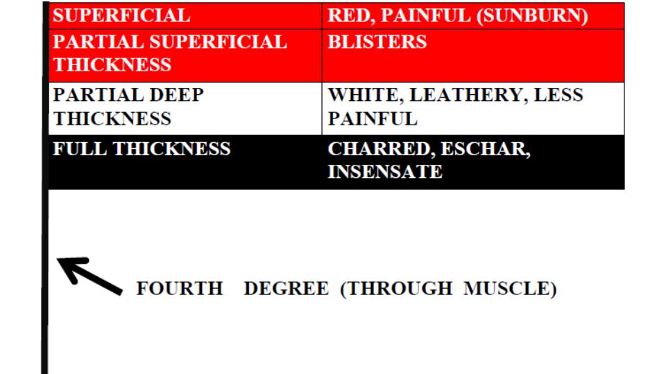
Burn Degree
Burn Degree

Fluid Resuscitation

The Parkland Formula is probably one of the most well known but both this formula and the modified Brooke formula have led to fluid “creep” or over-resuscitation, which may have lasting consequences. Major guidelines and Rosenalli approve another method, the “Rule of Tens.”

Rule of Tens

  • Estimate burn size (TBSA) to the nearest 10%.
  • Multiply %TBSA x 10 = Initial fluid rate in mL/hr (for adult patients weighing 40 kg to 80 kg).
  • For every 10 kg above 80 kg add 100 mL/hr to the rate
  • Limitation: Overestimates fluids 100 [Alvarado]

Consider Referral to Burn Center

Bad Burn

  • Partial thickness burns > 10% TBSA
  • Full thickness burns
  • Electrical burns or chemical burns
  • Inhalation injury

Bad Place – Burns in the sensitive places such as face or genitals or important areas such as hands or major joints .

Bad Protoplasm – Preexisting medical disorders that could complicate management or prolong recovery (such as immunocompromised HIV patients).

Generously Donated Rosh Review Questions

Question 1. [polldaddy poll=8816037]

Question 2. A 52-year-old woman is brought to the emergency department with burns from a house fire. Physical exam reveals superficial burns over her entire left arm and partial-thickness and full-thickness burns covering her entire right arm, her anterior right leg and anterior trunk. [polldaddy poll=8824315]

Answers

1. C. Burn classification is based on burn depth. Second-degree burns are classified into superficial and deep partial-thickness burns. Deep partial-thickness burns extend into thereticular dermis. Skin color is usually a mixture of red and blanched white, and capillary refill is slow. Blisters are thick-walled and commonly ruptured and the skin may appear leathery white. Two-point discrimination may be diminished, but pressure and pinprick applied to the burned skin can be felt. Whereas superficial partial-thickness burns usually re-epithelialize 7-10 days after injury; so the risk of hypertrophic scarring is very small. For deep partial-thickness burns, tissue may undergo spontaneous epithelialization from the few viable epithelial appendages at this deepest layer of dermis and heal within 3-6 weeks. Because these burns have less capacity for re-epithelializing, a greater potential for hypertrophic scar formation exists. In deep partial-thickness burns, treatment with topical antimicrobial dressings is necessary to prevent infection as the burn wound heals. Contraction across joints, with resulting limitation in range of motion, is a common sequela. Splash scalds often cause second-degree burns.

2. B. The extent of burn size in this patient is 36% total body surface area. A thorough and accurate estimation of burn size is essential to guide therapy and to determine when to transfer a patient to a burn center. The extent of burns is expressed as the total percentage of body surface area. Superficial burns are not included in the burn assessment. For adult assessment, the most expeditious method to estimate total percentage of body surface area is the Rule of Nines. This method only takes into account partial-thickness and full-thickness burns. Each leg represents 18% total percentage of body surface area; each arm represents 9% total percentage of body surface area; the anterior and posterior trunk each represent 18% total percentage of body surface area; and the head represents 9% total percentage of body surface area. This patient has partial-thickness and full-thickness burns covering her entire right arm, her anterior right leg and anterior trunk, which calculates to 36%. The superficial burn on her left arm is not included in the calculation. According to the Rule of Nines, the percentage in this patient can be calculated as follows: entire right arm = 9%; anterior right leg = 9%; anterior trunk 18%. 9+9+18= 36%.

References:

1. Mlcak RP, Suman OE, Herndon DN. Respiratory management of inhalation injury. Burns. 2007;33:(1)2-13. [pubmed]

2. Kashefi NS, Nathan JI, Dissanaike S. Does a Nebulized Heparin/N-acetylcysteine Protocol Improve Outcomes in Adult Smoke Inhalation? Plast Reconstr Surg Glob Open. 2014;2:(6)e165. [pubmed]

3. Elsharnouby NM, Eid HE, Abou Elezz NF, Aboelatta YA. Heparin/N-acetylcysteine: an adjuvant in the management of burn inhalation injury: a study of different doses. J Crit Care. 2014;29:(1)182.e1-4. [pubmed]

Episode 26 – The Spinal Cord

(ITUNES OR Listen Here)

The Free Open Access Medical Education (FOAM)

In January 2015, ACEP recommended against the use of long backboards by EMS, “Backboards should not be used as a therapeutic intervention or as a precautionary measure either inside or outside the hospital or for inter-facility transfers.”

We review the use of longboards and cervical collars for spinal immobilization using posts by Thomas D of ScanCrit (Curse of the Cervical Collar, Cervical Collar RIP,  Cervical Collars Slashed From Guidelines), a post by Dr. Minh Le Cong from PHARM, and this Medest118 post.

The bottom line:

  • The benefits of devices to aid in spinal immobilization such as cervical collars and long backboards are controversial.  Guidelines and protocols are continuing to recommend judicious use of these devices.  Examples include:
    • Clearing collars in obtunded blunt trauma patients with negative high quality CT [EAST]
    • Selective application of cervical collars [ILCOR]
    • No backboards and selective pre-hospital immobilizaiton [ACEP]

The Bread and Butter

We differentiate between spinal shock and neurogenic shock, cover the incomplete cord syndromes (anterior cord, central cord, Brown-Sequard Syndrome), and fly through some of the cover using Tintinalli (7e) Chapter 255; Rosen’s (8e) Chapter 43, 106  But, don’t just take our word for it.  Go enrich your fundamental understanding yourself.

Spinal shock – Reduced reflexes following think of this as a stunning of the spinal cord.

Neurogenic shock –  This is loss of sympathetic innervation from injury to the cervical or thoracic spine, typically from a cervical or upper thoracic spinal cord injury, resulting in bradycardia and hypotension.

  • Warm, peripherally vasodilated , and hypotensive  from loss of sympathetic arterial tone with a relative bradycardia from unopposed parasympathetic (vagal) tone
  • Typically presents within 30 minutes, can last 6 weeks
  • Diagnose only after excluding other sources of shock
  • Treatment: crystalloid, vasopressors

Incomplete Cord Syndromes

 Better prognosis than complete cord syndromes. Means there is some sensory or motor preserved distal to lesion (i.e. rectal tone or perineal sensation)

Anterior Cord Syndrome 

  • Complete loss of motor, pain, and temperature below but retain posterior columns (position and vibration)
  • Flexion injury or decreased perfusion (aortic surgery or injury)
  • Paralysis and hypalgesia below the level of injury with preservation of posterior column (position and vibration)

Central Cord Syndrome

  • Sensory and motor deficit, often associated with hyperextension injuries (think whiplash)
  • Affects arms>legs
  • Think MUDE (pronounced muddy): Motor, Upper, Distal, Extension (injury)

Brown-Sequard Syndrome

  • Classically associated with a stab wound
  • Loss of motor function and position and vibration on ipsilateral side with contralateral loss of pain and temperature (fibers cross)

 Reflex Review

C4 Spontaneous breathing: “3-4-5 keep the diaphragm alive”
C5 Shoulder shrug
C6 Flexion at elbow:  think flexing your elbow up to drink before…
C7 Extension at elbow: …extending it to set a drink down.
C8-T1 Flexion of fingers
T1-T12 Intercostal and abdominal muscles
L1-L2 Flexion at hip
L3 Adduction at hip
L4 Abduction at hip
L5 Dorsiflexion of foot
S1-S2 Plantar flexion of foot
S2-S4 Rectal sphincter tone: “2-3-4 keeps your junk off the floor”

Generously Donated Rosh Review Questions 

Question 1. A patient arrives to the ED 15 minutes after being involved in a MVC. He is conscious, and there is no obvious trauma. He is immobilized on a long spine board with a cervical collar in place. His BP is 60/40 mm Hg and HR is 60 bpm. His skin is warm.

[polldaddy poll=8775757]

Question 2. [polldaddy poll=8776250]

Answers

1.  A. Loss of deep tendon reflexes is expected. Neurogenic shock occurs after an injury to the spinal cord. Sympathetic outflow is disrupted resulting in unopposed vagal tone. The major clinical signs are hypotension and bradycardia. Patients are generally hypotensive with warm, dry skin because the loss of sympathetic tone impairs the ability to redirect blood flow from the periphery to the core circulation. The most commonly affected area is the cervical region, followed by the thoracolumbar junction, the thoracic region, and the lumbar region. The anatomic level of the injury to the spinal cord impacts the likelihood and severity of neurogenic shock. Injuries above the T1 level have the capability of disrupting the spinal cord tracts that control the entire sympathetic system leading to the loss of deep tendon reflexes.Neurogenic shock must be differentiated from “spinal” shock, which refers to neuropraxia (B) associated with incomplete spinal cord injuries. This state is transient (C) and resolves in 1 to 3 weeks. Alpha-1 vasopressors (e.g., phenylephrine), in addition to dopamine, norepinephrine, and epinephrine (D), should be used to maintain blood pressure and ensure organ perfusion.

2. C.  In the anterior spinal cord syndrome, just the posterior columns are preserved and so patients lose all pain and temperature sensation as well as motor function. Most cases of anterior cord syndrome follow aortic surgery, but it has also been reported in the setting of hypotension, infection, vasospasm, or anterior spinal artery ischemia or infarct. In trauma, typically hyperflexion of the cervical spine causes the injury to the spinal cord.

Loss of all motor and sensory function (B) occurs with a complete transection of the spinal cord. Most commonly this occurs after a significant trauma. Isolated motor function loss (A) is not a classic syndrome and would result from a small area of injury on the cord just involving the corticospinal tract. Upper greater than lower motor weakness occurs (D) with a central cord syndrome. Sensory involvement is variable although burning dysesthesias in the upper extremities may occur. Most commonly the syndrome occurs after a fall or motor vehicle accident.

FOAMcastini – Kappa

(ITUNES OR LISTEN HERE)

Reading papers is hugely important but learning how to read medical literature is an entirely different realm and many of us have statsphobia.

The Free Open Access Medical Education (FOAM)

Kappa – It’s Greek To Me

The Bread and Butter

Kappa – a coefficient indicating the degree of inter-rater reliability.  How reliability are people getting the same result for a certain test or evaluation?

  • For example, you would want two people looking at the same chest x-ray to agree on the presence or absence of an infiltrate. Sometimes, chance comes into play and kappa tries to account for this.  Similarly, clinical decision aids are often comprised of various historical and physical features.  It would be nice if different clinicians evaluating the same patient would turn up the same results (thereby yielding the decision aid consistent and reliable).

To see how to calculate kappa, check this out.

The value of kappa ranges from -1 (perfect disagreement that is not due to chance) to +1 (perfect agreement that IS due to chance).  A value of 0 means than any agreement is entirely due to chance [1-2].

kappa

People debate over what a “good” kappa is.  Some say 0.6,  some say 0.5 [1-2]. In the PECARN decision aid, for example, the authors only included variables with a kappa of 0.5 [3-4].

Limitations:

  • Prevalence – if prevalence is high, chance agreement is also high.  Kappa takes into account the prevalence index; however, raters may also be predisposed to not diagnose a rare condition, so that the prevalence index provides only an indirect indication of true prevalence, altered by rater behavior [6].
  • The raters – agreement may vary based on rater skill, experience, or education.  For example, when PECARN variables were looked at between nurses and physicians, the overall kappa for “low risk” by PECARN was 0.32, below the acceptable threshold as this number suggests much of the agreement may be due to chance [6].
  • Kappa is based on the assumption that ratings are independent (ie a rater does not know the category assigned by a prior rater).

References
1. Viera AJ, Garrett JM. Understanding interobserver agreement: the kappa statistic. Fam Med. 2005;37:(5)360-3.

2. McGinn T, Wyer PC, Newman TB, et al. Tips for teachers of evidence-based medicine: 3. Understanding and calculating kappa. CMAJ. 2004;171 (11)

3.Kuppermann N, Holmes JF, Dayan PS, et al. Identification of children at very low risk of clinically-important brain injuries after head trauma: a prospective cohort study. Lancet. 2009;374:(9696)1160-70. [pubmed]

4. Gorelick MH, Atabaki SM, Hoyle J, et al. Interobserver agreement in assessment of clinical variables in children with blunt head trauma. Acad Emerg Med. 2008;15:(9)812-8.

5. Nigrovic LE, Schonfeld D, Dayan PS, Fitz BM, Mitchell SR, Kuppermann N. Nurse and Physician Agreement in the Assessment of Minor Blunt Head Trauma. Pediatrics. 2013.

6. de Vet HC, Mokkink LB, Terwee CB, Hoekstra OS, Knol DL.  Clinicians are right not to like Cohen’s κ 2013;346:f2125

Episode 25 – Skin and Skin Structure Infections

(ITUNES OR LISTEN HERE)

The Free Open Access Medical Education (FOAM)

We review the post by Bryan Hayes, PharmD, FAACT on Academic Life in Emergency Medicine,  Sulfamethoxazole-Trimethoprim for Skin and Soft Tissue Infections: 1 or 2 Tablets BID?

The Take Home: Most abscesses do not need antibiotics after incision and drainage.  If the patient has systemic signs (fever, tachycardia), co-morbidities, or a concurrent cellulitis, they may need antibiotics.  Sulfamethoxazole-trimethoprim (SMX-TMP / Bactrim) is one of the most commonly used agents in this case, as it covers MRSA.

Dosing: Two double strength (DS) tablets twice daily is a commonly prescribed regimen; yet,1 DS tablet twice daily is sufficient in most cases with exceptions for patients >100 kg, immunocompromised, or in trauma [1]. ; however, this increases the likelihood of adverse events (nausea, hyperkalemia) without notable substantial positive return.

The Bread and Butter

We cover cellulitis and abscesses, necrotizing infections, and Erythema Multiforme/Stevens-Johnson Syndrome/Toxic Epidermal necrolysis using Tintinalli (7e) Chapter; Rosen’s (8e) Chapter 137 as well as the IDSA guidelines.  But, don’t just take our word for it.  Go enrich your fundamental understanding yourself.

Cellulitis/Abscess

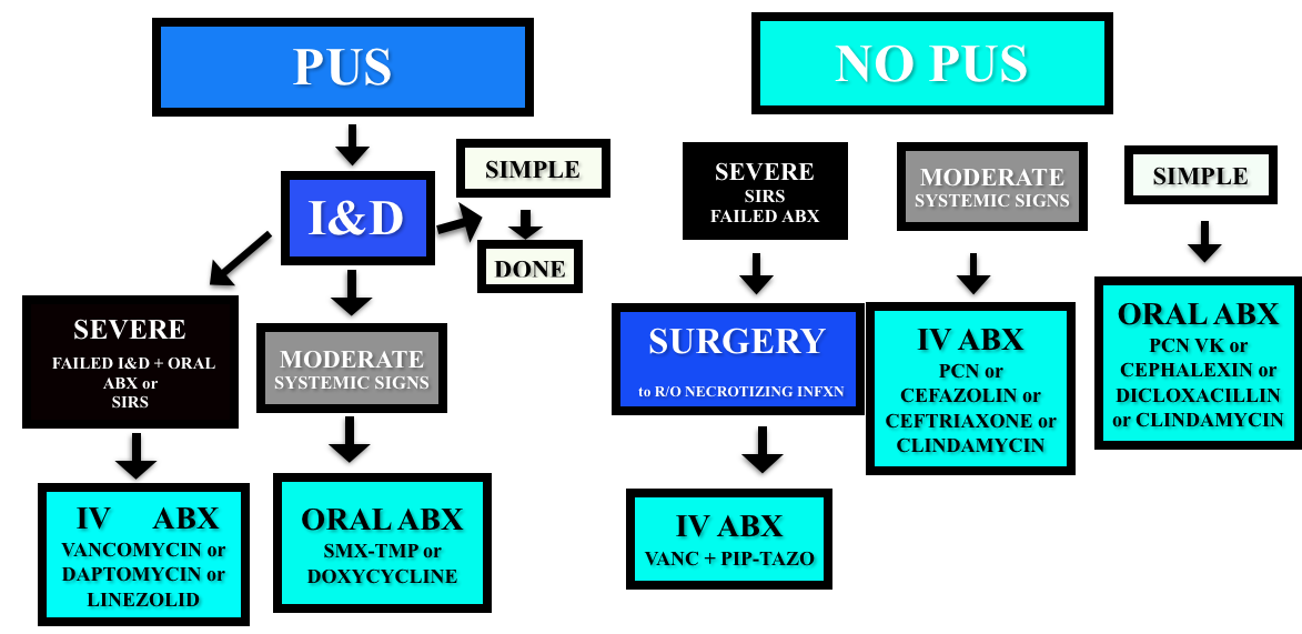
Non-purulent – cover for strep (penicillin, cephalexin/cefazolin).  Even in areas with high incidences of community MRSA, the recommendations for non-purulent skin infections is strep coverage. Five days of treatment is probably enough, and IDSA and Rosenalli approved (see this post)

Purulent – Incise & Drain (I&D).  Cover for MRSA in patients that failed initial I&D or those with systemic signs.

Adapted from the 2014 IDSA Guidelines

Necrotizing Infections

Presentation – pain out of proportion to exam findings, abnormal tachycardia (particularly out of proportion to degree of fever). Crepitus is not reliable (present in 13-30% of patients).

Diagnosis – clinical. X-rays, CT, and MRI have all been used but both x-rays and CT lack sensitivity and it’s probably not a good idea to send a sick patient to MRI.  The gold standard diagnosis is operative findings.

  • The LRINEC score was derived to aid in diagnosis using lab values (sodium, creatinine, white blood cell count, hemoglobin, glucose, CRP) but has not been sufficiently predictive in validation attempts [10].

Classification

  • Type I – polymicrobial. Common in diabetics, immunocompromised.
  • Type II – monomicrobial (often Group A strep or clostridia).
  • Type III? – vibrio, but apparently this is controversial.

Treatment

  • Intravenous fluid and general resuscitation, surgical consult, and antibiotics.

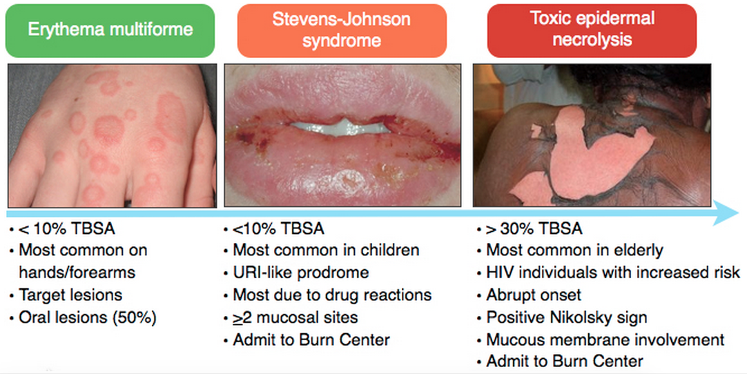
Erythema Multiforme/Stevens-Johnson/Toxic Epidermal Necrolysis (for more, see this Hippo EM podcast)

Courtesy of Rosh Review

Medications most associated with SJS/TEN:  Antibiotics (sulfa), Anti-epileptics, Nonsteroidals

Treatment of SJS/TEN:  Fluid resuscitation, Burn Center/ICU care, and IVIG

Generously Donated Rosh Review Questions 

Question 1. A 22-year-old woman presents to the emergency department with a painful rash. She has had several days of malaise, arthralgias, and low-grade fever, and today developed diffuse painful erythema across her body which is beginning to blister. She takes no medications, but reports completing a course of trimethroprim-sulfamethoxazole one week ago for a urinary tract infection. Examination reveals diffuse tender erythema over her trunk and extremities with multiple ill-defined large bullae, some of which have ruptured, leaving large areas of denuded skin behind. Oral ulcerations are also noted. [polldaddy poll=8689023]

Question 2.  A 54-year-old man with diabetes presents with severe leg pain. The pain has worsened over the last two days with increased swelling of the calf. He has no chest pain or shortness of breath. Vital signs are: T 101.8°F, BP 98/62, HR 118, RR 18. Physical examination is notable for erythema of the calf, mild tenderness, and crepitus. You initiate IV fluids and broad-spectrum antibiotics. [polldaddy poll=8689082]

Answers

1. This patient has toxic epidermal necrolysis (TEN). TEN is an acute inflammatory process characterized by tender erythema, painful bullae formation, and subsequent exfoliation. It often begins with prodromal symptoms such as fever, malaise, and myalgias. TEN is considered a dermatologic emergency and patients may appear toxic on presentation. Medications (within the first few months of administration) are the most common cause, with sulfa and penicillin antibiotics, anticonvulsants, and oxicam non-steroidal anti-inflammatory drugs commonly implicated. Management of TEN involves admission to a burn unit, fluid resuscitation, and prevention of secondary infection. Steroids are not an indicated treatment. Staphylococcal scalded skin syndrome (C) occurs primarily in infants and young children. Infection with exotoxin-producing Staphyloccus aureus leads to diffuse erythroderma and subsequent exfoliation. Mucous membranes are not usually involved. Treatment is fluid resuscitation and antibiotics. Infection with HSV-1 and HSV-2 results in localized skin infection, though in patients with underlying immunosuppression or malignancy it may lead to disseminated herpes simplex virus infection (A), characterized by diffuse vesicles and ulcerations and multisystem involvement. Photosensitive drug reactions (B) are characterized by confluent erythema, macules, papules, or sometimes vesicles in sun-exposed areas such as the face, neck, and arms, occurring within 1-3 weeks of the patient taking an offending agent.  Medications commonly associated with photosensitivity include sulfonamides, thiazides, furosemide, and fluoroquinolones

2. Necrotizing fasciitis is an infection of the subcutaneous tissue that spreads rapidly across the fascial planes and is often fatal even with aggressive treatment. Risk factors for necrotizing infections include diabetes, vascular insufficiency, and immunosuppression. Classically patients have pain out of proportion to examination. Later findings include diffuse swelling, erythema, induration and crepitus. The gold standard for diagnosis is direct visualization in the operating room by a surgeon. Surgeons may elect to perform a bedside biopsy prior to full exploration. Management includes aggressive IV hydration, broad-spectrum antibiotics, and surgical debridement. CT scan with intravenous contrast of the lower extremity (A) may demonstrate findings suggestive of a necrotizing infection including subcutaneous gas, stranding along the fascial planes or fluid collection. However, the negative predictive value of CT scan has not been quantified and is not yet considered the gold standard. Doppler ultrasound of the lower extremity (B) may be helpful in identifying venous thrombosis as a cause of edema and fullness of the leg. Additionally, sonography may visualize an area of deep fluid collection and may demonstrate artifact from a significant amount of subcutaneous air if present. Measurement of serum lactate and CPK (C) is helpful when positive but not sensitive enough to rule out the diagnosis of necrotizing fasciitis. The Laboratory Risk Indicator for Necrotizing Fasciitis (LRINEC) incorporates other laboratory markers (CRP, WBC, Hemoglobin, Sodium, Creatinine, Glucose) into a decision-rule however lacks sufficient sensitivity in larger studies.

References

1.Stevens DL, Bisno a. L, Chambers HF, et al. Practice Guidelines for the Diagnosis and Management of Skin and Soft Tissue Infections: 2014 Update by the Infectious Diseases Society of America. Clin Infect Dis. 2014.

2.  Shehab N1, Patel PR, Srinivasan A, Budnitz DS. Emergency department visits for antibiotic-associated adverse events. Clin Infect Dis. 2008 Sep 15;47(6):735-43.

3.  Bourgois FT, Mandl KD, Valim C et al.  Pediatric Adverse Drug Events in the Outpatient Setting: An 11-Year National Analysis. Pediatrics. Oct 2009; 124(4): e744–e750.
4. Goldman JL, Jackson MA, Herigon JC et al.  Trends in Adverse Reactions to Trimethoprim-Sulfamethoxazole.  Pediatrics. Jan 2013; 131(1): e103–e108.

5. Khow KS, Yong TY. Hyponatraemia associated with trimethoprim use. Curr Drug Saf. 2014;9:(1)79-82. [pubmed]

6.Antoniou T, Hollands S, Macdonald EM, et al. Trimethoprim-sulfamethoxazole and risk of sudden death among patients taking spironolactone. CMAJ. 2015.

7. Hepburn MJ, et al. Comparison of short-course (5 days) and standard (10 days) treatment for uncomplicated cellulitis.  Arch Intern med 2004; 164, 1669-1674

8. Hurley HJ, Knepper BC, Price CS, et al. Avoidable antibiotic exposure for uncomplicated skin and soft tissue infections in the ambulatory care setting. Am J Med. 2013;126(12):1099–106.

9. Pallin DJ, Binder WD, Allen MB et al.  Clinical Trial: Comparative Effectiveness of Cephalexin Plus Trimethoprim-Sulfamethoxazole Versus Cephalexin Alone for Treatment of Uncomplicated Cellulitis: A Randomized Controlled Trial.  Clin Infect Dis. (2013) 56 (12): 1754-1762.

10.Liao CI, Lee YK, Su YC et al. Validation of the laboratory risk indicator for necrotizing fasciitis (LRINEC) score for early diagnosis of necrotizing fasciitis. Tzu Chi Medical Journal (2012) 24(2):73-76

Episode 24 – Mild Traumatic Brain Injury/Concussion

(ITUNES OR LISTEN HERE)

The Free Open Access Medical Education (FOAM)

We cover the Taming the SRU podcast, “Ketamine Cagematch” (iTunes), a debate between Dr. Minh Le Cong and Dr. Chris Zammit.

Dogma persists that ketamine may increase intracranial pressure, which would be bad in traumatic brain injury (TBI) given the fixed space in the cranial vault.  These are largely from Yet, these patients often need sedation, for agitation or intubation, and drops in blood pressure are also deleterious (see EMCrit on neuroprotective intubation).

PRO (Le Cong): The literature doesn’t show clinically significant deleterious outcomes from ketamine use in the head injured patient.  Review in Annals on ketamine and ICP.  Deleterious effects of apnea may result from other sedative agents.

CON (Zammit): Studies showing that ketamine does not increase ICP confounded by the presence of other sedatives on board.  As a result, there may still be a risk to using ketamine in these patients.

The Bread and Butter

We cover key points on concussion and mild TBI from Tintinalli 254 but to be honest, Rosenalli is lacking on this topic so we’ve turned to the ACEP clinical policy, AAN guidelines, Ontario Pediatric Guidelines, and the  AAP guidelines on the topic.  But, don’t just take our word for it.  Go enrich your fundamental understanding yourself.

Mild TBI and concussion are often referred to interchangeably and the definition varies [1-3].  Unfortunately, concussion is often thought of only in the sports population and not all-comers to the Emergency Department (ED) so patients may not receive proper .

Definition: Essentially, any alteration in mental status following head injury. Per the CDC:  Head injury from blunt trauma or acceleration/deceleration with GCS 14-15 PLUS

  • observed or self-reported transient confusion, disorientation, or impaired consciousness
  • Amnesia around the time of injury
  • Signs of other neurologic or neuropsychological dysfunction
  • Any loss of consciousness (LOC) less than 30 minutes (!) [3]

For more on how reliable LOC is in these patients, see this post.

Causes: Falls and motor vehicle collisions are the most common causes in adults, whereas sports are the more common in kids.

  • The effects on the brain are largely a result of “secondary injury,” consistent of alterations in ion-channel, metabolic pathways, and electrochemical imbalances.  These are more functional than structural.

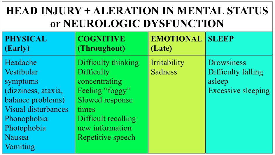
Symptoms: There’s a good bit of overlap with migraine, more significant head trauma, and symptoms that may be confused for behavioral issues in younger populations.

Screen Shot 2015-02-19 at 1.27.09 PM

Testing: Not all head injuries require imaging.  Certainly, patients with focal neurologic findings warrant imaging; yet, in other cases, validated decision aids exist to help determine which patients may or may not need imaging.  In adults, we like the Canadian CT Head tool , although the ACEP clinical policy uses the New Orleans Criteria.  For a solid review of the two, check out this SGEM episode.  In children, consider the use of the PECARN decision aid. Otherwise, a good neurologic exam and observation should suffice.

Something to consider – patients often believe that a “normal” CT scan of the head means that they don’t have a concussion.  As a result, they may not take concussion precautions seriously.  In this case, imaging may provide false reassurance.

There’s increased attention on biomarkers like GFAP, total tau, and S-100B but these are not ready for prime time and are not incredibly specific [3].  Further, standardized assessment tools such as SCAT3 and ACE may be useful, but are used predominantly on-site for sports related incidents.

Treatment

  • Education – this is one of the biggest areas in which ED providers may make a difference. Give patients or family members precautions for concussion, even if the injury or mechanism seems relatively mild.  Studies show that a majority of pediatric patients do not follow up as instructed after a concussion [4].  Perhaps communication of the the potential gravity of concussion and long-term implications may improve follow up.
  • Rest – This the is the mainstay of initial treatment for most mild TBI [1-3, 5].  It’s unclear how strict this rest needs to be, but a recent study in the pediatric literature found no benefit to strict rest versus standard instructions (1-2 days rest + graduated return) [6].
  • Graduated return to activity – after a period of rest, it’s recommended to slowly resume activities, spending at least 24 hours at each level of increased activity.  If an individual gets symptomatic, they should return to the level of activity at which they were asymptomatic (see the Zurich protocol).
  • Return to play – clearing people from the ED is NOT a good idea.  For those on the sidelines, a player should be removed from activity for the day after a suspected concussion.

Why do we care in the ED?

  • Quality of Life – Concussive symptoms can be quite disabling.  Giving patients a name for their symptoms, resources, and education may help them understand the process of recovery and available resources.
  • Second Impact Syndrome – A second head injury in a patient symptomatic from a concussion may experience diffuse cerebral edema, possibly from loss of cerebral autoregulation.* [2]
  • Post Concussion Syndrome (PCS) – a subset of the concussion population will have persistence of symptoms which, if they persist > 1 week to 3 months after the injury, is deemed PCS. *  This is thought to be more prevalent in individuals with a history of depression, anxiety, or migraines and in those with more severe ED presentations [2].
  • Bouncebacks – Patients may present to the ED with symptoms of concussion but may not give a history of significant trauma.  Remember that concussion may occur after a seemingly minor bump to the head and elderly may need extra support with activities of daily living or may bounce back with a more life threatening injury.
  • Lack of follow up.  While some may follow up with concussion specialists, the majority of our patient population lacks the ability for meaningful or specialist follow up.  We can’t assume that someone else is going to guide our patients through concussion recovery or that when we write “follow  up with _____” that it will happen.  Our care may be it, make it good.

*The literature on these is sparse and complicated by variable definitions, information, and standardized reporting.

 Generously Donated Rosh Review Questions (scroll for answers)

Question 1. A 33-year-old man with no past medical history presents with a headache 3 days after a closed head injury. The patient states that he stood up from kneeling and hit the top of his head on a wood cabinet. There was no loss of consciousness or seizure activity. In addition to the headache, he complains of difficulty concentrating at work and dizziness. His physical examination is unremarkable.[polldaddy poll=8663736]

Question 2. A 42-year-old man is brought to the ED after he tripped and fell while he and his wife were on a walk. His wife notes that she saw him hit his head on the pavement and that he did not respond to her for 45 seconds. When he started to respond, she says that he was very confused. In the ED, his vital signs are BP 135/75, HR 88, RR 14, and oxygen saturation 98% on room air. On exam, you note some minor lacerations on the patient’s upper extremities, face, and scalp and his GCS is 15. As the wife recounts what happened, the patient does not recall any of the events and continuously asks to repeat what happened to him. A CT scan of the brain is normal. The patient is diagnosed with a concussion and is ready to be discharged from the ED. [polldaddy poll=8663741]

References

1. Tavender EJ, Bosch M, Green S, et al. Quality and consistency of guidelines for the management of mild traumatic brain injury in the emergency department. Acad Emerg Med. 2011;18:(8)880-9. [pubmed]

2. Haydel M.  Management Of Mild Traumatic Brain Injury In The Emergency Department. Emergency Medicine Practice.  September 2012 Volume 14, Number 9

3. Jagoda AS, Bazarian JJ, Bruns JJ, et al. Clinical policy: neuroimaging and decisionmaking in adult mild traumatic brain injury in the acute setting. Ann Emerg Med. 2008;52:(6)714-48. [pubmed]

4.Hwang V, Trickey AW, Lormel C, et al. Are pediatric concussion patients compliant with discharge instructions? J Trauma Acute Care Surg. 2014;77(1):117–22; discussion 122.

5. Brown NJ, Mannix RC, O’Brien MJ, Gostine D, Collins MW, Meehan WP. Effect of cognitive activity level on duration of post-concussion symptoms. Pediatrics. 2014;133(2):e299–304.

6.Thomas DG, Apps JN, Hoffmann RG, McCrea M, Hammeke T. Benefits of strict rest after acute concussion: a randomized controlled trial. Pediatrics. 2015;135:(2)213-23. [pubmed]

Answers

 1.  D.  The patient presents with minor head trauma and complaints consistent with a concussion and should have neurology follow up arranged. A concussion is a minor TBI that is often seen in MVCs and collision sports (football, hockey). It is typically caused by a rotational injury or an acceleration-deceleration injury. Patients will present with a number of non-specific symptoms including headaches, dizziness, confusion, amnesia, difficulty concentrating, and blurry vision but do not have focal neurologic findings. Despite the absence of severe intracranial injury, patients can have chronic and debilitating symptoms from concussions. Neurology referral is recommended, as patients should have functional testing and tracking of their symptoms for resolution. It is vital to counsel patients to avoid contact sports or activities that increased the risk of recurrent injury as these patients are at risk for more severe injury with second impact.In the absence of focal neurologic findings, absence of antiplatelet or anticoagulant use and minor trauma, imaging is not needed (A, B, C).

2. Cerebral concussions are clinically characterized by headaches, confusion, dizziness, and amnesia for the event. Concussions are characterized by a transient loss of consciousness that occurs immediately following blunt, nonprenetrating head trauma, caused by impairment of the reticular activating system. Concussions present without focal neurologic deficits, and CT and MRI show no acute abnormalities. Although not commonly performed for concussions, functional imaging, such a PET scan, may show changes in blood flow and glucose uptake. It is critical to inform the patient and his or her family of the second impact syndrome (SIS), which occurs when a patient suffers a second concussion after being symptom-free from the first. Although SIS is more common in sports based injuries, especially among teenagers, the risk of serious sequelae following a second concussion is immense. Due to neurochemical and autoregulatory changes that may still be present, a second concussion soon after a first generally produces a rapid neurologic decline that proves fatal. Patients should be told to avoid activities that could cause falls or trauma for at least 1 week after the patient is completely asymptomatic from the first concussion. Most patients with a concussion can be discharged from the emergency department and advised to follow-up with a primary care physician within 1 week (B). Although some patients have only transient symptoms from a concussion, others may experience persistent symptoms termed postconcussive syndrome (PCS).  PCS symptoms most often include headache as well as memory, sensory, sleep, and concentration disturbances. It is important to consider PCS in all patients with a concussion, but a primary care physician is generally able to care for these patients. It is unnecessary to obtain an MRI (C).  Although patients should avoid trauma and falls for at least 1 week after being completely asymptomatic after the first concussion, a change insleeping position (D) is unnecessary.

Episode 23 – SBO and Mesenteric Ischemia

(ITUNES OR LISTEN HERE)

The Free Open Access Medical Education (FOAM)

This week we’re covering Dr. Jacob Avila’s post on ultrasound for small bowel obstruction (SBO)  located at Ultrasound of the week.  He has an accompanying video on 5minSono.

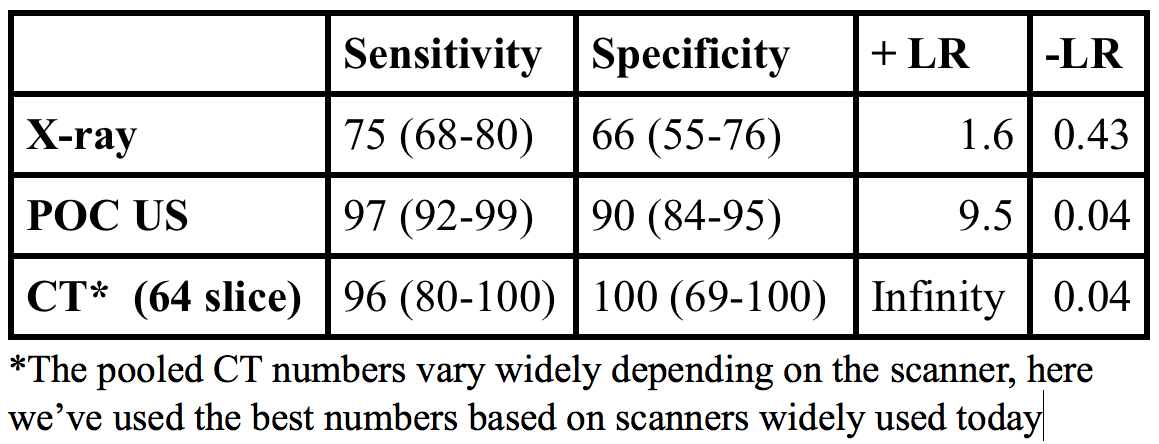
Point of care ultrasound has good operating characteristics for diagnosis of SBO with a LR+ 9.5, LR- 0.04, far better than abdominal x-ray [1].

Screen Shot 2015-01-25 at 10.20.44 AM

What to look for:

  • Dilated loops of bowel > 2.5 cm in diameter
  • Additional clues:  “To and fro” peristalsis
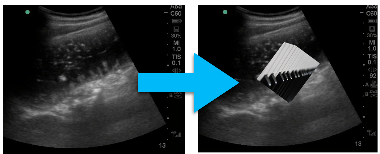
  • The piano key sign, Tanga sign
Piano Key Sign
Piano Key Sign

Problems with abdominal x-ray:

  • Rosen’s: Abdominal x-rays are “diagnostic in approximately 50 to 60% of cases of SBO, equivocal in 20 to 30%, and normal, nonspecific, or misleading in 10 to 20%” [2].
  • American College of Radiology: they can “prolong the evaluation period … while often not obviating the need for additional examinations, particularly CT” [3].

Limitations:

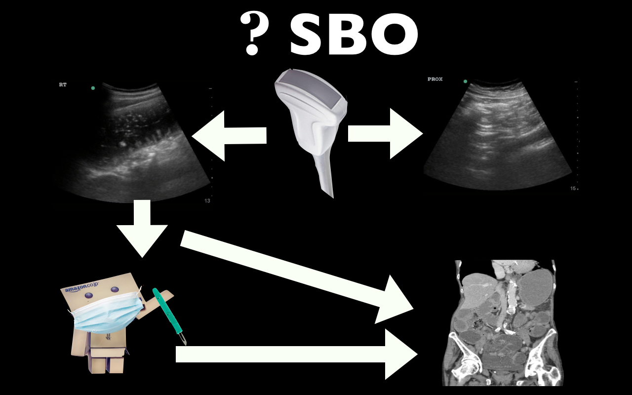
  • While ultrasound can diagnose SBO, there is little evidence to suggest that we can identify transition points or strangulation/necrosis.  As such, there can still be a role for CT scan, particularly in first time SBO to identify a transition point.
  • The EAST guidelines acknowledge the utility of ultrasound yet this practice is far from accepted in the surgical community.  Surgical colleagues will likely still want concrete imaging such as an x-ray or CT; however, ultrasound performed concurrent with the history and physical may speed up patient’s disposition to definitive care/imaging.
Possible algorithm for use of US in SBO

More FOAM on the topic:

The Bread and Butter

We cover key points on SBO and Acute Mesenteric Ischemia from Rosenalli, that’s Tintinalli (7e) Chapter 86; Rosen’s (8e) Chapter 92.  But, don’t just take our word for it.  Go enrich your fundamental understanding yourself.

Small Bowel Obstruction

Etiology of intestinal obstruction: “HANG IV.” Hernia, Adhesions (most common cause), Neoplasm, Gallstone ileus, Intussusception, Volvulus

Treatment

  • Intravenous fluids – resuscitate the patient!
  • Antiemetics.  If a patient is compromising their airway, an aspiration risk, or vomiting despite antiemetics, consider the use of a nasogastric tube.  Shockingly, “use of nasogastric decompression is considered dogma by many emergency physicians and surgeons, its effect in decreasing the duration of SBO has scant support in the medical literature” [2, 5].  This point demonstrates that SBO is not a monolithic disease entity but a spectrum of pathology with variable treatments depending on patient’s sickness.
  • Antibiotics that cover gram-negative and anaerobic organisms
  • Admit. Most of these patients will likely go to the surgical service; however,

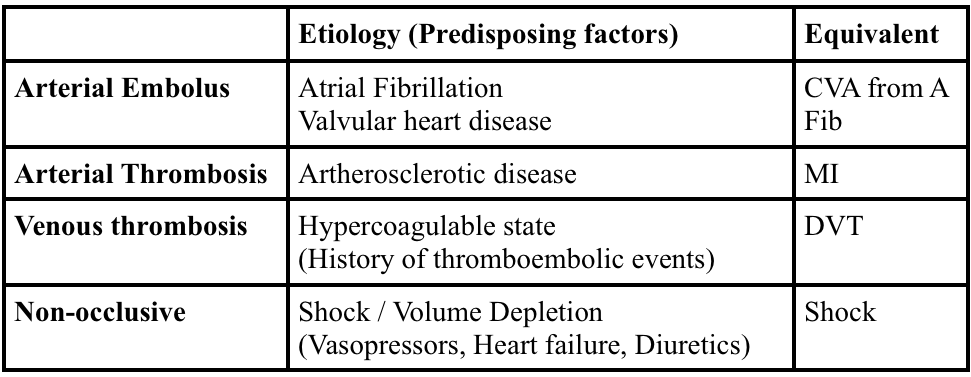
Acute Mesenteric Ischemia

Screen Shot 2015-01-24 at 7.18.02 PM

 Generously Donated Rosh Review Questions (scroll for answers)

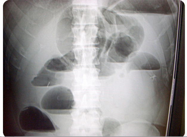
Question 1. A 73-year-old man presents with vomiting and abdominal pain for 2 days. The patient has a remote history of cholecystectomy and appendectomy. Examination reveals a markedly distended abdomen and absent bowel sounds. Lab studies show an elevated WBC count and a lactate of 4.3 mmol/L. An abdominal radiograph is obtained that is shown below. [polldaddy poll=8607202]

Screen Shot 2015-01-26 at 8.40.19 AM

Question 2. An 87-year-old woman presents with worsening abdominal pain over the last 24 hours. She has minimal tenderness on examination but an elevated lactic acid. An abdominal CT Scan demonstrates mesenteric ischemia. [polldaddy poll=8607198]

References:

1. Taylor MR, Lalani N. Adult small bowel obstruction. Acad Emerg Med. 2013;20(6):528–44.

2. Roline CE, Reardon RF.  “Disorders of the Small Intestine.  Rosen’s Emergency Medicine.  8th ed. pp 1216-1224.e2.

3. Ros PR, Huprich JE. ACR Appropriateness Criteria on suspected small-bowel obstruction. J Am Coll Radiol. 2006;3:(11)838-41.

4. Vicario SJ, Price TG.  “Bowel Obstruction and Volvulus.” Tintinalli’s Emergency Medicine: A Comprehensive Study Guide.  7th ed. pp 581-583.

5.  Fonseca AL, Schuster KM, Maung AA, Kaplan LJ, Davis KA. Routine nasogastric decompression in small bowel obstruction: is it really necessary? Am Surg. 2013;79:(4)422-8.

Answers.

1. D. This patient presents with a high-grade small bowel obstruction (SBO) with evidence of bowel ischemia (elevated lactate). Mortality has fallen in the last century with aggressive surgical treatment (from 60% to 5%). The abdominal radiograph above shows multiple air-fluid levels consistent with an SBO. Radiographs are abnormal in 50-60% of cases and are more likely to demonstrate abnormality when the obstruction is high-grade versus partial. Two views (upright and supine or supine and decubitus) should be obtained. Mechanical obstruction refers to the presence of a physical barrier to the flow of intestinal contents. In a simple obstruction, the intestinal lumen is partially or completely obstructed causing intestinal distension proximally but does not cause compromise of the vascular supply. In a closed-loop obstruction, a segment of bowel is obstructed at two sequential sites usually by twisting on a hernia opening or adhesive band leading to compromise of blood flow eventually resulting in bowel ischemia. Ischemia may only be seen on CT scan or occasionally, on laparoscopy or laparotomy. However, an elevated lactate in the setting of an SBO is highly suggestive of intestinal ischemia. The presence of blood in stool (either gross blood or guaiac positive stools) also suggests the presence of ischemia or infarction. When compromise of the vascular supply is suspected, the patient should have an emergent surgical consultation for operative management. Immediate management should also include placement of a nasogastric tube for decompression of the proximal parts of the intestines, intravascular volume resuscitation and intravenous antibiotics when vascular compromise is suspected or confirmed. CT scan of the abdomen and pelvis (A) is considered complimentary to plain films and is more sensitive and specific. Additionally, CT scan can reveal the site and cause of obstruction. However, surgical evaluation of a high-grade SBO should not be delayed for advanced imaging. Colonoscopy (B) is not indicated in small bowel obstruction. There is an increased risk of perforation. An enema and polyethylene glycol (C) is the treatment for constipation, and may worsen the outcome in patients with high-grade bowel obstruction.

2. Arterial emboli account for more than 50% of cases of mesenteric ischemia. The classic presentation of mesenteric ischemia is abdominal pain out of proportion to examination. Most commonly, thrombi develop in the left ventricle or atrium and embolize into the aorta. From the aorta, the emboli pass into one of the branches supplying the circulation to the gut. Thesuperior mesenteric artery is the most common site of embolization because of its large diameter and narrow angle of takeoff from the aorta. Mesenteric ischemia usually involves the small intestine and sometimes the right colon. The large intestine has significantly more collateral flow and is not as susceptible to ischemia. Aortic dissection (A) may lead to mesenteric ischemia depending on the location of the dissection. It is also possible to have a primary dissection of the mesenteric blood supply (e.g. SMA).Primary arterial thrombosis (C) of the mesentery is much less common and arises from progression of underlying atherosclerotic disease. Patients will often have a history of intestinal “angina” or chronic mesenteric ischemia during which symptoms occur after eating when the gut requires additional blood supply which is limited by the atherosclerotic changes. Venous thrombosis (D) is the least common etiology of mesenteric ischemia and most commonly affects the superior mesenteric vein.

FOAMcastini – Reflections on ACEP tPA Clinical Policy Update Draft

(iTunes or Listen Here)

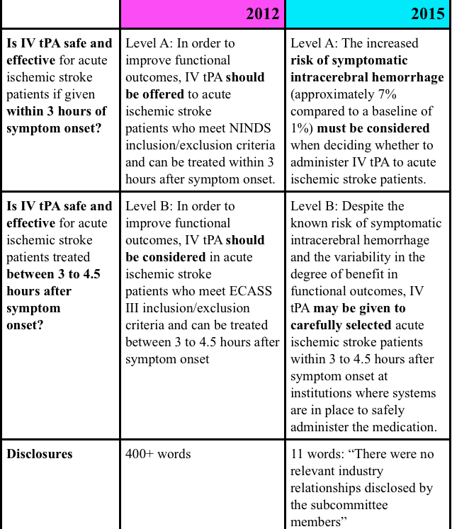
As detailed in this FOAMcastini, ACEP just released a draft of an updated clinical policy on tPA for acute ischemic stroke.  This came in the wake of years of controversy over the aggressive position taken in the 2012 clinical policy.

While FOAMcast is not an interview style podcast, we felt compelled to get some perspective on Emergency Physicians a little more experienced than ourselves.  Here we interview:

Dr. Ryan Radecki (@emlitofnote), Assistant Professor, University of Texas – Houston

  • We don’t know who best benefits from tPA so elucidating which patients are “carefully selected” may get hard.
  • See his response to the policy on his blog here

Dr. David Newman (#draftnewman), Associate Professor of Emergency Medicine, Mount Sinai Hospital

  • The process for ACEP clinical policy creation seems to work.  The constituency expressed concern and the college listened and went back and re-created the policy from the bottom up.
  • This policy reflects a move from content expert to methodologists which better reflects the evidence compared with opinions (and is the standard per USPSTF).

Dr. Anand Swaminathan (@EMSwami), Assistant Professor of Emergency Medicine, NYU

  • Inclusion of more rigorous methodology and review of evidence.
  • May be perceived as too “soft” by tPA supporters.  This policy may not overtly change practice but may open up avenue of conversations.

Dr. Ken Milne (@thesgem), Chief of Staff at South Huron Hospital

  • When looking at “carefully selected” patients, as noted in the policy, remember to use the Evidence Based Medicine trifecta of evidence, patient values, and clinical expertise.  In isolation, one component is not sufficient.
  • Previous discussions of NINDS, and the Cochrane tPA article

FOAMcastini – ACEP tPA Clinical Policy Update

(ITUNES OR LISTEN HERE)

The Free Open Access Medical Education (FOAM)

Comment on the 2015 ACEP tPA Clinical Policy Draft here

ACEP tPA Clinical Policy 2012 – This policy has been entrenched in controversy since it was published.  Why?  Well, largely because the evidence was given a stronger level recommendation than the data supported, conflicts of interest abounded, and the data (mostly from NINDS (The SGEM review), ECASS, and IST-3) were problematic.  This has been well covered by these reviews of the clinical policy:

What changed in the 2015 draft?

Screen Shot 2015-01-13 at 10.22.04 AM

Episode 22 – The Knee

(ITUNES OR LISTEN HERE)

The Free Open Access Medical Education (FOAM)

This week we’re covering a post from the incredible pediatric resource, Don’t Forget the Bubbles, “Knee X-ray Interpretation” by Dr. Tessa Davis.  We use a systematic approach to assessing chest x-rays, so why not knee x-rays?

  •  Know the anatomy
  •  Look at:
    • Effusion
    • Main bones
    • Tibiofemoral alignment
    • Tibial plateaus
    • Intercondylar eminence
    • Patellar tendon disruption
    • Patellar fracture

The Bread and Butter

We summarize some key topics from Rosenalli, that’s Tintinalli (7e) Chapter s271, 281; Rosen’s (8e) Chapters 57, 136.  But, don’t just take our word for it.  Go enrich your fundamental understanding yourself.

Knee Dislocation

  • Anterior is most common (40%), posterior (33%)
  • Approximately 50% of knee dislocations may be relocated upon presentation to the hospital (this does not reduce risk of badness)
  • Most worrisome sequelae = popliteal artery disruption.  Of patients with popliteal disruption, the amputation rate rises to 90% 8 hours after the injury without surgical intervention.
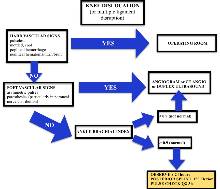
  • Workup may depend on your institution (ex: angiogram vs. CT angio vs. ultrasound) but all patients will need an ABI + 24 hour of pulse checks per current standards.
Screen Shot 2015-01-08 at 1.10.48 PM
Algorithm (adopted from Rosen’s)

Septic Arthritis

  • Most Common Organisms: S. aureus, N. gonorrhea
  • Hematogenous spread
  • Most Common Location: knee, hip

Risk factors such as immunocompromised hosts and use of steroids are risk factors for septic arthritis but the ones with the highest likelihood ratio (LR+ >10 is ideal):

  • Skin infection overlying prosthetic joint (LR+ 15)
  • Joint surgery within the preceding 3 months (LR+ 6.9)
  • Age > 80 (LR+ 3.5)

Diagnosis:  In the red, hot, swollen, painful joint, think septic arthritis.  Clinical and laboratory indicators aren’t great. Synovial fluid analysis, particularly the culture exists as the gold standard.  Arthrocentesis Trick of the Trade from ALiEM. Here are the operating characteristics from Margaretten et al:

  • Fever: Sensitivity 57%
  • Lab tests: White Blood Cell count (WBC), sedimentation rate (ESR), and c-reactive protein don’t perform well
    • WBC LR+  1.4 (1.1-1.8); LR- 0.28 (0.07-1.10)
    • Erythrocyte sedimentation rate 1.3 (1.1-1.8); LR- 0.17 (0.20-1.30)
    • C-reactive protein  1.6 (1.1-2.5); LR- 0.44 (0.24-0.82)
  • Synovial fluid gram stain and culture is the “gold standard.”

Treatment: Intravenous antibiotics and washout of the joint by orthopedics in the operating room

 Generously Donated Rosh Review Questions 

Question 1. A 67-year-old man with a history of gout presents with atraumatic left knee pain. Physical examination reveals an effusion with overlying warmth and erythema. There is pain with passive range of motion. He reports a history of gout in this joint in the past. [polldaddy poll=8568492]

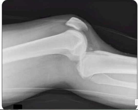
Question 2.  A 27-year-old woman presents with severe left knee pain after an MVC where she was the front passenger. She states her knee hit the dashboard. An X-ray of the patient’s knee is shown below. After reduction, the physical examination reveals swelling of the knee and an Ankle-Brachial Index (ABI) of 0.8. [polldaddy poll=8569540]

Screen Shot 2015-01-12 at 5.13.06 PM

Answers.

1. D. Septic arthritis is a bacterial or fungal infection of a joint typically spread hematogenously unless there is direct bacterial contamination. The synovium is highly vascular and lacks a basement membrane making it susceptible to bacterial seeding. Certain conditions predispose individuals to septic arthritis including diabetes, sickle cell disease, immunocompromise, alcoholism or pre-existing joint disease like rheumatoid arthritis or gout. Fever is present in less than half of cases of septic arthritis so with clinical suspicion an arthrocentesis is indicated. The knee is the most common joint affected and patients have pain (especially on passive range of motion) and decreased range of motion often accompanied by warmth, erythema and fever. This patient may have an acute gouty flare, but the clinician must exclude an infection. On joint fluid analysis, the white blood cell count of a septic joint is typically > 50,000. Indomethacin (B) is a non-steroidal anti-inflammatory agent commonly used in the treatment of acute gout. Gout is an arthritis caused by deposition of monosodium urate monohydrate crystals in the joint space. Acute flares involve a monoarticular arthritis with a red, hot, swollen and tender joint. Acute episodes of gout result from overproduction or decreased secretion of uric acid. However, measurement of serum uric acid (C) does not correlate with the presence of absence of an acute flare. A radiograph of the knee (D) may show chronic degenerative changes associated with gout but will not help to differentiate a gouty arthritis versus septic arthritis.

2. C. Obtain Angiography. This patient presents with a knee dislocation and signs of a popliteal artery injury requiring angiography for diagnosis. A knee dislocation refers to a dislocation of the tibia in relation to the femur and not a patellofemoral dislocation. A tibiofemoral dislocation is a limb-threatening emergency due to the high rate of popliteal artery injury. The neurovascular bundle (popliteal artery, popliteal vein and common peroneal nerve) runs posteriorly in the popliteal fossa. The popliteal artery is tethered to the femur and tibia by a fibrous tunnel and is inherently immobile making it susceptible to injury during dislocation. Knee dislocations typically occur in major trauma. An MVC where the knee strikes the dashboard is a common scenario. The dislocation is usually clinically obvious and should be emergently reduced regardless of the presence of confirmatory X-rays. The leg should rapidly be assessed for any “hard” signs of vascular injury including an absence of pulse, limb ischemia, rapidly expanding hematoma, the presence of a bruit or thrill and pulsatile bleeding. Neurologic status should also be assessed prior to and after reduction. After reduction, all patients should have ankle-brachial index (ABI) performed. A normal ABI is > 0.9. Any patient with an ABI less than this should be further investigated for a popliteal injury with angiography. Splint and elevation (D) may be appropriate once a vascular injury is ruled out. The patient should not be discharged home (A) with an abnormal ABI. Observation and repeat ABI (B) is indicated if the initial ABI is normal.

Episode 22 – The Knee

(ITUNES OR LISTEN HERE)

The Free Open Access Medical Education (FOAM)

This week we’re covering a post from the incredible pediatric resource, Don’t Forget the Bubbles, “Knee X-ray Interpretation” by Dr. Tessa Davis.  We use a systematic approach to assessing chest x-rays, so why not knee x-rays?

  •  Know the anatomy
  •  Look at:
    • Effusion
    • Main bones
    • Tibiofemoral alignment
    • Tibial plateaus
    • Intercondylar eminence
    • Patellar tendon disruption
    • Patellar fracture

The Bread and Butter

We summarize some key topics from Rosenalli, that’s Tintinalli (7e) Chapter s271, 281; Rosen’s (8e) Chapters 57, 136.  But, don’t just take our word for it.  Go enrich your fundamental understanding yourself.

Knee Dislocation

  • Anterior is most common (40%), posterior (33%)
  • Approximately 50% of knee dislocations may be relocated upon presentation to the hospital (this does not reduce risk of badness)
  • Most worrisome sequelae = popliteal artery disruption.  Of patients with popliteal disruption, the amputation rate rises to 90% 8 hours after the injury without surgical intervention.
  • Workup may depend on your institution (ex: angiogram vs. CT angio vs. ultrasound) but all patients will need an ABI + 24 hour of pulse checks per current standards.
Screen Shot 2015-01-08 at 1.10.48 PM
Algorithm (adopted from Rosen’s)

Septic Arthritis

  • Most Common Organisms: S. aureus, N. gonorrhea
  • Hematogenous spread
  • Most Common Location: knee, hip

Risk factors such as immunocompromised hosts and use of steroids are risk factors for septic arthritis but the ones with the highest likelihood ratio (LR+ >10 is ideal):

  • Skin infection overlying prosthetic joint (LR+ 15)
  • Joint surgery within the preceding 3 months (LR+ 6.9)
  • Age > 80 (LR+ 3.5)

Diagnosis:  In the red, hot, swollen, painful joint, think septic arthritis.  Clinical and laboratory indicators aren’t great. Synovial fluid analysis, particularly the culture exists as the gold standard.  Arthrocentesis Trick of the Trade from ALiEM. Here are the operating characteristics from Margaretten et al:

  • Fever: Sensitivity 57%
  • Lab tests: White Blood Cell count (WBC), sedimentation rate (ESR), and c-reactive protein don’t perform well
    • WBC LR+  1.4 (1.1-1.8); LR- 0.28 (0.07-1.10)
    • Erythrocyte sedimentation rate 1.3 (1.1-1.8); LR- 0.17 (0.20-1.30)
    • C-reactive protein  1.6 (1.1-2.5); LR- 0.44 (0.24-0.82)
  • Synovial fluid gram stain and culture is the “gold standard.”

Treatment: Intravenous antibiotics and washout of the joint by orthopedics in the operating room

 Generously Donated Rosh Review Questions 

Question 1. A 67-year-old man with a history of gout presents with atraumatic left knee pain. Physical examination reveals an effusion with overlying warmth and erythema. There is pain with passive range of motion. He reports a history of gout in this joint in the past. [polldaddy poll=8568492]

Question 2.  A 27-year-old woman presents with severe left knee pain after an MVC where she was the front passenger. She states her knee hit the dashboard. An X-ray of the patient’s knee is shown below. After reduction, the physical examination reveals swelling of the knee and an Ankle-Brachial Index (ABI) of 0.8. [polldaddy poll=8569540]

Screen Shot 2015-01-12 at 5.13.06 PM

Answers.

1. D. Septic arthritis is a bacterial or fungal infection of a joint typically spread hematogenously unless there is direct bacterial http://www.mindanews.com/buy-imitrex/ contamination. The synovium is highly vascular and lacks a basement membrane making it susceptible to bacterial seeding. Certain conditions predispose individuals to septic arthritis including diabetes, sickle cell disease, immunocompromise, alcoholism or pre-existing joint disease like rheumatoid arthritis or gout. Fever is present in less than half of cases of septic arthritis so with clinical suspicion an arthrocentesis is indicated. The knee is the most common joint affected and patients have pain (especially on passive range of motion) and decreased range of motion often accompanied by warmth, erythema and fever. This patient may have an acute gouty flare, but the clinician must exclude an infection. On joint fluid analysis, the white blood cell count of a septic joint is typically > 50,000. Indomethacin (B) is a non-steroidal anti-inflammatory agent commonly used in the treatment of acute gout. Gout is an arthritis caused by deposition of monosodium urate monohydrate crystals in the joint space. Acute flares involve a monoarticular arthritis with a red, hot, swollen and tender joint. Acute episodes of gout result from overproduction or decreased secretion of uric acid. However, measurement of serum uric acid (C) does not correlate with the presence of absence of an acute flare. A radiograph of the knee (D) may show chronic degenerative changes associated with gout but will not help to differentiate a gouty arthritis versus septic arthritis.

2. C. Obtain Angiography. This patient presents with a knee dislocation and signs of a popliteal artery injury requiring angiography for diagnosis. A knee dislocation refers to a dislocation of the tibia in relation to the femur and not a patellofemoral dislocation. A tibiofemoral dislocation is a limb-threatening emergency due to the high rate of popliteal artery injury. The neurovascular bundle (popliteal artery, popliteal vein and common peroneal nerve) runs posteriorly in the popliteal fossa. The popliteal artery is tethered to the femur and tibia by a fibrous tunnel and is inherently immobile making it susceptible to injury during dislocation. Knee dislocations typically occur in major trauma. An MVC where the knee strikes the dashboard is a common scenario. The dislocation is usually clinically obvious and should be emergently reduced regardless of the presence of confirmatory X-rays. The leg should rapidly be assessed for any “hard” signs of vascular injury including an absence of pulse, limb ischemia, rapidly expanding hematoma, the presence of a bruit or thrill and pulsatile bleeding. Neurologic status should also be assessed prior to and after reduction. After reduction, all patients should have ankle-brachial index (ABI) performed. A normal ABI is > 0.9. Any patient with an ABI less than this should be further investigated for a popliteal injury with angiography. Splint and elevation (D) may be appropriate once a vascular injury is ruled out. The patient should not be discharged home (A) with an abnormal ABI. Observation and repeat ABI (B) is indicated if the initial ABI is normal.