Episode 39 – Likelihood Ratios

(ITUNES OR LISTEN HERE)

The Free Open Access Medical Education (FOAM)

We cover Dr. Rory Spiegel’s blog EMNERD, covering an article in Chest 2015 by Pivetta et al, discussing the ways lung ultrasound (US) may be far more helpful than the brain natriuretic peptide (BNP) in determining heart failure in the dyspneic patient.  

  • BNP: + LR 2, – LR 0.2
  • Lung US: + LR 22, – LR 0.03

Core Content – Likelihood Ratios

Likelihood Ratios can help us use diagnostic maneuvers to determine whether a patient has a disease process.  The calculations (as promised on the podcast):

  • + LR = Sensitivity/(1-Specificity)
  • -LR = 1- Sensitivity/(Specificity)

Interpreting LRs really involves only 3 numbers: 1, 10, 0.1. 

The utility of likelihood ratios also depends on our pre-test probability. This is essentially our assessment of a patient. Pretend that a patient comes in and states she’s 18 weeks pregnant and she has an intrauterine pregnancy on bedside ultrasound with a fetal heart rate of 150. Your pretest probability that this patient is pregnant is 100%. As such, no test will really be able to move that needle. Similarly, a male comes in with abdominal pain and a normal genital exam.  What’s your pre-test probability that the patient is pregnant? Somewhere around 0%. Again, a test will not help you here, regardless of the LR of a pregnancy test.  Another 28 year old female patient may come in with abdominal pain and last menstrual cycle 3 weeks ago. What’s your pretest probability that she’s pregnant? Probably in that uncertain but possible range – 20-50%. Here, a test may be useful if it has a good LR.  If the +LR of the HCG is high the patient is very likely pregnant and, conversely a low -LR meaning that if the test is negative, the patient is nearly certainly not pregnant.

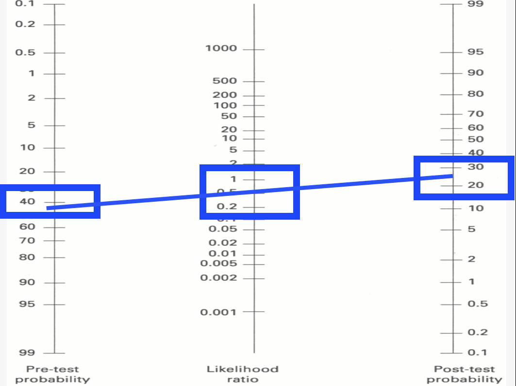
LR near 1 is useless.  Using the Fagan nomogram, one can see that if the pre-test probability is in the “I’m not sure range”, a LR near 1 moves the needle slightly up but to the “I’m still not sure range.”  This means that the diagnostic test will not be much help in our post-test probability.

LRs near 1 are useless

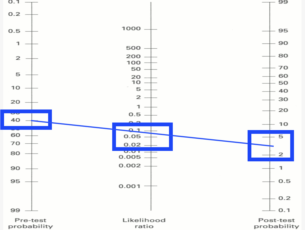
-LRs are helpful once they’re in the 0.1 range.  Using this nomogram we can see that in a patient that we’re not sure about, a test with a -LR of 0.1 can reduce the likelihood that the patient has the disease in question to the low single digits (whether or not that’s enough depends on the disease process in question).

Screen Shot 2015-12-08 at 10.35.38 AM
-LRs of 0.1 and below are very useful

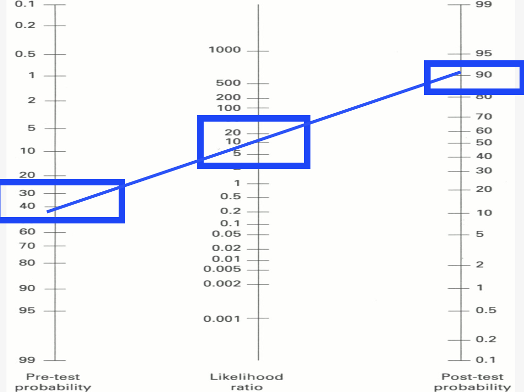
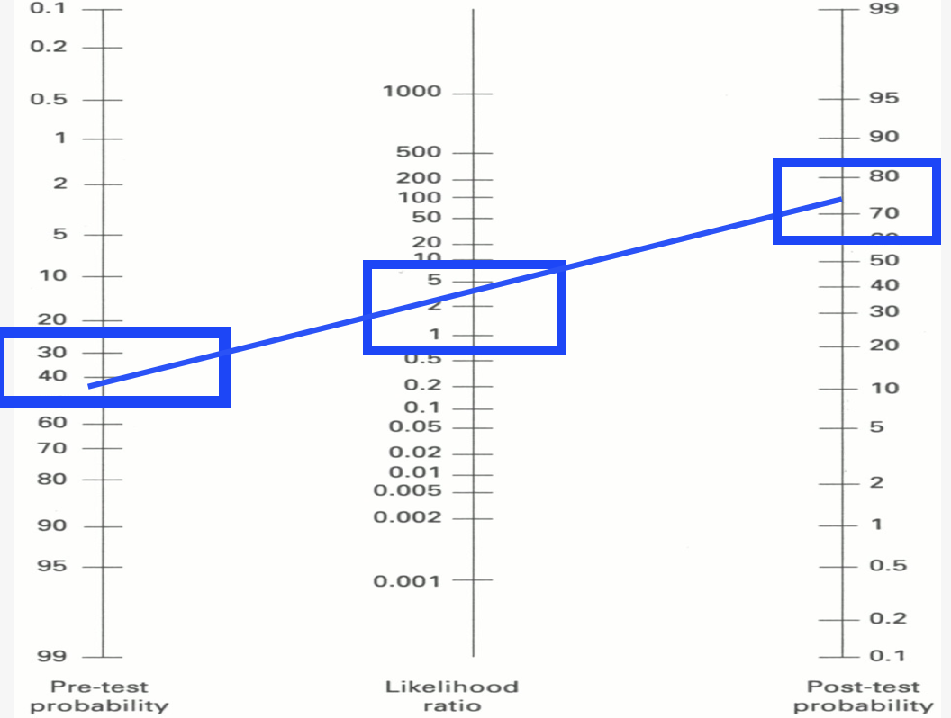
+ LRs near 10 are very useful as, if the test is positive, the patient likely has the disease. In a patient that one says “maybe they have X disease?”, a pre-test probability of say, 40%, a positive test with a +LR of 10 means that there’s a 90-something percent probability that the patient does have the disease. We can be much more certain.


+LR of 10 very useful

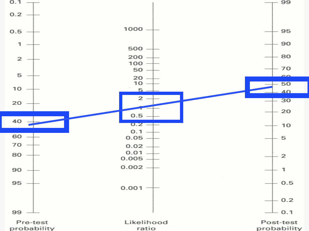
+LRs from 0 to 5 are not very useful. They may shift the probability from a pre-test probability of “maybe?” to a post-test probability of “maybe.”

Screen Shot 2015-12-10 at 6.09.26 AM

 -LRs from 1 to 0.2 are not very useful. They may shift the post-test probability slightly but not much.

+LR 0-5 and -LR 1-0.2 with minimal utility

More resources:

Deeks JJ. Diagnostic tests 4: likelihood ratios. BMJ. 329(7458):168-169. 2004. [article]

BoringEM on Likelihood Ratios.

Episode 22 – The Knee

(ITUNES OR LISTEN HERE)

The Free Open Access Medical Education (FOAM)

This week we’re covering a post from the incredible pediatric resource, Don’t Forget the Bubbles, “Knee X-ray Interpretation” by Dr. Tessa Davis.  We use a systematic approach to assessing chest x-rays, so why not knee x-rays?

  •  Know the anatomy
  •  Look at:
    • Effusion
    • Main bones
    • Tibiofemoral alignment
    • Tibial plateaus
    • Intercondylar eminence
    • Patellar tendon disruption
    • Patellar fracture

The Bread and Butter

We summarize some key topics from Rosenalli, that’s Tintinalli (7e) Chapter s271, 281; Rosen’s (8e) Chapters 57, 136.  But, don’t just take our word for it.  Go enrich your fundamental understanding yourself.

Knee Dislocation

  • Anterior is most common (40%), posterior (33%)
  • Approximately 50% of knee dislocations may be relocated upon presentation to the hospital (this does not reduce risk of badness)
  • Most worrisome sequelae = popliteal artery disruption.  Of patients with popliteal disruption, the amputation rate rises to 90% 8 hours after the injury without surgical intervention.
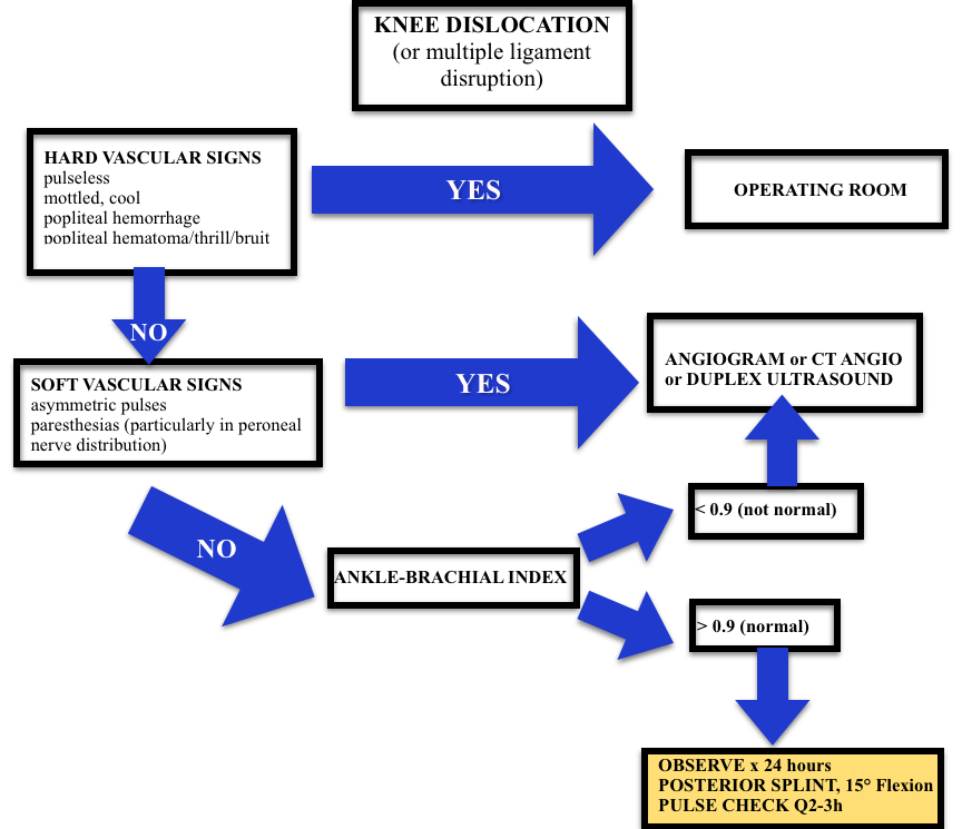
  • Workup may depend on your institution (ex: angiogram vs. CT angio vs. ultrasound) but all patients will need an ABI + 24 hour of pulse checks per current standards.
Screen Shot 2015-01-08 at 1.10.48 PM
Algorithm (adopted from Rosen’s)

Septic Arthritis

  • Most Common Organisms: S. aureus, N. gonorrhea
  • Hematogenous spread
  • Most Common Location: knee, hip

Risk factors such as immunocompromised hosts and use of steroids are risk factors for septic arthritis but the ones with the highest likelihood ratio (LR+ >10 is ideal):

  • Skin infection overlying prosthetic joint (LR+ 15)
  • Joint surgery within the preceding 3 months (LR+ 6.9)
  • Age > 80 (LR+ 3.5)

Diagnosis:  In the red, hot, swollen, painful joint, think septic arthritis.  Clinical and laboratory indicators aren’t great. Synovial fluid analysis, particularly the culture exists as the gold standard.  Arthrocentesis Trick of the Trade from ALiEM. Here are the operating characteristics from Margaretten et al:

  • Fever: Sensitivity 57%
  • Lab tests: White Blood Cell count (WBC), sedimentation rate (ESR), and c-reactive protein don’t perform well
    • WBC LR+  1.4 (1.1-1.8); LR- 0.28 (0.07-1.10)
    • Erythrocyte sedimentation rate 1.3 (1.1-1.8); LR- 0.17 (0.20-1.30)
    • C-reactive protein  1.6 (1.1-2.5); LR- 0.44 (0.24-0.82)
  • Synovial fluid gram stain and culture is the “gold standard.”

Treatment: Intravenous antibiotics and washout of the joint by orthopedics in the operating room

 Generously Donated Rosh Review Questions 

Question 1. A 67-year-old man with a history of gout presents with atraumatic left knee pain. Physical examination reveals an effusion with overlying warmth and erythema. There is pain with passive range of motion. He reports a history of gout in this joint in the past. [polldaddy poll=8568492]

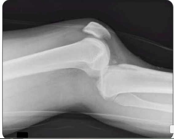
Question 2.  A 27-year-old woman presents with severe left knee pain after an MVC where she was the front passenger. She states her knee hit the dashboard. An X-ray of the patient’s knee is shown below. After reduction, the physical examination reveals swelling of the knee and an Ankle-Brachial Index (ABI) of 0.8. [polldaddy poll=8569540]

Screen Shot 2015-01-12 at 5.13.06 PM

Answers.

1. D. Septic arthritis is a bacterial or fungal infection of a joint typically spread hematogenously unless there is direct bacterial http://www.mindanews.com/buy-imitrex/ contamination. The synovium is highly vascular and lacks a basement membrane making it susceptible to bacterial seeding. Certain conditions predispose individuals to septic arthritis including diabetes, sickle cell disease, immunocompromise, alcoholism or pre-existing joint disease like rheumatoid arthritis or gout. Fever is present in less than half of cases of septic arthritis so with clinical suspicion an arthrocentesis is indicated. The knee is the most common joint affected and patients have pain (especially on passive range of motion) and decreased range of motion often accompanied by warmth, erythema and fever. This patient may have an acute gouty flare, but the clinician must exclude an infection. On joint fluid analysis, the white blood cell count of a septic joint is typically > 50,000. Indomethacin (B) is a non-steroidal anti-inflammatory agent commonly used in the treatment of acute gout. Gout is an arthritis caused by deposition of monosodium urate monohydrate crystals in the joint space. Acute flares involve a monoarticular arthritis with a red, hot, swollen and tender joint. Acute episodes of gout result from overproduction or decreased secretion of uric acid. However, measurement of serum uric acid (C) does not correlate with the presence of absence of an acute flare. A radiograph of the knee (D) may show chronic degenerative changes associated with gout but will not help to differentiate a gouty arthritis versus septic arthritis.

2. C. Obtain Angiography. This patient presents with a knee dislocation and signs of a popliteal artery injury requiring angiography for diagnosis. A knee dislocation refers to a dislocation of the tibia in relation to the femur and not a patellofemoral dislocation. A tibiofemoral dislocation is a limb-threatening emergency due to the high rate of popliteal artery injury. The neurovascular bundle (popliteal artery, popliteal vein and common peroneal nerve) runs posteriorly in the popliteal fossa. The popliteal artery is tethered to the femur and tibia by a fibrous tunnel and is inherently immobile making it susceptible to injury during dislocation. Knee dislocations typically occur in major trauma. An MVC where the knee strikes the dashboard is a common scenario. The dislocation is usually clinically obvious and should be emergently reduced regardless of the presence of confirmatory X-rays. The leg should rapidly be assessed for any “hard” signs of vascular injury including an absence of pulse, limb ischemia, rapidly expanding hematoma, the presence of a bruit or thrill and pulsatile bleeding. Neurologic status should also be assessed prior to and after reduction. After reduction, all patients should have ankle-brachial index (ABI) performed. A normal ABI is > 0.9. Any patient with an ABI less than this should be further investigated for a popliteal injury with angiography. Splint and elevation (D) may be appropriate once a vascular injury is ruled out. The patient should not be discharged home (A) with an abnormal ABI. Observation and repeat ABI (B) is indicated if the initial ABI is normal.

Episode 22 – The Knee

(ITUNES OR LISTEN HERE)

The Free Open Access Medical Education (FOAM)

This week we’re covering a post from the incredible pediatric resource, Don’t Forget the Bubbles, “Knee X-ray Interpretation” by Dr. Tessa Davis.  We use a systematic approach to assessing chest x-rays, so why not knee x-rays?

  •  Know the anatomy
  •  Look at:
    • Effusion
    • Main bones
    • Tibiofemoral alignment
    • Tibial plateaus
    • Intercondylar eminence
    • Patellar tendon disruption
    • Patellar fracture

The Bread and Butter

We summarize some key topics from Rosenalli, that’s Tintinalli (7e) Chapter s271, 281; Rosen’s (8e) Chapters 57, 136.  But, don’t just take our word for it.  Go enrich your fundamental understanding yourself.

Knee Dislocation

  • Anterior is most common (40%), posterior (33%)
  • Approximately 50% of knee dislocations may be relocated upon presentation to the hospital (this does not reduce risk of badness)
  • Most worrisome sequelae = popliteal artery disruption.  Of patients with popliteal disruption, the amputation rate rises to 90% 8 hours after the injury without surgical intervention.
  • Workup may depend on your institution (ex: angiogram vs. CT angio vs. ultrasound) but all patients will need an ABI + 24 hour of pulse checks per current standards.
Screen Shot 2015-01-08 at 1.10.48 PM
Algorithm (adopted from Rosen’s)

Septic Arthritis

  • Most Common Organisms: S. aureus, N. gonorrhea
  • Hematogenous spread
  • Most Common Location: knee, hip

Risk factors such as immunocompromised hosts and use of steroids are risk factors for septic arthritis but the ones with the highest likelihood ratio (LR+ >10 is ideal):

  • Skin infection overlying prosthetic joint (LR+ 15)
  • Joint surgery within the preceding 3 months (LR+ 6.9)
  • Age > 80 (LR+ 3.5)

Diagnosis:  In the red, hot, swollen, painful joint, think septic arthritis.  Clinical and laboratory indicators aren’t great. Synovial fluid analysis, particularly the culture exists as the gold standard.  Arthrocentesis Trick of the Trade from ALiEM. Here are the operating characteristics from Margaretten et al:

  • Fever: Sensitivity 57%
  • Lab tests: White Blood Cell count (WBC), sedimentation rate (ESR), and c-reactive protein don’t perform well
    • WBC LR+  1.4 (1.1-1.8); LR- 0.28 (0.07-1.10)
    • Erythrocyte sedimentation rate 1.3 (1.1-1.8); LR- 0.17 (0.20-1.30)
    • C-reactive protein  1.6 (1.1-2.5); LR- 0.44 (0.24-0.82)
  • Synovial fluid gram stain and culture is the “gold standard.”

Treatment: Intravenous antibiotics and washout of the joint by orthopedics in the operating room

 Generously Donated Rosh Review Questions 

Question 1. A 67-year-old man with a history of gout presents with atraumatic left knee pain. Physical examination reveals an effusion with overlying warmth and erythema. There is pain with passive range of motion. He reports a history of gout in this joint in the past. [polldaddy poll=8568492]

Question 2.  A 27-year-old woman presents with severe left knee pain after an MVC where she was the front passenger. She states her knee hit the dashboard. An X-ray of the patient’s knee is shown below. After reduction, the physical examination reveals swelling of the knee and an Ankle-Brachial Index (ABI) of 0.8. [polldaddy poll=8569540]

Screen Shot 2015-01-12 at 5.13.06 PM

Answers.

1. D. Septic arthritis is a bacterial or fungal infection of a joint typically spread hematogenously unless there is direct bacterial contamination. The synovium is highly vascular and lacks a basement membrane making it susceptible to bacterial seeding. Certain conditions predispose individuals to septic arthritis including diabetes, sickle cell disease, immunocompromise, alcoholism or pre-existing joint disease like rheumatoid arthritis or gout. Fever is present in less than half of cases of septic arthritis so with clinical suspicion an arthrocentesis is indicated. The knee is the most common joint affected and patients have pain (especially on passive range of motion) and decreased range of motion often accompanied by warmth, erythema and fever. This patient may have an acute gouty flare, but the clinician must exclude an infection. On joint fluid analysis, the white blood cell count of a septic joint is typically > 50,000. Indomethacin (B) is a non-steroidal anti-inflammatory agent commonly used in the treatment of acute gout. Gout is an arthritis caused by deposition of monosodium urate monohydrate crystals in the joint space. Acute flares involve a monoarticular arthritis with a red, hot, swollen and tender joint. Acute episodes of gout result from overproduction or decreased secretion of uric acid. However, measurement of serum uric acid (C) does not correlate with the presence of absence of an acute flare. A radiograph of the knee (D) may show chronic degenerative changes associated with gout but will not help to differentiate a gouty arthritis versus septic arthritis.

2. C. Obtain Angiography. This patient presents with a knee dislocation and signs of a popliteal artery injury requiring angiography for diagnosis. A knee dislocation refers to a dislocation of the tibia in relation to the femur and not a patellofemoral dislocation. A tibiofemoral dislocation is a limb-threatening emergency due to the high rate of popliteal artery injury. The neurovascular bundle (popliteal artery, popliteal vein and common peroneal nerve) runs posteriorly in the popliteal fossa. The popliteal artery is tethered to the femur and tibia by a fibrous tunnel and is inherently immobile making it susceptible to injury during dislocation. Knee dislocations typically occur in major trauma. An MVC where the knee strikes the dashboard is a common scenario. The dislocation is usually clinically obvious and should be emergently reduced regardless of the presence of confirmatory X-rays. The leg should rapidly be assessed for any “hard” signs of vascular injury including an absence of pulse, limb ischemia, rapidly expanding hematoma, the presence of a bruit or thrill and pulsatile bleeding. Neurologic status should also be assessed prior to and after reduction. After reduction, all patients should have ankle-brachial index (ABI) performed. A normal ABI is > 0.9. Any patient with an ABI less than this should be further investigated for a popliteal injury with angiography. Splint and elevation (D) may be appropriate once a vascular injury is ruled out. The patient should not be discharged home (A) with an abnormal ABI. Observation and repeat ABI (B) is indicated if the initial ABI is normal.

Episode 21 – Acute Kidney Injury

(ITUNES OR LISTEN HERE)

The Free Open Access Medical Education (FOAM)

Dr. Josh Farkas of the PulmCrit blog has produced a couple of blog posts on the importance of renal protection in sepsis, Renoresuscitation: Sepsis resuscitation designed to avoid long-term complications and Renal microvascular hemodynamics in sepsis: a new paradigm.  Much of this is theoretical and certainly not something that is standard practice, rathery a theory extrapolated from subgroups of several trials.

Suggested renoresuscitation measures:

(1) Avoid renal failure – avoid nephrotoxins (many antibiotics, NSAIDs, ace-inhibitors), avoid hyperchloremic metabolic acidosis.

(2) Avoid volume overload – treating decreased urine output by flooding a patient with fluids is not necessarily the best move.

(3) Protect the glycocalyx of the endothelium – this suggestion proffers more questions than answers. Steroids? Albumin? Certain vasopressors?  Stay tuned, as we’re not really certain what this entails.

The Bread and Butter

We summarize some key topics from Rosenalli, that’s Tintinalli (7e) Chapter 91; Rosen’s (8e) Chapter 97.  But, don’t just take our word for it.  Go enrich your fundamental understanding yourself.

Acute Kidney Injury – typically a creatinine 1.5-2x the patient’s baseline is classified as acute kidney injury.  Urine output can be increased initially but determine whether a patient is making urine and how much, as urine output <0.5 mL/kg/h qualifies as AKI.

RIFLE criteria
RIFLE criteria

Importance – AKI is associated with worse outcomes, although it’s unclear as to whether this is merely a marker of

  • Found in 35-65% of admissions to the intensive care unit, in 5-20% of hospital admissions.  Furthermore, AKI is associated with higher mortality.
  • Renal failure can also cause significant problems for the patient such as electrolyte abnormalities (hyperkalemia the most worriesome, but also hyperphosphatemia) and pulmonary edema.

Etiology – many causes of AKI are reversible or amenable to treatment.

Prerenal – this is one of the most common causes of acute kidney injury and basically is caused by decreased blood flow to the kidney.  Associated with a high BUN/creatinine ratio, increased urine osmolality, a urine sodium concentration less than 20 mEq/L, and FENa less than 1% (this is why getting urine sodium and a concurrent chemistry panel is key).

  • Hypovolemia – volume depleted, hemorrhage, intravascular volume depletion from congestive heart failure or cirrhosis.
  • Hypotension – poor cardiac output (heart failure, valvular problems), shock
  • Decreased flow through the renal artery disease – Nonsteroidal anti-inflammatories: inhibit prostaglandins in the afferent arteriole.  ACE inhibitors prevent the conversion of angiotensin I to angiotensin II, leading to decreased levels of angiotensin II, which when absent decreases the GFR because of dilatation of the efferent arteriole.

Post Renal (Obstructive) – Check out Episode 2 on urologic emergencies.

  • Benign prostatic hypertrophy (BPH) is the most common cause but medications such as anticholinergics and pseudoephedrine. Trauma, stones, strictures, and malignancy can also cause obstruction.

Intrinsic acute renal failure divided into: tubular disease (most common), glomerular disease, vascular disease and interstitial disease.

  • Least common form of AKI in the ED, more common in inpatients.
  • Acute Tubular Necrosis (ATN) most common cause – via nephrotoxins such as aminoglycosides and contrast.
  • Granular “muddy brown” casts – think of necrosis from the “N” in ATN and necrosis tends to be dark.

Indications for emergent dialysis – AEIOU

A- Acidosis

E- Electrolyte emergencies (hyperkalemia!)

I-  Intoxication with dialyzable toxins (ethylene glycol)

O- Overloaded with volume

U- Uremia

 Generously Donated Rosh Review Questions 

Question 1. A 72-year-old man is brought to the ED from a nursing home for evaluation of oliguria. He is found to have an acutely elevated BUN and plasma creatinine from baseline. A Foley catheter is placed; his urine sodium (UNa) is measured below 20 mEq/L and fractional excretion of sodium (FENa) below 1%. [polldaddy poll=8545511]

Question 2.  A 54-year-old man presents to the ED in acute renal failure (ARF). [polldaddy poll=8545512]

Answer 1.  D. This patient’s oliguria with acutely elevated BUN and plasma creatinine suggest that he is in acute renal failure (ARF). His UNa <20 mEq/L and FENa <1% indicate that he has intact reabsorptive function and is able to conserve sodium. This is consistent with prerenal azotemia as the cause for his ARF.

Acute tubular necrosis (ATN) (A), loop diuretics (e.g., furosemide) (B), and osmotic diuresis (e.g., mannitol) (C)all lead to UNa >20 mEq/L and FENa >1% because there is impairment in the ability to concentrate the urine. In such cases, a high-sodium load is excreted.

Answer 2. A.   Acute tubular necrosis (ATN) is a severe form of impairment of tubular epithelial cells caused by ischemia or toxic injury. It is a leading cause of ARF. One of its hallmarks is the presence of brown granular casts on urinalysis. These contain cellular debris rich in cytochrome pigments. In contrast, hyaline casts (B) are usually nonspecific but present after exercise; red cell casts (C) are indicative of glomerular hematuria (e.g., glomerulonephritis); and white cell casts (D) imply renal parenchymal inflammation (e.g., acute interstitial nephritis, pyelonephritis).

Episode 17 – The Spleen!

(ITUNES OR LISTEN HERE)

The Free Open Access Medical Education (FOAM)

We review Dr. Scott Weingart’s episode 133 on pre-hospital REBOA (resuscitative endovascular balloon occlusion of the aorta). Weingart interviews Dr. Gareth Davies about the encounter, underscoring the increasing use of REBOA.

For a quick REBOA refresher, check out Episode 121.

REBOA (Review of REBOA) – First described in 1954 in the Korean War, this is a form of hemorrhage control below the level of the chest without having to do a thoracotomy with aortic cross clamping, which has sparse mortality benefit and can be dangerous to providers.  Most of the REBOA literature is from swine models and case-series, although there are currently larger trials underway.

  • Outline of procedure – Obtain arterial access through the common femoral artery, pass a vascular sheath, float a balloon catheter to the appropriate section of the aorta, and inflate the balloon to occlude blood flow. The aorta is divided into three zones so that balloon occlusion is performed in Zone 1 for abdominal injuries or Zone 3 for pelvic injuries, while Zone 2 is a proposed no-occlusion zone.

The Bread and Butter

Rosen’s Chapter 46, 134 and Tintinalli Chapter 260.

What DOES the spleen do?

Answer: More than most appreciate. The spleen filters the blood, removing abnormal or old red blood cells (RBCs), debris, and antibody coated bacteria from the bloodstream.  It also serves as a reservoir for RBCs and platelets and synthesizes antibodies, opsonins, etc.

Splenic Trauma – EAST Guidelines (their podcast)

Diagnosis – suspect spleen trauma clinically, with hypotension, left upper quadrant abdominal pain, or even minimal trauma, especially after infectious mononucleosis.

  • CT with IV contrast (hemodynamically stable patients).  While FAST and DPL can detect peritoneal free fluid, they can’t detect subcapsular bleeds.
  • Unstable patients – operating room versus interventional radiology

Grading – 1 is 1 (<1 cm laceration depth), 3 is 3 (>3 cm laceration depth). Everything else is somewhere in between.   Higher grades typically result in more aggressive interventions. Historically these get operative intervention

  • Grade 1: < 1 cm laceration depth or<10% subcapsular hematoma
  • Grade 2: 1-3 cm laceration or 10-50% subcapsular
  • Grade 3: > 3 cm laceration depth or >50% subcapsular hematoma
    • Grade 3 or higher should be considered for angiography with embolization (Level II, III – EAST)
  • Grade 4: partially devascularized spleen or contrast blush
  • Grade 5:  a very battered, devascularized spleen

There’s slightly more to spleen grading, check out this post from Dr. McGonigal

Trivia:  The punctate extravasation sometimes seen on angiogram after blunt trauma?

Answer: The Seurat Spleen (pubmed), named after the pointillist painter.

Treatment –

  • Unstable patients: Operating room or Angiographic Intervention (IR) (Level II – EAST)
    • Note: Board exam? Send the patient to the OR.  In reality, there is some practice variation. Many would still argue the patient belongs in the OR, some places have combined OR/IR suites, and some opt for IR.
    • Post splenectomy patients will need immunizations for the encapsulated bacteria
  • Stable patients: Nonoperative management, which often comprises in-hospital monitoring, serial abdominal exams and hematocrits, etc is becoming increasingly common as first line for higher grade splenic injuries.  The key here is that the team must be able to take the patient to the OR or IR should the situation change.  Angioembolization has also gained momentum as management

Post-Splenectomy Sepsis (Review)– Most common in the first years after splenectomy and in children.

Presentation – Patients may present with a vague flu-like illness or gastroenteritis but may go on to develop septic shock, DIC, and multiorgan dysfunction. In addition, meningitis without overwhelming infection or shock is a common presentation of pneumococcal infection in asplenic patients.

Etiology – encapsulated bacteria (Streptococcus pneumoniae, Haemophilus influenza, and Neisseria meningitidis), capnocytophaga canimorsus (dog bites), and parasites such as babesiosis (endemic in New England), malaria, and ehrlichiosis.

Management – Labs, blood cultures, antibiotics (typically ceftriaxone)

Splenic Sequestration – Second most common cause of death in kids with Sickle Cell Disease <5 years of age.

Classic presentation – LLL: LUQ, lethargy, lightness (pallor).

Labs: 3 point drop in hemoglobin, increased reticulocyte count, and thrombocytopenia.

Generously Donated Rosh Review Questions (Scroll for Answers)

Question 1.  [polldaddy poll=8376275]

Question 2.  A 23-year-old man presents with a stab wound to the abdomen. His vital signs are HR 132, BP 88/45. He has a positive FAST.[polldaddy poll=8376283]


References:

Chapter 46, 134. Rosen’s Emergency Medicine, 8e.

Chapter 260. Tintinalli’s Emergency Medicine: A Comprehensive Study Guide, 7e. New York, NY: McGraw-Hill; 2011


Answers.

1.  C.  Splenic artery aneurysms are the most common type of visceral arterial aneurysms, accounting for up to 60% of cases. Etiologies include arterial fibrodysplasia, portal hypertension, and increased splenic AV shunting in pregnancy. Clinical presentation is vague with left upper quadrant pain with radiation to the left shoulder or subscapular area. Most of the aneurysms are <2 cm in diameter; only 2% result in life-threatening rupture. Treatment is surgical resection if the patient is symptomatic. Otherwise, asymptomatic patients can undergo transcatheter embolization. Of those aneurysms that rupture, up to 95% occur in young pregnant women.

Hepatic artery aneurysms (A) represents 20% of visceral artery aneurysms and are caused by atherosclerosis, infection, and abdominal trauma. Clinical presentation can mimic cholecystitis. Inferior mesenteric artery aneurysms(B) are uncommon. Superior mesenteric artery aneurysms (D) are the 3rd-most common visceral aneurysms. IV drug abusers are at increased risk.

2. D. This patient presents with hemorrhagic shock from a penetrating abdominal trauma and should be immediately transferred to the operating room for an exploratory laparotomy. Stab wounds are the most common form of penetrating trauma. About 70% of anterior stab wounds penetrate the peritoneum. It is difficult to predict the specific organ injured based on the external location of the wound. Initial management should focus on securing the airway, assessing and supporting the patients breathing and circulation. IV access and supplemental oxygen should be provided. In hypotensive trauma patients, early blood transfusion should be initiated and consideration should be made for massive transfusion protocol. Concomitant with the primary and secondary survey, a Focused Assessment with Sonography for Trauma (FAST) exam should be performed. The speed and accuracy of the FAST has almost completely replaced the need for diagnostic peritoneal lavage. In a FAST exam, images are obtained of the splenorenal space, hepatorenal space (Morrison’s pouch), heart and bladder (pouch of Douglas). A FAST exam has high sensitivity in detecting as little as 100 ml of fluid. A FAST examination showing free fluid in any of the abdominal views in the presence of hypotension should lead to the patient being transported to the operating room for exploratory laparotomy according to the Advanced Trauma Life Support (ATLS) algorithm. Angiographic embolization (A) is useful in patients with pelvic fractures and bleeding from pelvic vessels. CT scan of the abdomen and pelvis (B) can be performed in trauma patients who are stable to further assess for injuries.Diagnostic peritoneal lavage (C) does not play a role in management of penetrating trauma patients with hypotension and positive FAST examination.
 

Episode 9 – Pregnancy Emergencies

Episode 9 (iTunes or listen here)

The Free Open Access Medical education (FOAM)

We review Dr. Scott Weingart’s Practical Evidence Podcast #3 – ACEP 2012 Management of Early Pregnancy, in which he summaries the ACEP 2012 Clinical Policy on this topic.

  • The discriminatory zone is out. Get ultrasounds in pregnant patients, regardless of the quantitative beta-hCG.
  • A certain beta-hCG level can not be used to rule in or rule out ectopic pregnancy or viable intrauterine pregnancy (IUP), get the ultrasound and ensure you identify the uterus.
  • If an ultrasound (including radiology’s formal ultrasound) is indeterminate for ectopic versus IUP, that patient should have a repeat ultrasound and follow up with OB within 48 hours.

A good FOAM ectopic rule out pathway

The Bread and Butter

We summarize some key topics from the following readings, Tintinalli (7e) Chapters 102-105; Rosen’s 8(e) Chapter 178 but, the point isn’t to just take our word for it. Go enrich your fundamental understanding yourself!

Bleeding after the First Trimester

The two things we worry about the most are placental abruption and placenta previa.

Placental Abruption – premature separation of the placenta from the uterine lining.

  • Classic presentation: painful vaginal bleeding.
  • Actual presentation: 30% do not have vaginal bleeding, uterine pain/tenderness, back pain, hypotension, nausea.
  • Risk factors: hypertension, previous abruption, uterine scar, cocaine, smoking, blunt trauma, older age
  • Diagnosis: Ultrasound, clinical suspicion, MRI. Note – Ultrasound is specific but NOT sensitive.
  • Management: Intravenous access, hemoglobin, type and screen, coagulation panel, resuscitation, ultrasound, likely call OB.

Placenta Previa – implantation of the placenta over the cervical os.

  • Classic presentation: painless vaginal bleeding
  • Diagnosis: Ultrasound
  • Management is the same as for abruption but do NOT do a speculum or cervical exam unless there is no OB service.

Abdominal Pain in Pregnancy

Pregnant women are at risk for the same abdominal emergencies as everyone else – appendicitis, cholecystitis, and pyelonephritis but they’re also at risk for some special pregnancy related issues.

Septic Abortion – evidence of infection with any type of abortion, often due to unsanitary abortions or retained products of conception

  • Presentation: fever, uterine tenderness at under 20 weeks of gestation
  • Treatment: Ampicillin 3g IV, gentamicin 1-2 mg/kg IV, stat OB consult for source control

Chorioamnionitis – infection or inflammation of the placenta and fetal membranes, often after 16 weeks of gestation

  • Presentation: fever, uterine tenderness, fever/maternal tachycardia, sepsis
  • Risk factors: preterm labor
  • Treatment: Ampicillin 3g IV, gentamicin 1-2 mg/kg IV, stat OB consult for source control

HELLP syndrome – hemolysis, elevated liver enzymes, low platelets.

  • Diagnostics: CBC with schistocytes, Platelets <100,000, Elevated transaminases, Normal or elevated BUN/creatinine, abnormal coagulation profile. CT abdomen/pelvis if patient hemodynamically stable.
  • Complications: liver hematoma, splenic or liver rupture.
  • Management: Magnesium, control of blood pressure (labetalol, hydralazine), correction of coagulopathy, delivery of the fetus.

Pre-Eclampsia/Eclampsia

New onset hypertension (BP >140/90) after 20 weeks of gestation in a previously normotensive patient plus one of the following: proteinuria or end organ dysfunction (pulmonary edema, renal dysfunction, visual changes, etc). Eclampsia is a sequelae of pre-eclampsia, like HELLP syndrome, characterized by seizures and coma.

  • Control seizures with magnesium
  • Control BP if DBP >105 mmHg
  • Check for organi injury (CBC/platelet count, transaminases, BUN/Creatinine)

Generously donated Rosh Review questions (scroll for answers)

Question 1. A 27-year-old woman 32 weeks pregnant presents with bright-red vaginal bleeding for 1 day. The patient denies any pain and is not tender on abdominal exam. Her vital signs are BP 115/70, HR 90, and RR 16. [polldaddy poll=8210380]

Question 2. A 26-year-old woman presents with abdominal cramping after a positive home pregnancy test. Her vitals are T 98.7°F, HR 94, BP 110/66, RR 18, oxygen saturation 97%. Her exam is unremarkable. Labs reveal a serum beta HCG of 1000 mIU and she is Rh positive. She states that the pregnancy is wanted. An ultrasound is performed as seen below.[polldaddy poll=8206329]

More FOAM on vaginal bleeding:

Answers.

1. A. Placenta previa is characterized by painless, fresh vaginal bleeding in late pregnancy. Placenta previa occurs in 1% of pregnancies and is defined as a placenta that extends near, partially over, or completely over the cervical os. These patients are at an increased risk for life-threatening hemorrhage. As a result, the first step in management of placental previa is to obtain intravenous access in anticipation of fluid resuscitation and possible transfusion. Obstetrical consultation is also advised.

2. D. This patient presents with abdominal pain and a positive pregnancy test raising the concern for an ectopic pregnancy. Ectopic pregnancy complicates about 1.5 – 2.0% of pregnancies and is potentially life threatening. There are a number of risk factors for ectopic pregnancy including pelvic inflammatory disease, prior tubal surgery and previous ectopic pregnancy. This patient has an early pregnancy based on the low beta hCG. The transvaginal ultrasound shows an early gestational sac without a yolk sac or fetal pole within the uterus. This ultrasound does not rule out the diagnosis of an ectopic pregnancy as an ectopic pregnancy can cause a decidual reaction in the uterus, which appears similar to an early gestational sac. The definitive ultrasound finding for an intrauterine pregnancy would be the presence of a yolk sac or fetal pole. It is expected that above the discriminatory hCG zone of 1500-2500 mIU, a definitive IUP should be identified. Patients with a beta hCG below the discriminatory zone without a definitive IUP can be managed conservatively with repeat hCG level in 48 hours (the level should double every 48 hours) and repeat ultrasound.