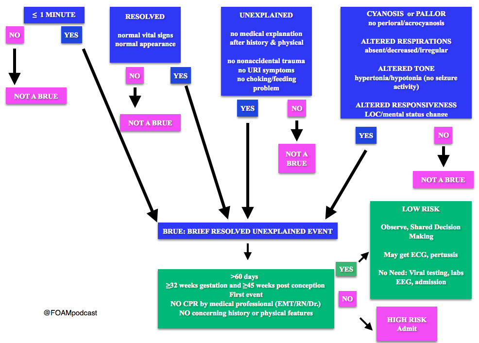
Note: Some have voiced concerns that these guidelines potentially downplay the event. The concern is that the yield of these workups and admissions may be low, but possibly worthwhile. At FOAMcast, we are not qualified to critique these guidelines but there are helpful tables and charts in them to realize that these recommendations really are only for specific events and children AFTER thorough history and physical.
Generously Donated Rosh Review Question
A 6-week-old boy is brought to the emergency room because of cyanosis. He was sleeping comfortably in a supine position right after a feeding when he suddenly choked, became limp and lips turned blue. The mother witnessed the event and blew to the face of the boy. The whole episode lasted for about two minutes. EMS was called and upon arrival at the house, the boy was back to his usual self. At the ER, he boy has normal vital signs with normal physical examination findings.
A. Admit for cardiorespiratory monitoring. The boy in the vignette had an apparent life-threatening event (ALTE) which is not a specific diagnosis but a description of an acute, unexpected episode that is frightening to the caretaker. ALTE includes one or more of the following features: apnea, color change (may be cyanotic, pallid, erythematous or plethoric), marked change in muscle tone (limpness or rigidity) and choking or gagging. A specific cause for ALTE can be identified in over one-half of patients after a careful history, physical examination, and appropriate laboratory evaluation. The remaining cases are considered idiopathic if no cause can be identified after a thorough assessment. Common etiologies for ALTE include gastroesophageal reflux, neurologic problems (such as seizures), and respiratory infection. The history of an ALTE must be taken seriously, even if the infant appears entirely well by the time he or she is evaluated. In-hospital observation with cardiorespiratory monitoring is indicated for infants whose initial evaluation suggests physiologic compromise. Hospital admission may provide important clinical information where additional episodes may be witnessed by medical personnel during the observation period. In addition, serious underlying medical conditions may become apparent. Discharging the patient after reassuring the parents (B) and observing the patient for four hours in the ER (C) are not appropriate management strategies for the infant in the vignette who needs admission for cardiorespiratory monitoring. Requesting for complete blood count (D) is not routinely done in the evaluation of an ALTE and would not aid in the management for the infant in the vignette.
We cover two bits of FOAM, one from Emergency Medicine Literature of note on the use of meclizine for vertigo and an EMcrit episode on the HiNTs exam.
Emergency Medicine Literature of Note – Dr. Ryan Radecki – Treating what you believe is peripheral vertigo?Using meclizine? So are most people.But this is not an evidence based practice. While meclizine is Rosen approved, Tintinalli recommends transdermal scopolamine as the first line treatment [1,2]. Following a recent recall of meclizine (oddly because iron bottles contained meclizine instead of iron), Dr. Radecki probes into why we use meclizine for vertigo.
Meclizine is an anti-histamine and has been thought to have anti-emetic properties.
A 1968 paper compared 16 anti-emetics/combinations and did NOT conclude that meclizine was the best. In fact, scopolamine and amphetamine performed best. Promethazine (phenergan) is also a good choice based on this paper [3].
EMcrit – Dr. Scott Weingart – The HiNTs exam has taken off, particularly in the FOAM world, as a means of disguising between central and peripheral causes of vertigo. In 2010, an EMcrit episode popularized this in the FOAM world. See this video demonstrating the HiNTs exam.
Head Impulse – rapid head rotation by the examiner with the subject’s vision fixed on an object (i.e. examiner’s nose). The examiner rapidly rotates the head towards midline and the patient’s eyes should remain fixed on the target.
Abnormal (loss of fixation on target and movement of eyes away from target followed by correct saccade as patient fixates on target) = peripheral (vestibulo-ocular reflex impaired)
Normal = central
Nystagmus
Horizontal nystagmus with a unidirectional fast phase (away from affected side) = probably peripheral
Patients with horizontal nystagmus may have central pathology but may have direction-changing nystagmus (i.e. fast phase beating in one direction when looking to right and the opposite direction when looking left).
Vertical or nystagmus = central pathology
Test of Skew Patients should fixate on a target while the provider alternately covers each eye. As the cover is moved from one eye to the other, the uncovered eye must correct for the misalignment and will look up or down to focus back on the target. This slight correction is observed repeatedly as the cover is moved from one eye to the other.
Skew deviation/misalignment = probably central, often in posterior fossa abnormalities
No skew deviation= peripheral
INFARCT – Impulse Normal, Fast-phase Alternating nystagmus, and Refixation on Cover Test
Issues with HiNTs
Can only be performed on patients with continuous vertigo.
External validity is a major issue with HiNTs.
Providers – Of the 4 studies have examined the operating characteristics of HiNTs, none have used emergency providers and instead have examined how the exam performs in the hands of two neuro-ophthalmologists, neuro-otologists, and neurologists with 4 hours of specialized training in the exam. It’s unclear whether HiNTs would be reliable or valid when performed by emergency providers [4-6].
Patients – The patients examined in many of these studies have other indicators of badness on neurologic exam. In one study, patients had to have gait instability and/or truncal ataxia to enroll. Then, 76/101 (76%) of those patients had a central cause. These patients were sick and not the undifferentiated vertiginous patients we see primarily as emergency providers [4-6].
In the words of leading HiNTs expert Dr. Newman-Toker, HiNTs “requires expertise not routinely available in emergency departments.” As such, his team is piloting quantitative video-oculography to aid in diagnosis using HiNTs [7]. An Annals of Emergency Medicine review also warned that HiNTs may not be ready for emergency provider use [9].
We delve into core content on vertigo using Rosen’s Medicine (8e) Chapter 19,and Tintinalli’s Emergency Medicine: A Comprehensive Study Guide(7e) Chapter 164 “Vertigo and Dizziness.”
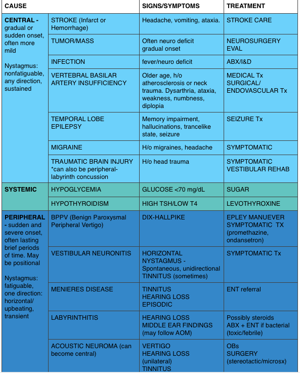
Vertigo is often characterized by the sensation of spinning and falls into the broad and frustrating category of “dizziness.” Often, when a dizzy patient presents we perseverate on characterizes what the patient means by “dizzy.” However, some argue that this is not an appropriate approach as a study found 50% of patients changed the character of their dizziness when questioned again after 10 minutes [9]. Additionally, the clinical characteristics differentiating peripheral from central causes of vertigo are not entirely reliable. Despite these limitations, it is expected that we are familiar with “classic” presentations.
A 50-year-old man presents with episodic severe vertigo lasting hours, with associated symptoms of unilateral tinnitus, fluctuating low-frequency hearing loss, and aural fullness. [polldaddy poll=9245427]
A 20-year-old woman presents with an acute onset of dizziness. The patient describes the sensation that the room is spinning when she turns her head to the left and it is accompanied by nausea and vomiting. The symptoms resolve with turning her head away from that side. Examination reveals nystagmus elicited by deviating the eyes to the left and no other neurologic findings. [polldaddy poll=9245971]
Answers
1. B. Meniere’s disease is characterized by episodic severe vertigo lasting hours, with associated symptoms of unilateral tinnitus, fluctuating low-frequency hearing loss, and aural fullness. Typical onset is in the fifth decade of life. The cause is uncertain but is speculated to result from allergic, infectious, or autoimmune injury. The histopathologic finding includes endolymphatic hydrops, which is thought to be caused by either overproduction or underresorption of endolymph in the inner ear. Meniere’s disease is a clinical diagnosis mostly based on history. Testing may be obtained to support the diagnosis and rule out other disorders. Audiometry often demonstrates a low-frequency sensorineural hearing loss. An FTA-ABS test may be obtained to rule out syphilis. Electronystagmography (ENG) may demonstrate a unilateral peripheral vestibular weakness on caloric testing. When the diagnosis is uncertain, a brain MRI with contrast is obtained to evaluate for a retrocochlear lesion. The differential diagnosis of Meniere’s disease includes acute labyrinthitis, neurosyphilis, labyrinthine fistula, autoimmune inner ear disease, vestibular neuronitis, and migraine-associated vertigo.The most common cause of peripheral vestibular vertigo in adults is benign paroxysmal positional vertigo (A). BPPV occurs in all age groups but more often between ages 50 and 70 but is not associated with hearing loss and made worse with movement. In a perilymph fistula (C) rapid changes in air pressure (barotrauma), otologic surgery, violent nose blowing or sneezing, head trauma, or chronic ear disease may cause leakage of perilymph fluid from the inner ear into the middle ear and result in episodes of vertigo. Associated signs and symptoms are variable but can include a sudden pop in the ear followed by hearing loss, vertigo, and sometimes tinnitus. Acute vertigo associated with nausea and vomiting (but without neurologic or audiologic symptoms) that originates in the vestibular nerve is known as vestibular neuronitis (D). Vestibular neuronitis can occur spontaneously or can follow viral illness.
2. B. This patient presents with peripheral vertigo most consistent with benign paroxysmal peripheral vertigo (BPPV) and should be treated with an Epley maneuver. Vertigo is defined as the sensation of disorientation in space combined with a sensation of motion. Patients typically describe a “room-spinning” sensation or the feeling of sea sickness. Vertigo can be divided into two types: central and peripheral. Central vertigo are those disorders arising from the central nervous system and include ischemic stroke, vertebrobasilar insufficiency and infectious causes (meningitis, mastoiditis, syphilis). Central vertigo is characterized by longer duration of symptoms, minimal change with position, gradual onset and multidirectional nystagmus. Peripheral vertigo includes BPPV, Meniere’s disease, Labyrinthitis and vestibular neuritis. Peripheral vertigo may have intermittent symptoms (BPPV) or continuous symptoms but should not be associated with other neurologic deficits or changes and should have unidirectional nystagmus. The symptoms in BPPV are elicited by specific movements of the head and relieved by returning the head to a neutral position. The symptoms should be acute in onset and of a short duration. In BPPV, the symptoms are cause by the presence of an otolith in one of the semicircular canals. Although pharmacologic intervention may be necessary in the acute setting with meclizine or benzodiazepines, the best treatment for BPPV is the Epley maneuver. The Epley maneuver is a series of positions that the clinician takes the patient through that leads to expulsion of the otolith from the semicircular canal and relief of symptoms. Imaging with a non-contrast head CT (C) is not indicated in peripheral vertigo of any cause as the patient’s pathology is in the inner ear and not the brain. If a central cause is suspected, MRI of the brain (A) is the best test for diagnosis as the causative lesion will likely be in the posterior fossa, which is not seen well on CT scan. Steroid treatment (D) is the indicated management for vestibular neuritis but does not play a role in the treatment of BPPV.
References:
Chang AK, Olshaker AS. Dizziness and Vertigo. In: Marx JA, Hockberger RS, Walls RM eds. Rosen’s Emergency Medicine, 8th e.
Goldman B. Chapter 164. Vertigo and Dizziness. In: Tintinalli JE, Stapczynski J, Ma O, Cline DM, Cydulka RK, Meckler GD, T. eds.Tintinalli’s Emergency Medicine: A Comprehensive Study Guide, 7e. New York, NY: McGraw-Hill; 2011.
Cohn B. Can Bedside Oculomotor (HINTS) Testing Differentiate Central From Peripheral Causes of Vertigo? Annals of Emergency Medicine. 64(3):265-268. 2014.
Strupp M, Zingler VC, Arbusow V et al. Methylprednisolone, Valacyclovir, or the Combination for Vestibular Neuritis. N Engl J Med. 351(4):354-361. 2004.
Sleep through 2015? We picked the minds of some brilliant Emergency Medicine folk and came up with this short list of important happenings.
TRAUMA
FAST (Focused Assessment using Sonography in Trauma) guided resuscitative – thoracotomy. (Recommended by Haney Mallemat, Rob Orman).
Bottom Line: If a trauma code comes in and has neither cardiac activity nor pericardial effusion on FAST, the odds of survival are essentially nil. Inaba and colleagues found the following:
Population: 187 patients at LA/USC deemed “appropriate” for thoracotomy (at this institution: penetrating trauma patients with absent vital signs and blunt trauma patients with a loss of vital signs en route or in the resuscitation bay).
Intervention: Emergency medicine resident performed FAST before/concurrent with thoracotomy
Outcome: Of the 126 patients without cardiac activity on FAST, none survived.
Many of the patients with cardiac activity did not survive, as well. This paper gives individuals guidance to make the decision to crack the chest but has stirred up a debate as to whether this would lessen educational opportunities for a potentially heroic procedure.
Say NO to long backboards (Recommended by Lauren Westafer). In January 2015, ACEP recommended against the use of long backboards. Many state and local protocols shifted away from moving this some time ago and even more since. The FOAM community has been up in arms about the inefficacies and harms of backboards for quite some time, again echoing that FOAM can serve as a forecaster for change to make it less cognitively distressing when time to change our practice.
RESUSCITATION
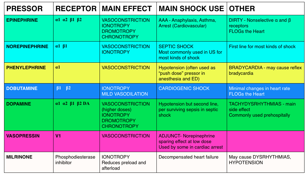
Peripheral Vasopressors (Recommended by Haney Mallemat, Rob Orman) – We detail the literature underlying the use of peripheral vasopressors in this podcast.
They think that the combination of a systematic review by Loubani et aland The Cardenas-Garcia study, peripheral vasopressors may be safely run through large bore peripheral IVs proximal to the antecubital fossa. Note: These should be closely monitored (protocolized is best) and short durations (<6 h) have been associated with minimal complicaitons.
SEPSIS
Intravenous fluids in sepsis (Recommended by Haney Mallemat). The PROMISE trial was published in early 2015, adding to ProCESS and ARISE. In these studies, patients typically got 2 L of crystalloid upfront and then 2L in the first 6 hours. Over 3 days after enrollment, most got just under 4 L. Most patients received under 6L IVF. There has been a movement for more judicious use of fluids in sepsis rather than dumping 4-6 L of IVF upfront. Marik articulately explained this in this article.
Mallemat challenges us: Before giving a fluid bolus use ultrasound and ask these questions, “Does the LV need it, and can the RV take it?”
CMS Core Measure (Recommended by Jeremy Faust). The National Quality Forum has been pushing for Measure 0500 See this EMcrit podcast on this topic.
Sexual intercourse 3-4 times per week may aid in expulsion of distal kidney stones. This year, two large studies by Pickard et al and Furyk et al demonstrated no benefit in stone passage for ureterolithiasis (particularly in stones <5 mm). Then, a paper by Dolouglu et al excited many folks, if for entertainment value. Since tamsulosin doesn’t seem to help, what about sexual intercourse, 3-4 times per week, in male patients with partners?
In this study the mean expulsion time did not differ significantly between groups.
MEDICAL EDUCATION
Merging of FOAM resources. (Recommended by Michelle Lin). Blogs and podcasts are growing and often supplement one another. Dr. Lin predicts the future will be in these conglomerates (ex: merging of EMcrit and PulmCrit and massive undertakings such as ALiEM and CandiEM).
Merging of FOAM with traditional journals. (Recommended by Michelle Lin). Projects such as the Skeptic’s Guide to Emergency Medicine have merged with Academic Emergency Medicine and the Canadian Journal of Emergency Medicine (ex: SGEM HOP, journal paper) and massive FOAM resource ALiEM has also collaborated with Annals of Emergency Medicine (Ex:Journal club, paper). The merging of FOAM with paid, traditional resources is the future, per Dr. Lin.
OVERDIAGNOSIS
Overdiagnosis is a problem, and people are starting to rage against it. (Recommended by Lauren Westafer). An Overdiagnosis conference exists and JAMA Internal Medicine has a series of articles, “Less is More,” frequently detailing evidence of overdiagnosis.
One of 2015’s prominent articles for emergency physicians was the Hutchinson et al study. In this study, CTPA scans read as positive for pulmonary embolism underwent review by 3 chest radiologist who adjudicated that, actually, 25.9% of the “positive” scans (n=45) did not actually have pulmonary embolisms. The harms from this exist beyond the risk of anticoagulation (think about how an ED approach for a myriad of complaints differs for a patient with a history of thromboembolism).
The FOAM community swelled with appreciation and respect for the late Dr. John Hinds. Please watch his SMACC talk, “Crack the Chest, Get Crucified,” in which his excellence in medical education shines, delivering pearls for nearly anyone.
FOAMcast brings you pearls from conferences we attend and presently it’s the American College of Emergency Physicians annual meeting, ACEP15 in Boston.
On this episode we cover the following topics:
Foodborne Illnesses – Dr. David Pigott
Wound Management – Dr. Benjamin Lawner
Irrigation – Need 50-100 cc per centimeter of wound. To get adequate PSI, take an 18g angiocath on a 30 cc syringe = 4-15 PSI
Some predictors of poor healing and infection
Location – head and neck more vascular so less likely to get infected
Length – >5 cm more likely to have problems
Diabetics – microvascular damange makes healing more problematic
Tips for Presentations – Dr. Haney Mallemat (@CriticalCareNow)
Avoid bullet points
Be brief
Use a sans serif font (and only one font throughout)
FOAMcast brings you pearls from conferences we attend and presently it’s the American College of Emergency Physicians annual meeting, ACEP15 in Boston. On this episode we cover the following topics:
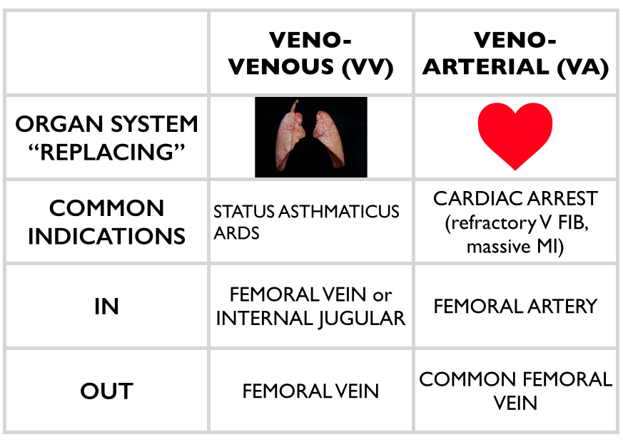
Extracorporeal Membrane Oxygenation (ECMO) – Dr. Haney Mallemat (@CriticalCareNow)
ECMO is promising in certain devestating disease processes – essentially heart or lung failure. For example, in the CHEER trial, the investigators had a 54% rate of neuro-intact survival after cardiac arrest with ECMO []. Yet, ECMO can be confusing. Dr. Mallemat simplified this for the emergency physician (see this site for more complete explanations)
Stop the Madness: Diagnostic Imaging in Nephrolithiasis – Workshop with Drs. Eddy Lang, Rebecca Smith-Bindman, Grant Innes, and Lauren Westafer
FOAMcast brings you pearls from conferences we attend and presently it’s the American College of Emergency Physicians annual meeting, ACEP15 in Boston.
Jeremy Hoffman and Rick Bukata summarize recent, relevant literature each year (recently known as #hofkata). This year, our top three favorite
Dr. Jeff Kline (@klinelab) spoke on pulmonary embolism.
High Risk PE? Consider lysis (this is controversial, we are simply reporting Dr. Kline’s talk)
Size and location. Massive and proximal= bad
SBP <90 for more than 15 min OR 40mmHg drop from baseline
Signs of RV strain – echocardiography showing RV dilation OR hypokinesis?
Elevated troponin or BNP
ECG findings suggestive of cardiac strain: sinus tachycardia, incomplete right bundle branch block, complete right bundle branch block, T-wave inversion in leads V1 – V4.
Kline also participated in a knowledge translation workshop where he argued that sub-segmental PEs, without DVT on ultrasound, are NOT a real thing. This is controversial but he also argued that treating these is associated with harm [Carrier et al]
This is an exciting week of primary literature, particularly as many large critical care trials were published in major journals despite being “negative studies.” We are excited by this as, too often, we see “negative studies” discarded. Further, these studies examined things some practices we seem to believe in: balanced crystalloids, apneic oxygenation (see Dr. Scott Weingart’s podcast on the FELLOW study), treating fever. We love that the Free Open Access Medical Education (FOAM) community and study authors are examining beloved practice and open to questioning the very things we believe in. Well done.
We cover two core content papers out by Dr. Paul Young (@dogICUma) in JAMA and NEJM this week. His trials are as clever as his Twitter handle.
SPLIT – The FOAM world has sung the praises of balanced fluids given they have more physiologic composition. The thought, as detailed in this post, is that 0.9% NaCl contains an ABnormally large amount of chloride which may cause a hyperchloremic metabolic acidosis. Prior literature suggests an increased incidence of kidney injury with saline compared with balanced solutions. Thus, Dr. Paul Young and colleagues sought to study this with the best trial, to date, on this topic.
Multicenter, blinded, cluster-randomized, double-crossover trial of adult ICU patients receiving crystalloids randomizing patients to 0.9%NaCl or Plasma-lyte (balanced solution).
Sites used one fluid for seven weeks and then crossed over to the other fluid (labeled Fluids A and B).
Primary outcome: AKI according to the RIFLE criteria within 90 days – no difference between groups.
9.6% in Plasma-lyte group vs 9.2% in the saline group (absolute difference 0.4% [95% CI, −2.1%-2.9%]; RR, 1.04 [95% CI, 0.80-1.36]; P = .77)
Secondary outcomes: No difference in renal replacement therapy, ICU days, mechanical ventilation, or mortality
A few things to keep in mind:
70% of patients were admitted to the ICU from the OR (mostly cardiac surgery) and only ~15% from the ED
Patients got a median of 2L (1L -3.5L) of the study fluid, that’s it. These were not large volume resuscitations.
90% of patients received fluids prior to enrollment, 60% got balanced crystalloid and only 30% 0.9% NaCl.
HEAT– We can’t help treating fever. We like the numbers euboxic, elevated temp? It must be bad! Doctors, parents, nurses treat fever reflexively. Yet, there’s a thought that fever may be evolutionary and could potentially be protective. Treating pain or discomfort? That’s one thing, but here the authors sought to determine if there was a clinically important benefit to treating the number in ICU patients.
Randomised controlled, double blinded study of n=690 ICU patients with T>38F + suspected infection randomized to receive either 1 g paracetamol (acetaminophen/APAP) or placebo every 6 hours.
Primary outcome: median ICU-free days to day 28 – no difference 23 (IQR 13-25) in paracetamol group vs 22 in placebo group (IQR 12-25); P=0.07
No difference in secondary outcomes of mortality at 28 and 90 days
Limitations: ~30% of patients in both arms received open label APAP after the course of the study drug
Bottom Line Pearls:
It appears we may be SPLITting hairs over fluid choices. Giving a couple of liters? Fluid choice may not matter. SPLIT does not provide literature for larger volume resuscitations.
Treating fever in ICU patients with suspected infection doesn’t have an effect on ICU free days. Treat discomfort and pain with APAP but don’t expect to save lives or ICU beds by doing so.
“Negative studies” are important. So is examining our practice.
The blog Brown Coat Nation (University of Illinois, Chicago) has a new series entitled “Inconceivable.” The idea is to expose medical terminology that we tend to use incorrectly. The first installment is focused on the misuse of the term “left shift,” and it’s the focus of this FOAMcastini.
The Core Content
The correct use of the term “left shift” refers to the presence of banded (immature) neutrophils in the blood. It does not refer to an elevated white blood cell count with a high percentage of neutrophils. An elevated white blood cell count with an abnormally high percentage of neutrophils should be called “neutrophillic leukocytosis.” Only the presence of immature neutrophils in the periphery (including bands) can accurately be called a “left shift.”
The term “left shift” is derived from the diagrams of the six stages of neutrophil development in the bone marrow. On the far left, you see the most basic precursor: the myeloblast. On the far right of the diagram one finds the mature segmented neutrophil (also known as the “polymorphonuclear leukocyte, or PMN). But just to the left of that is the “banded” neutrophil (the 5th stage of neutrophil development in which the large band of nuclear material has not yet “disbanded” into segments).
When an infection runs rampant, sometimes the bone marrow runs out of mature neutrophils to send to the periphery. So, the marrow panics and releases immature banded neutrophils that normally would not be considered “ready for prime time.”
Here’s some relevant spaced repetition on SIRS: Along with leukocytosis (>12,000) and leukopenia (<4,000), Bandemia >10% is one of the SIRS criteria (Systemic Inflammatory Response Syndrome). Temperature (>38C or <36C), Heart rate >90 (“sirs-ycardia”), tachypnea (RR>20) or PCO2 <32) are the other SIRS criteria. The presence of at least two these categories constitutes “positive SIRS”. In pediatrics, the temperature, heart rate, and respiratory rates should be age-adjusted.
Check out our previous episode on appendicitis for a reminder on whether leukocytosis (with or without neutrophillic predominance) is useful in risk stratification.
This week we cover a post from Dr. Rory Spiegel, author of EMnerd, on initial nonoperative management of acute appendicitis based on an article by Salminen et al in JAMA 2015.
530 patients with CT confirmed acute, uncomplicated appendicitis were randomized to operative intervention (n=273 receiving open laparotomies) or non-operative intervention (n=257 receiving antibiotics).
27.3% (n=70, CI 22-33.2%) of patients who received medical management (ertapenem x 3 days then 5 days of levofloxacin) had an appendectomy by the 1 year mark
7 patients (2.7%) in medical management group had complicated appendicitis at one year, 0 had abscesses
45 patients (20.5%) in the operative group had surgical complications
This is a non-inferiority study where the intent is to demonstrate that an experimental treatment (antibiotics alone) is not substantially worse than a control treatment (immediate surgery). The authors set the non-inferiority margin at 24%, which means that a failure rate (appendectomy by 1 year) >24% would render medical management inferior.
Authors Conclusion: “Among patients with CT-proven, uncomplicated appendicitis, antibiotic treatment did not meet the prespecified criterion for noninferiority compared with appendectomy.”
Spiegel’s Conclusion: “there is a great deal to be determined before this non-invasive strategy can be considered mainstream practice…in what was once considered an exclusively surgical disease, the majority of patients can effectively be managed conservatively. Despite not meeting their own high standards for non-inferiority, the authors demonstrated that for most patients with acute appendicitis, when treated conservatively with antibiotics we can avoid surgical intervention without complications of delays to definitive care.”
More FOAM on non-operative treatment of appendicitis: The SGEM
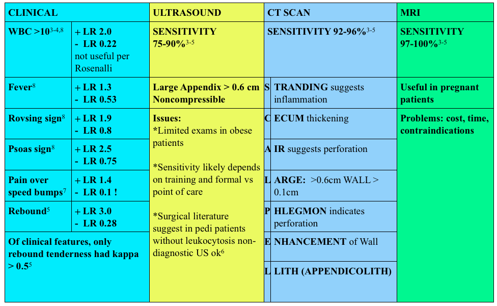
Use of contrast enhanced CT scans controversial. Rosenalli and the American College of radiology concur that oral contrast is probably not needed but does increase the emergency department length of stay [3-5].
Treatment:
Surgical consult
Antibiotics:
Broad spectrum beta-lactams: ampicillin-sulbactam 3g IV (75 mg/kg IV in peds) piperacillin-tazobactam 4.5g IV, cefoxitin 2g IV (40 mg/kg IV in peds) OR metronidazole 500 mg IV + ciprofloxacin 400 mg IV
Other things to consider in special populations in right lower quadrant:
1. A 22-year-old man presents with abdominal pain followed by vomiting for 1 day. His examination is significant for right lower quadrant tenderness to palpation. He has a negative Rovsing sign. [polldaddy poll=9026936]
2. A 22-year-old woman presents with lower abdominal pain and vaginal discharge. She is sexually active with men with inconsistent barrier protection. Her vitals are normal other than temperature of 101°F. On examination, there is yellow cervical discharge, no cervical motion tenderness, but uterine and left adnexal tenderness. An ultrasound does not show any evidence of tubo-ovarian abscess. [polldaddy poll=9026939]
Answers.
1. B. Sensitivity or the true positive rate measures the proportion of actual positives that are correctly identified as such. It is determined by dividing the number of true positives of the test by the number of true positives + false negatives. Tests with a high sensitivity are good for ruling out disease as the test has very few false negatives. A test with high sensitivity is advantageous as a screening tool as it misses very few people with the disease. The onset of pain before vomiting has been found to be as high as 100% sensitive in diagnosing acute appendicitis.Rovsing’s sign (D) (indirect tenderness) describes pain felt in the right lower quadrant upon palpation of the left lower quadrant. This sign signifies the presence of peritoneal irritation and has a sensitivity of 58%. Right lower quadrant pain (C) has a sensitivity of 81% and fever (A) has a sensitivity of 67%.
2.This patient presents with signs and symptoms consistent with pelvic inflammatory disease (PID) and should be treated with ceftriaxone 250 mg IM and 2 weeks of doxycycline. PID is an ascending infection beginning in the cervix and vagina and ascending to the upper genital tract. Neisseria gonorrhoeae and Chlamydia trachomatis are most commonly implicated. It can present with a myriad of symptoms although lower abdominal pain is the most common. Other symptoms include fever, cervical or vaginal discharge and dyspareunia. Pelvic examination reveals cervical motion tenderness (CMT), adnexal tenderness and vaginal or cervical discharge. Inadequately treated PID can lead to tubo-ovarian abscess, chronic dyspareunia and infertility. Due to the variable presentation and serious sequelae, the CDC recommends empiric treatment of all sexually active women who present with pelvic or abdominal pain and have any one of the following: 1) CMT, 2) adnexal tenderness or 3) uterine tenderness. Treatment should cover the most common organisms and typically consists of a third generation cephalosporin (ceftriaxone) and a prolonged course of doxycycline. Patients with systemic manifestations or difficulty tolerating PO should be admitted for management.Ceftriaxone and azithromycin (A) are used in the treatment of cervicitis or urethritis. Clindamycin (C) and metronidazole (D) are used in the treatment of bacterial vaginosis. References:
1.Salminen P, Paajanen H, Rautio T, et al. Antibiotic Therapy vs Appendectomy for Treatment of Uncomplicated Acute Appendicitis: The APPAC Randomized Clinical Trial. JAMA. 2015;313(23):2340
This week we cover posts from the Wessex ICS site, The Bottom Line, which is an excellent source for breakdown of recent and important trials. This site is great for reviews of high impact trials in critical care. We cover their post on a systematic review of peripheral pressor complications and then we delve into a recent prospective trial by Cardenas-Garcia and colleagues that came up at SMACC.
Systematic review of the literature 1946-Jan 2014 (does not include most recent trial)
Outcome – local tissue injury or extravasation: 325 separate events, 318/325 peripheral pressors
Signal that distal lines are not ideal for running pressors: 204 events (local tissue injury) were distal to the antecubital fossa/popliteal fossa (90% of events)
Signal that duration of pressors running peripherally may impact likelihood of adverse event. Increasing number of events were reported at the 6-12 hour mark (n=9) then 12-24 hour (n=18) and then almost all >24 hour
ICU fellows and attendings determined if peripheral pressors were warranted and then initiated the following protocol:
Vein diameter >4 mm measured with ultrasonography and PIV confirmed with US before pressors started
Upper extremity only, contralateral to the blood pressure cuff
IV size 20 gauge or 18 gauge
No hand, wrist, or antecubital fossa PIV access position
Blood return from the PIV access prior to VM administration
Assessment of PIV access function q 2h as per nursing protocol
Immediate alert by nursing staff to the medical team if line extravasation, with prompt initiation of local treatment
72 hours maximum duration of PIV access use
N=734 patients
19/783 peripheral vasopressor administrations with infiltration of site (2%) with no events of local tissue injury
The take home: If a patient needs vasopressors, you can start them through a good, proximal peripheral IV. Sometimes patient or situation factors delay central lines, this doesn’t mean it needs to delay patient’s therapy. Know what to do in the event of infiltration (see this EMCrit post).
Core Content
Tintinalli (7e) Chapter 24
Panchal et al – Phenylephrine bolus dosing in peri-intubation period
Answer. C. Norepinephrine is considered the vasopressor of choice for treatment of septic shock. Norepinephrine acts primarily as an α-adrenergic agonist, causing vasoconstriction that results in an increase in blood pressure. It also has β-adrenergic properties, which causes an increase in cardiac output and heart rate. The combination of α-adrenergic and β-adrenergic properties benefits patients who have septic shock. Norepinephrine also has a short duration of action, which allows for rapid adjustment of dosing in response to changes in a patient’s hemodynamic status. Dopamine (A) was once widely used in the treatment of septic shock, but studies have shown that it has no advantage over norepinephrine and its use is associated with a higher death rate. Epinephrine (B) has both α-adrenergic and β-adrenergic properties and has a greater affinity for alpha- and beta-receptors than norepinephrine. Its use is associated with a higher rate of cardiac dysrhythmias and a decrease in splanchnic blood flow. Phenylephrine (D) is a pure α-adrenergic agent that causes vasoconstriction and impairment of tissue blood flow throughout the body, most notably in the splanchnic circulation.