Episode 43 – Alcohols

(ITUNES OR LISTEN HERE)

The Free Open Access Medical Education (FOAM)

The Skeptic’s Guide to Emergency Medicine Episode 144, “That Smell of Isopropyl Alcohol for Nausea in the Emergency Department.” This podcast reviews an article by Beadle et al, an RCT on the use of inhalational isopropyl alcohol for nausea.

Population – Adults in the Emergency Department

Intervention – Nasal inhalation of an isopropyl alcohol pad for ~ 60 seconds (time 0 and, if still nauseated, at 2 minutes and 4 minutes)

Comparison – Nasal inhalation of an identical pad soaked in saline.

Outcome -Nausea score at 10 minutes post treatment using an 11-point verbal numeric response scale (0:“no nausea”; 10:“worst nausea imaginable”)

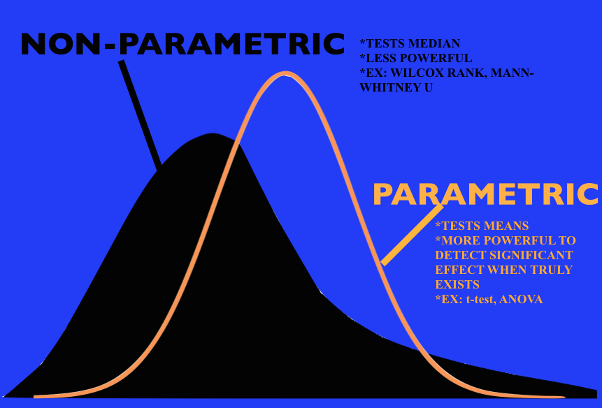
  • Lower in the isopropyl group (Median score of 3 vs. 6 on an 11 point scale, p<0.001). This gave an effect size of 3 (95% CI 2 to 4).

Limitations – single center, convenience sample, unclear blinding as isopropyl likely has a particular smell to it, outcome nausea score, not vomiting

Eagerton-Warburton et al conducted an RCT of intravenous ondansetron 4mg, metoclopramide 20mg, or saline (placebo) of adult patients with nausea and found  no significant difference in nausea scores on a visual analog scale between groups (n=270).

Core Content

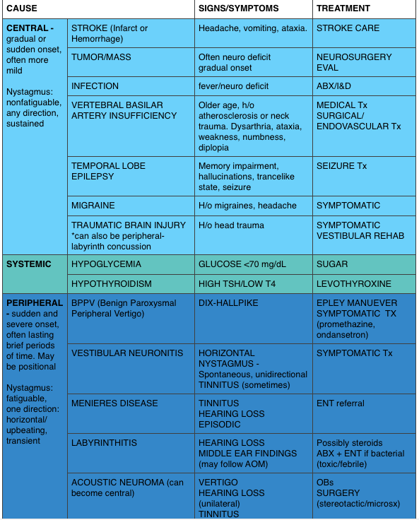
We delve into core content on vertigo using Rosen’s Medicine (8e),  Chapter 155 “Toxic Alcohols”  and Tintinalli’s Emergency Medicine: A Comprehensive Study Guide  (7e) Chapter 179“Alcohols.”

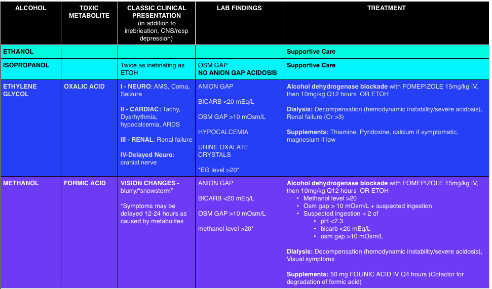
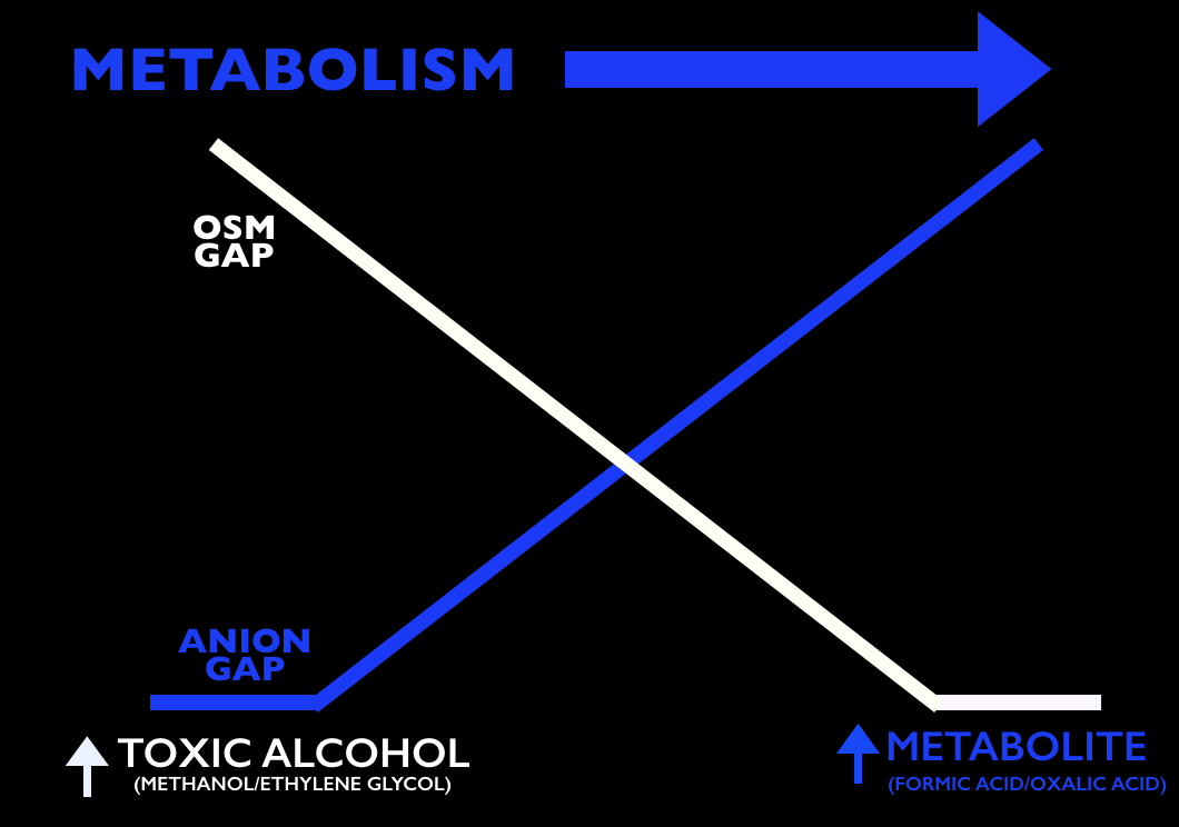
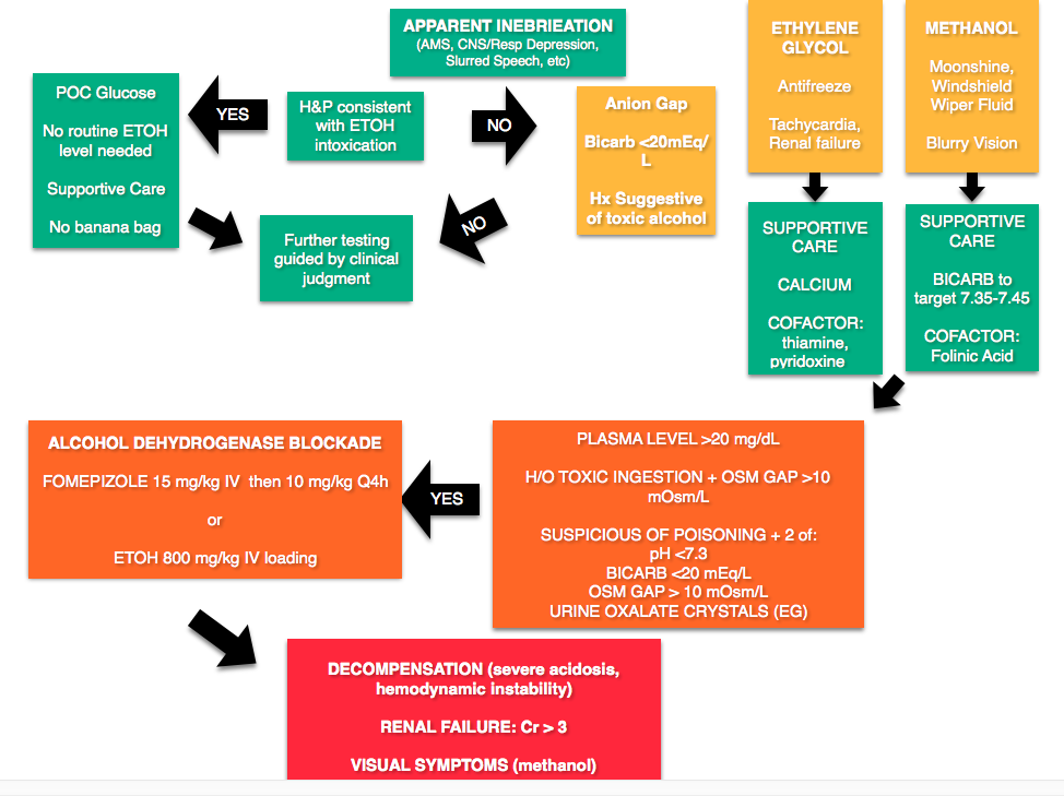
Any alcohol can be “toxic,” the ramifications depend on the dose. Toxic alcohols typically refer to ethylene glycol (EG) and methanol.  In these cases, the parent compound (EG or methanol) is inebriating but not particularly toxic. These compounds are metabolized, like any alcohol, but unfortunately the metabolites (oxalic acid – EG, formic acid – methanol) have unique toxic properties. For example, the oxalic acid produced from EG metabolism combines with calcium.  These deposit into tissues causing renal failure and neurologic sympotms. The formic acid has a predilection for the retina, causing visual symptoms.

Screen Shot 2016-02-19 at 7.01.02 PM

Toxic Alcohols

Osmolal Gap (calculator) – One of the touted features of toxic alcohols is the elevated anion gap, that is, the difference between the measured serum osmolality and the calculated osmolarity.

Calculated Osmolarity: 2[Sodium] + [Glucose]/18 + [BUN]/2.8 + [ETOH]/4.6    (Note: Some references use ETOH/3.7)

  • Problems:  A normal osm gap does not exclude the presence of toxic alcohols as the osm gap decreases as the alcohol is metabolized.  Also, other disease processes can increase the osm gap, including shock, diabetic ketoacidosis, etc [2,3].
Osm Gap vs Anion Gap As Toxic Alcohols Metabolize

A common pearl exists that urine can be placed under a Wood’s lamp for fluorescence due to the fluorescein in ethylene glycol antifreeze (to detect leaks). Unfortunately this is neither sensitive nor specific [4,5].

Treatment

Screen Shot 2016-02-17 at 9.04.49 PM

Rosh Review Questions

Question 1. [polldaddy poll=9311166]

Question 2. [polldaddy poll=9311159]

Answers

1. A. Hypocalcemia. Most laboratories do not have the capability to measure serum levels of toxic alcohols in a timely fashion. As a result, physicians must use other clinical markers to determine the presence of these unmeasured toxins. The classic laboratory abnormality is an elevated serum osmolality with an osmolar gap since the actual toxic alcohol is not measured in its own quantity but contributes to the osmolality of the blood. When calculating the osmolar gap, it is important to have a measured serum ethanol level as this is a large contributor to the final osmolality.Ethylene glycol ingestion leads to the formation of calcium oxalate crystals. As a result, serum calcium levels will decrease and hypocalcemia is a surrogate marker for ethylene glycol toxicity.Hypoglycemia (B) is not characteristic of a toxic alcohol ingestion. Hypoglycemia is often seen in chronic alcoholics due to poor nutritional intake. In children, ethanol may induce hypoglycemia. Other toxic ingestions which may lead to hypoglycemia include beta-blockers, sulfonylureas and insulin. Hypokalemia (C) is not associated with any toxic alcohol ingestions. As patients become more acidemic from alcohol ingestions, hyperkalemia may develop. Hyponatremia (D) is not associated with toxic alcohol ingestions. Evidence of hyponatremia on laboratory analysis should prompt further investigation for other underlying causes.

2.C.  Glycolic acid level greater than 8 mmol/L. A glycolic acid level greater than 8 mmol/L is an indication for emergent hemodialysis following an acute ethylene glycol overdose. Ethylene glycol is found in automobile coolants, antifreeze, hydraulic brake fluid, de-icing agents and industrial solvents. Symptoms progress through 4 stages: inebriation and intoxication, cardiopulmonary symptoms (tachycardia, hypertension, pulmonary edema), renal symptoms (acute renal failure), and delayed neurologic symptoms (cranial neuropathy). Treatment includes 1.) Alcohol dehydrogenase (ADH) enzyme blockade with fomepizole or ethanol to prevent toxic and acidic metabolite production, 2.) Correction of the metabolic acidosis with sodium bicarbonate, and 3.) Hemodialysis for removal of the parent compound and toxic metabolites. Although somewhat controversial with the advent of fomepizole other indications for hemodialysis following ethylene glycol ingestion include severe metabolic acidosis, renal failure, deterioration despite intensive care, and electrolyte disturbances.Other indications for emergent hemodialysis following an acute ethylene glycol overdose include: ethylene glycol level greater than 50 mg/dL (B), anion gap greater than 20 mmol/L (A)and initial pH less than 7.3 (D).

References

  1.  Beadle KL, Helbling AR, Love SL, April MD, Hunter CJ. Isopropyl Alcohol Nasal Inhalation for Nausea in the Emergency Department: A Randomized Controlled Trial. Annals of emergency medicine. 2015. [pubmed]
  2. Chapter 155.  “Toxic Alcohols. ” In: Marx JA, Hockberger RS, Walls RM eds.  Rosen’s Emergency Medicine, 8th e.
  3. Chapter 179. Alcohols. In: Tintinalli JE, Stapczynski J, Ma O, Cline DM, Cydulka RK, Meckler GD, T. eds. Tintinalli’s Emergency Medicine: A Comprehensive Study Guide, 7e.New York, NY: McGraw-Hill; 2011.
  4. Tobe TJM, Braam GB, Meulenbelt J, et al: Ethylene glycol poisoning mimicking Snow White. Lancet 2002; 359: pp. 444
  5. Wallace KL, Suchard JR, Curry SC, et al: Diagnostic use of physicians’ detection of urine fluorescence in a simulated ingestion of sodium fluorescein–containing antifreeze. Ann Emerg Med 2001; 38: pp. 49

Episode 42 – End of Life

(ITUNES OR LISTEN HERE)

The Free Open Access Medical Education (FOAM)

We cover an EMcrit episode on Semantics of End of Life Discussions with Dr. Ashley Shreves as well as pearls from another favorite episode with her, Episode 93 – Critical Care Palliation.  We can’t do these episodes justice summarizing them so listen to them. (Update:  Apparently these podcasts were taken down due to personal reasons. From what we understand you may be able to obtain a copy by contacting the EMcrit site).

Screen who to have “the conversation” with looking for patients with signs of instability:

  • 85 y/o and older
  • dementia/frailty
  • advanced cancer/disease

Key conversational points:

  • Lead with, ” I’m so worried about (the patient).”
  • “What do you think your (mom/dad/etc) would say about how she is now?”
  • “Got it.”  Whether a family member agrees or disagree, let them know you heard them.

If a patient/family member are overly optimistic about the patient getting better one can try, “Many people find it helpful to talk about what would we should do if (the patient) doesn’t get better.”

Respect patient/family values that differ from yours.  If the patient’s family just wants the heart beating regardless, and that’s ok.  Per Dr. Shreves, this population is 5-10% and may be called “vitalists.”

Another key point Dr. Shreves emphasizes is that palliative care, comfort care, and allowing a natural death often mean escalating care – ensuring the patient is comfortable, clean, etc.

Core content 

We delve into core content on vertigo using Rosen’s Medicine (8e) electronic chapter, “End of Life,”  and Tintinalli’s Emergency Medicine: A Comprehensive Study Guide  (7e) Chapter 297 “Death and Dying.”

DNAR (Do Not Attempt Resuscitation) –  technically only speak to a patient’s wishes to receive CPR [3].  Problematic for several reasons, including:

  • Issue lies in the word “resuscitate,” which may be used to include fluids, antibiotics, vasopressors, advanced means of ventilation or, at the extreme, CPR.  The AHA guidelines have moved to DNAR from DNR but even this language isn’t clear.
  • Major societies are moving towards the language AND, Allow Natural Death [4].
    • AND is preferred because it describes what happens and is more clear, is kinder language laden with reduced guilt
  • The TRIAD II-IV studies surveyed EMS personnel, physicians, and medical students respectively and provided the participants with an advance care directive as well as case scenarios.  The participants then indicated whether a patient was a DNR or full code and the appropriate action.  Both physicians and EMS providers performed poorly and variably, indicating that the directives were not clear [5-7].

Palliative care and hospice are not interchangeable.  Palliative care has a more broad definition and

  • Palliative care: “An approach that improves quality of life of patients and their families facing the problem associated with life-threatening illness, through the prevention and relief of suffering by means of early identification and impeccable assessment and treatment of pain and other problems, physical, psychosocial and spiritual ” [1].  There is no time limit in this scenario.
  • Hospice care: subset of patients that a doctor has estimated likely has 6 months or less to live [1].

Opioid Equivalents

Many terminally ill patients or those at the end of their life are on chronic opioids.  While the opioid epidemic is a problem, this is not the appropriate population from which we should withhold appropriate analgesia. It can be difficult to convert between dosages to adequately treat a patient’s pain.  Here’s a free calculatorScreen Shot 2016-01-21 at 8.04.21 AM

References

  1. Online Chapter. End of Life. In: Marx JA, Hockberger RS, Walls RM eds.  Rosen’s Emergency Medicine, 8th e.
  2. Chapter 297. Death and Dying. In: Tintinalli JE, Stapczynski J, Ma O, Cline DM, Cydulka RK, Meckler GD, T. eds. Tintinalli’s Emergency Medicine: A Comprehensive Study Guide, 7e. New York, NY: McGraw-Hill; 2011.
  3. Dugdale DC. .Do Not Resuscitate Orders.”  MedlinePlus Medical Encyclopedia.
  4. Breault JL. DNR, DNAR, or AND? Is Language Important? Ochsner J. 2011;11(4):302
  5. Mirarchi FL, Kalantzis S, Hunter D, McCracken E, Kisiel T. TRIAD II: do living wills have an impact on pre-hospital lifesaving care? J Emerg Med. 2009;36(2):105–15. doi:10.1016/j.jemermed.2008.10.003.
  6. Mirarchi FL, Costello E, Puller J, Cooney T, Kottkamp N. TRIAD III: nationwide assessment of living wills and do not resuscitate orders. J Emerg Med. 2012;42(5):511–20. doi:10.1016/j.jemermed.2011.07.015.
  7.  Mirarchi FL, Ray M, Cooney T.  TRIAD IV: Nationwide Survey of Medical Students’ Understanding of Living Wills and DNR Orders. J Patient Saf. 2014 Feb 27.
  8. Gurwitz JH, Lessard DM, Bedell SE, Gore JM. Do-Not-Resuscitate Orders in Patients Hospitalized With Acute Myocardial Infarction. 2014;164.
  9. Adams DH, Snedden DP. How misconceptions among elderly patients regarding survival outcomes of inpatient cardiopulmonary resuscitation affect do-not-resuscitate orders. J Am Osteopath Assoc. 2006;106(7):402–4.

Episode 41 – Vertigo

(ITUNES OR LISTEN HERE)

The Free Open Access Medical Education (FOAM)

We cover two bits of FOAM, one from Emergency Medicine Literature of note on the use of meclizine for vertigo and an EMcrit episode on the HiNTs exam.

Emergency Medicine Literature of Note – Dr. Ryan Radecki – Treating what you believe is peripheral vertigo?  Using meclizine? So are most people.  But this is not an evidence based practice.  While meclizine is Rosen approved, Tintinalli recommends transdermal scopolamine as the first line treatment [1,2]. Following a recent recall of meclizine (oddly because iron bottles contained meclizine instead of iron), Dr. Radecki probes into why we use meclizine for vertigo.

  • Meclizine is an anti-histamine and has been thought to have anti-emetic properties.
  • A 1968 paper compared 16 anti-emetics/combinations and did NOT conclude that meclizine was the best. In fact, scopolamine and amphetamine performed best. Promethazine (phenergan) is also a good choice based on this paper [3].

EMcrit – Dr. Scott Weingart – The HiNTs exam has taken off, particularly in the FOAM world, as a means of disguising between central and peripheral causes of vertigo. In 2010, an EMcrit episode popularized this in the FOAM world. See this video demonstrating the HiNTs exam.

  • Head Impulse – rapid head rotation by the examiner with the subject’s vision fixed on an object (i.e. examiner’s nose). The examiner rapidly rotates the head towards midline and the patient’s eyes should remain fixed on the target. 
    • Abnormal (loss of fixation on target and movement of eyes away from target followed by correct saccade as patient fixates on target) = peripheral (vestibulo-ocular reflex impaired)
    • Normal = central
  • Nystagmus 
    • Horizontal nystagmus with a unidirectional fast phase (away from affected side) = probably peripheral
      • Patients with horizontal nystagmus may have central pathology but may have direction-changing nystagmus (i.e. fast phase beating in one direction when looking to right and the opposite direction when looking left).
    • Vertical or nystagmus = central pathology
  • Test of Skew Patients should fixate on a target while the provider alternately covers each eye. As the cover is moved from one eye to the other, the uncovered eye must correct for the misalignment and will look up or down to focus back on the target. This slight correction is observed repeatedly as the cover is moved from one eye to the other.
    • Skew deviation/misalignment = probably central, often in posterior fossa abnormalities
    • No skew deviation= peripheral

INFARCT – Impulse Normal, Fast-phase Alternating nystagmus, and Refixation on Cover Test

Issues with HiNTs

  • Can only be performed on patients with continuous vertigo.
  • External validity is a major issue with HiNTs.
    • Providers – Of the 4 studies have examined the operating characteristics of HiNTs, none have used emergency providers and instead have examined how the exam performs in the hands of two neuro-ophthalmologists, neuro-otologists, and neurologists with 4 hours of specialized training in the exam. It’s unclear whether HiNTs would be reliable or valid when performed by emergency providers [4-6].
    • Patients – The patients examined in many of these studies have other indicators of badness on neurologic exam. In one study, patients had to have gait instability and/or truncal ataxia to enroll.  Then, 76/101 (76%) of those patients had a central cause. These patients were sick and not the undifferentiated vertiginous patients we see primarily as emergency providers [4-6].
    • In the words of leading HiNTs expert Dr. Newman-Toker, HiNTs “requires expertise not routinely available in emergency departments.” As such, his team is piloting quantitative video-oculography to aid in diagnosis using HiNTs [7]. An Annals of Emergency Medicine review also warned that HiNTs may not be ready for emergency provider use [9].

More FOAM on HiNTs: EMJclub, EMNerd

Core content 

We delve into core content on vertigo using Rosen’s Medicine (8e) Chapter 19,  and Tintinalli’s Emergency Medicine: A Comprehensive Study Guide  (7e) Chapter 164 “Vertigo and Dizziness.”

Vertigo is often characterized by the sensation of spinning and falls into the broad and frustrating category of “dizziness.”  Often, when a dizzy patient presents we perseverate on characterizes what the patient means by “dizzy.” However, some argue that this is not an appropriate approach as a study found 50% of patients changed the character of their dizziness when questioned again after 10 minutes [9].  Additionally, the clinical characteristics differentiating peripheral from central causes of vertigo are not entirely reliable. Despite these limitations, it is expected that we are familiar with “classic” presentations.

Screen Shot 2016-01-13 at 8.32.04 AM
*”Classic” presentations

Generously Donated Rosh Review Questions 

  1. A 50-year-old man presents with episodic severe vertigo lasting hours, with associated symptoms of unilateral tinnitus, fluctuating low-frequency hearing loss, and aural fullness. [polldaddy poll=9245427]
  2. A 20-year-old woman presents with an acute onset of dizziness. The patient describes the sensation that the room is spinning when she turns her head to the left and it is accompanied by nausea and vomiting. The symptoms resolve with turning her head away from that side. Examination reveals nystagmus elicited by deviating the eyes to the left and no other neurologic findings. [polldaddy poll=9245971]

Answers

1. B. Meniere’s disease is characterized by episodic severe vertigo lasting hours, with associated symptoms of unilateral tinnitus, fluctuating low-frequency hearing loss, and aural fullness. Typical onset is in the fifth decade of life. The cause is uncertain but is speculated to result from allergic, infectious, or autoimmune injury. The histopathologic finding includes endolymphatic hydrops, which is thought to be caused by either overproduction or underresorption of endolymph in the inner ear. Meniere’s disease is a clinical diagnosis mostly based on history. Testing may be obtained to support the diagnosis and rule out other disorders. Audiometry often demonstrates a low-frequency sensorineural hearing loss. An FTA-ABS test may be obtained to rule out syphilis. Electronystagmography (ENG) may demonstrate a unilateral peripheral vestibular weakness on caloric testing. When the diagnosis is uncertain, a brain MRI with contrast is obtained to evaluate for a retrocochlear lesion. The differential diagnosis of Meniere’s disease includes acute labyrinthitis, neurosyphilis, labyrinthine fistula, autoimmune inner ear disease, vestibular neuronitis, and migraine-associated vertigo.The most common cause of peripheral vestibular vertigo in adults is benign paroxysmal positional vertigo (A). BPPV occurs in all age groups but more often between ages 50 and 70 but is not associated with hearing loss and made worse with movement. In a perilymph fistula (C) rapid changes in air pressure (barotrauma), otologic surgery, violent nose blowing or sneezing, head trauma, or chronic ear disease may cause leakage of perilymph fluid from the inner ear into the middle ear and result in episodes of vertigo. Associated signs and symptoms are variable but can include a sudden pop in the ear followed by hearing loss, vertigo, and sometimes tinnitus. Acute vertigo associated with nausea and vomiting (but without neurologic or audiologic symptoms) that originates in the vestibular nerve is known as vestibular neuronitis (D). Vestibular neuronitis can occur spontaneously or can follow viral illness.

2. B. This patient presents with peripheral vertigo most consistent with benign paroxysmal peripheral vertigo (BPPV) and should be treated with an Epley maneuver. Vertigo is defined as the sensation of disorientation in space combined with a sensation of motion. Patients typically describe a “room-spinning” sensation or the feeling of sea sickness. Vertigo can be divided into two types: central and peripheral. Central vertigo are those disorders arising from the central nervous system and include ischemic stroke, vertebrobasilar insufficiency and infectious causes (meningitis, mastoiditis, syphilis). Central vertigo is characterized by longer duration of symptoms, minimal change with position, gradual onset and multidirectional nystagmus. Peripheral vertigo includes BPPV, Meniere’s disease, Labyrinthitis and vestibular neuritis. Peripheral vertigo may have intermittent symptoms (BPPV) or continuous symptoms but should not be associated with other neurologic deficits or changes and should have unidirectional nystagmus. The symptoms in BPPV are elicited by specific movements of the head and relieved by returning the head to a neutral position. The symptoms should be acute in onset and of a short duration. In BPPV, the symptoms are cause by the presence of an otolith in one of the semicircular canals. Although pharmacologic intervention may be necessary in the acute setting with meclizine or benzodiazepines, the best treatment for BPPV is the Epley maneuver. The Epley maneuver is a series of positions that the clinician takes the patient through that leads to expulsion of the otolith from the semicircular canal and relief of symptoms. Imaging with a non-contrast head CT (C) is not indicated in peripheral vertigo of any cause as the patient’s pathology is in the inner ear and not the brain. If a central cause is suspected, MRI of the brain (A) is the best test for diagnosis as the causative lesion will likely be in the posterior fossa, which is not seen well on CT scan. Steroid treatment (D) is the indicated management for vestibular neuritis but does not play a role in the treatment of BPPV.

References:

  1. Chang AK, Olshaker AS. Dizziness and Vertigo. In: Marx JA, Hockberger RS, Walls RM eds.  Rosen’s Emergency Medicine, 8th e.
  2. Goldman B. Chapter 164. Vertigo and Dizziness. In: Tintinalli JE, Stapczynski J, Ma O, Cline DM, Cydulka RK, Meckler GD, T. eds. Tintinalli’s Emergency Medicine: A Comprehensive Study Guide, 7e. New York, NY: McGraw-Hill; 2011.
  3. Wood CD, Graybiel A. Evaluation of sixteen anti-motion sickness drugs under controlled laboratory conditions. Aerospace medicine. 39(12):1341-4. 1968.
  4. Newman-Toker DE, Kattah JC, Alvernia JE, Wang DZ. Normal head impulse test differentiates acute cerebellar strokes from vestibular neuritis. Neurology. 70(24 Pt 2):2378-85. 2008.
  5. Kattah et al. HINTS to Diagnose Stroke in the Acute Vestibular Syndrome Three-Step Bedside Oculomotor Examination More Sensitive Than Early MRI Diffusion-Weighted Imaging. Stroke. 2009; 40: 3504-3510
  6. Newman-Toker et al. HINTS Outperforms ABCD2 to Screen for Stroke in Acute Continuous Vertigo and Dizziness. Academic Emergency Medicine. Volume 20, Issue 10, pages 986–996, October 2013
  7. Newman-Toked DE, Saber Tehran AS, Mantokoudis G et al. Quantitative video-oculography to help diagnose stroke in acute vertigo and dizziness: toward an ECG for the eyes. Stroke. 44(4):1158-61
  8. Cohn B. Can Bedside Oculomotor (HINTS) Testing Differentiate Central From Peripheral Causes of Vertigo? Annals of Emergency Medicine. 64(3):265-268. 2014. 
  9. Edlow JA. Diagnosing Dizziness: We Are Teaching the Wrong Paradigm!. Acad Emerg Med. 20(10):1064-1066. 2013.
  10. Newman-Toker DE, Cannon LM, Stofferahn ME, Rothman RE, Hsieh YH, Zee DS. Imprecision in patient reports of dizziness symptom quality: a cross-sectional study conducted in an acute care setting. Mayo Clinic proceedings. 82(11):1329-40. 2007.
  11. Strupp M, Zingler VC, Arbusow V et al. Methylprednisolone, Valacyclovir, or the Combination for Vestibular Neuritis. N Engl J Med. 351(4):354-361. 2004. 
  12. CFishman JM, Burgess C, Waddell A. Corticosteroids for the treatment of idiopathic acute vestibular dysfunction (vestibular neuritis). The Cochrane database of systematic reviews. 2011. 

Best Pearls and Biggest Trends of 2015

(ITUNES OR LISTEN HERE)

Sleep through 2015?  We picked the minds of some brilliant Emergency Medicine folk and came up with this short list of important happenings.

TRAUMA

FAST (Focused Assessment using Sonography in Trauma) guided resuscitative –  thoracotomy. (Recommended by Haney Mallemat, Rob Orman).

Bottom Line: If a trauma code comes in and has neither cardiac activity nor pericardial effusion on FAST, the odds of survival are essentially nil.  Inaba and colleagues found the following:

  • Population: 187 patients at LA/USC deemed “appropriate” for thoracotomy (at this institution: penetrating trauma patients with absent vital signs and blunt trauma patients with a loss of vital signs en route or in the resuscitation bay).
  • Intervention: Emergency medicine resident performed FAST before/concurrent with thoracotomy
  • Outcome:  Of the 126 patients without cardiac activity on FAST, none survived.

Many of the patients with cardiac activity did not survive, as well.  This paper gives individuals guidance to make the decision to crack the chest but has stirred up a debate as to whether this would lessen educational opportunities for a potentially heroic procedure.

Say NO to long backboards (Recommended by Lauren Westafer). In January 2015, ACEP recommended against the use of long backboards. Many state and local protocols shifted away from moving this some time ago and even more since.  The FOAM community has been up in arms about the inefficacies and harms of backboards for quite some time, again echoing that FOAM can serve as a forecaster for change to make it less cognitively distressing when time to change our practice.

RESUSCITATION

Peripheral Vasopressors (Recommended by Haney Mallemat, Rob Orman) – We detail the literature underlying the use of peripheral vasopressors in this podcast.

They think that the combination of a systematic review by Loubani et al and The Cardenas-Garcia study, peripheral vasopressors may be safely run through large bore peripheral IVs proximal to the antecubital fossa. Note: These should be closely monitored (protocolized is best) and short durations (<6 h) have been associated with minimal complicaitons.

SEPSIS

Intravenous fluids in sepsis (Recommended by Haney Mallemat). The PROMISE trial was published in early 2015, adding to ProCESS and ARISE. In these studies, patients typically got 2 L of crystalloid upfront and then 2L in the first 6 hours. Over 3 days after enrollment, most got just under 4 L.  Most patients received under 6L IVF.  There has been a movement for more judicious use of fluids in sepsis rather than dumping 4-6 L of IVF upfront.  Marik articulately explained this in this article.

  • Mallemat challenges us: Before giving a fluid bolus use ultrasound and ask these questions, “Does the LV need it, and can the RV take it?”

CMS Core Measure (Recommended by Jeremy Faust).  The National Quality Forum has been pushing for Measure 0500 See this EMcrit podcast on this topic.

ANALGESIA

Pain control in acute low back pain is tricky, and opioids may not help (Recommended by David Newman).

RENAL

Sexual intercourse 3-4 times per week may aid in expulsion of distal kidney stones. This year, two large studies by Pickard et al and Furyk et al demonstrated no benefit in stone passage for ureterolithiasis (particularly in stones <5 mm).  Then, a paper by Dolouglu et al excited many folks, if for entertainment value. Since tamsulosin doesn’t seem to help, what about sexual intercourse, 3-4 times per week, in male patients with partners?

In this study the mean expulsion time did not differ significantly between groups.

MEDICAL EDUCATION

Merging of FOAM resources. (Recommended by Michelle Lin). Blogs and podcasts are growing and often supplement one another. Dr. Lin predicts the future will be in these conglomerates (ex: merging of EMcrit and PulmCrit and massive undertakings such as ALiEM and CandiEM).

Merging of FOAM with traditional journals. (Recommended by Michelle Lin). Projects such as the Skeptic’s Guide to Emergency Medicine have merged with Academic Emergency Medicine and the Canadian Journal of Emergency Medicine (ex: SGEM HOP, journal paper) and massive FOAM resource ALiEM has also collaborated with Annals of Emergency Medicine (Ex:Journal club, paper). The merging of FOAM with paid, traditional resources is the future, per Dr. Lin.

OVERDIAGNOSIS

Overdiagnosis is a problem, and people are starting to rage against it. (Recommended by Lauren Westafer).  An Overdiagnosis conference exists and JAMA Internal Medicine has a series of articles, “Less is More,” frequently detailing evidence of overdiagnosis.

  • One of 2015’s prominent articles for emergency physicians was the Hutchinson et al study.  In this study, CTPA scans read as positive for pulmonary embolism underwent review by 3 chest radiologist who adjudicated that, actually, 25.9% of the “positive” scans (n=45) did not actually have pulmonary embolisms. The harms from this exist beyond the risk of anticoagulation (think about how an ED approach for a myriad of complaints differs for a patient with a history of thromboembolism).

The FOAM community swelled with appreciation and respect for the late Dr. John Hinds.  Please watch his SMACC talk, “Crack the Chest, Get Crucified,” in which his excellence in medical education shines, delivering pearls for nearly anyone.

Episode 40 – Femoral Nerve Blocks & Compartment Syndrome

(ITUNES OR LISTEN HERE)

The Free Open Access Medical Education (FOAM)

We cover one of Dr. Ken Milne’s podcasts on The Skeptic’s Guide to Emergency Medicine on regional anesthesia for hip fractures.  They cover a systematic review by Ritcey et al that found a reduction in pain scores in adult patients with femoral neck or hip fractures receiving one of the following means of regional anesthesia:

Why may this be important?

  • Better pain control for uncomfortable patients. Many patients with hip and femoral neck fractures are elderly. As such, opioids may be underdosed or pain medications may be used sparingly in these patients. Further, patients may still have pain with transfers.
  • Reduction in opioids. Opioids relieve pain but often have deleterious side effects. In addition to hypotension and allergic reactions, opioids may cause delirium.

Why don’t we do it? As a knowledge translation project, Dr. Milne’s podcast aims to propagate the best available, clinically relevant information to practitioners to mitigate the knowledge translation gap as much of the other world provides these blocks routinely to suspected fractures. A study by Haslam and colleagues from Canada suggests that while emergency providers know regional anesthesia is good in this scenario there are other barriers to adoption in North America [2].  Systemic barriers exist and include consultant worries about compartment syndrome, which is largely unfounded [3].  See this post for a more in-depth exploration.

Core Content

Compartment Syndrome – Tintinalli (7e) Chapter 275, “Compartment Syndrome.” Rosen’s  (8e) Chapter 49, “General Principles of Orthopedic Injuries.”

Compartment syndrome is typically caused by too much pressure in a confined space. Compartments are often surrounded by fascia and tissue with limited ability to stretch.  When the volume in the compartment increases or external forces compresses the compartment, blood flow in and out of the compartment are compromised.  Certain areas of the body are more predisposed to compartment syndrome (classics are the lower leg in tibia fractures and the forearm compartments).

Causes: Most cases of compartment syndrome are caused by fractures (~75%) but certainly not all.

Screen Shot 2015-12-23 at 8.04.41 AM

Diagnosis:

  • Clinical.  Classically this was taught as the 5 P’s (pain, parethesias, pallor, pulselessness, poikilothermia).  Yet, many of these perform poorly in real life.
  • Compartment pressures.  Many commercial devices exist to measure compartment pressures, yet these readings have poor specificity for compartment syndrome.
    • Historically, compartment pressures >30 mmHg or a delta pressure (Diastolic pressure minus the compartment pressure) <20-30 mmHg has indicated compartment syndrome.
    • Studies have measured compartment pressures on long bone fracture patients NOT suspected of having compartment syndrome and found that acting based on pressures would have resulted in 24-35% overdiagnosis (depending on a delta pressure cut-off of 20 mmHg and 30 mmHg respectively) [4-5]

Treatment:

  • Fasciotomy. Suspect compartment syndrome? Call surgery. In the the meantime, remove constrictive bands or clothing, give analgesia, place the limb in a dependent position, assess for rhabdomyolysis.

More FOAM on compartment syndrome: CoreEM

Generously Donated Rosh Review Questions 

1. A 24-year-old long distance athlete was brought to the emergency room complaining of severe leg pain in his left anterior lower leg. The pain became worse while running his ultra marathon yesterday and subsided after he finished. However he went for a run today and the pain returned. He describes the pain as a burning, tight pain that is 10/10. On physical exam he is in exquisite discomfort. There are no signs of trauma or broken bones. The pain is worsened on passive stretching of the leg. On palpation of his legs there is a firm wooden feeling. Distal pulses are palpated however his left leg does appear very pale. He has diminished 2-point sensory discrimination in his left leg compared to his right leg. [polldaddy poll=9241018]

2. You obtain a radiograph of a patient who was in a MVC. His GCS is 15. While being observed in the ED, the patient requests increasing doses of pain medication and is complaining of a deep, burning, unrelenting pain to his left lower extremity. He also states that he now feels tingling in his calf. [polldaddy poll=9241033]

Answers.

  1. Compartment syndrome is a serious emergency complication that should be considered whenever pain and paresthesias occur in an extremity after a fracture within an enclosed osseofascial space. It is caused by increased pressure within the compartment space that prevents adequate tissue perfusion. Compartment syndrome is most commonly associated with closed long-bone fractures of the tibia, but it can occur with isolated soft tissue trauma and even in open fractures. It has been described in a variety of situations such as prolonged procedures in the lithotomy position, the tuck position (knees tucked to chest for lumbar surgery), bedridden patients, from a spontaneous hemorrhage, and even the application of excessive traction in the reduction of a fracture. Compression dressings (A) and external wrappings should be avoided; increased compression will worsen perfusion. Elevating the limb (B) results in reduction in the local arteriovenous gradient and may be counterproductive and exacerbate compartment syndrome. It is best to keep the extremity level or slightly elevated (<10 degrees). Pressures <30 mmg Hg (C) generally do not produce compartment syndrome. The best measure of adequate limb perfusion, however, is not absolute compartment pressure but rather the differential between diastolic blood pressure and absolute compartment pressure. A pressure differential <30 mm Hg is considered by most to be an indication for emergent fasciotomy. However, with strong clinical suspicion, a fasciotomy may be required at any pressure differential as this is largely a clinical diagnosis. The initial presentation of compartment syndrome (E) usually begins with pain with passive stretching of the muscle groups, paresthesias with decreased sensation, and pain that is out of proportion to exam. Pallor and the loss of pulses are late and ominous findings.
  2. Compartment syndrome is a serious emergency complication that should be considered whenever pain and paresthesias occur in an extremity after a fracture within an enclosed osseofascial space. It is caused by increased pressure within the compartment space that prevents adequate tissue perfusion. Compartment syndrome is most commonly associated with closed long-bone fractures of the tibia, but it can occur with isolated soft tissue trauma and even in open fractures. It has been described in a variety of situations such as prolonged procedures in the lithotomy position, the tuck position (knees tucked to chest for lumbar surgery), bedridden patients, from a spontaneous hemorrhage, and even the application of excessive traction in the reduction of a fracture. Compression dressings (A) and external wrappings should be avoided; increased compression will worsen perfusion. Elevating the limb (B) results in reduction in the local arteriovenous gradient and may be counterproductive and exacerbate compartment syndrome. It is best to keep the extremity level or slightly elevated (<10 degrees). Pressures <30 mmg Hg (C) generally do not produce compartment syndrome. The best measure of adequate limb perfusion, however, is not absolute compartment pressure but rather the differential between diastolic blood pressure and absolute compartment pressure. A pressure differential <30 mm Hg is considered by most to be an indication for emergent fasciotomy. However, with strong clinical suspicion, a fasciotomy may be required at any pressure differential as this is largely a clinical diagnosis. The initial presentation of compartment syndrome (E) usually begins with pain with passive stretching of the muscle groups, paresthesias with decreased sensation, and pain that is out of proportion to exam. Pallor and the loss of pulses are late and ominous findings.

References:

  1. “Hip and Femur Injuries.” Chapter 270. Tintinalli’s Emergency Medicine: A Comprehensive Study Guide.
  2. Haslam L, Lansdown A, Lee J, et al. Survey of Current Practices: Peripheral Nerve Block Utilization by ED Physicians for Treatment of Pain in the Hip Fracture Patient Population. Canadian geriatrics journal : CGJ. 2013;16(1):16–21.
  3.  Karagiannis G, Hardern R. Best evidence topic report. No evidence found that a femoral nerve block in cases of femoral shaft fractures can delay the diagnosis of compartment syndrome of the thigh. Emerg Med J. 2005 Nov;22(11):814.Bhalla MC, Dick-Perez R. Exercise Induced Rhabdomyolysis with Compartment Syndrome and Renal Failure. Case Reports in Emergency Medicine. 2014:1-3. 2014.
  4. Whitney A, O’Toole RV, Hui E. Do one-time intracompartmental pressure measurements have a high false-positive rate in diagnosing compartment syndrome? The journal of trauma and acute care surgery. 76(2):479-83. 2014.
  5. Nelson JA. Compartment pressure measurements have poor specificity for compartment syndrome in the traumatized limb. The Journal of emergency medicine. 44(5):1039-44. 2013.

Episode 39 – Likelihood Ratios

(ITUNES OR LISTEN HERE)

The Free Open Access Medical Education (FOAM)

We cover Dr. Rory Spiegel’s blog EMNERD, covering an article in Chest 2015 by Pivetta et al, discussing the ways lung ultrasound (US) may be far more helpful than the brain natriuretic peptide (BNP) in determining heart failure in the dyspneic patient.  

  • BNP: + LR 2, – LR 0.2
  • Lung US: + LR 22, – LR 0.03

Core Content – Likelihood Ratios

Likelihood Ratios can help us use diagnostic maneuvers to determine whether a patient has a disease process.  The calculations (as promised on the podcast):

  • + LR = Sensitivity/(1-Specificity)
  • -LR = 1- Sensitivity/(Specificity)

Interpreting LRs really involves only 3 numbers: 1, 10, 0.1. 

The utility of likelihood ratios also depends on our pre-test probability. This is essentially our assessment of a patient. Pretend that a patient comes in and states she’s 18 weeks pregnant and she has an intrauterine pregnancy on bedside ultrasound with a fetal heart rate of 150. Your pretest probability that this patient is pregnant is 100%. As such, no test will really be able to move that needle. Similarly, a male comes in with abdominal pain and a normal genital exam.  What’s your pre-test probability that the patient is pregnant? Somewhere around 0%. Again, a test will not help you here, regardless of the LR of a pregnancy test.  Another 28 year old female patient may come in with abdominal pain and last menstrual cycle 3 weeks ago. What’s your pretest probability that she’s pregnant? Probably in that uncertain but possible range – 20-50%. Here, a test may be useful if it has a good LR.  If the +LR of the HCG is high the patient is very likely pregnant and, conversely a low -LR meaning that if the test is negative, the patient is nearly certainly not pregnant.

LR near 1 is useless.  Using the Fagan nomogram, one can see that if the pre-test probability is in the “I’m not sure range”, a LR near 1 moves the needle slightly up but to the “I’m still not sure range.”  This means that the diagnostic test will not be much help in our post-test probability.

LRs near 1 are useless

-LRs are helpful once they’re in the 0.1 range.  Using this nomogram we can see that in a patient that we’re not sure about, a test with a -LR of 0.1 can reduce the likelihood that the patient has the disease in question to the low single digits (whether or not that’s enough depends on the disease process in question).

Screen Shot 2015-12-08 at 10.35.38 AM
-LRs of 0.1 and below are very useful

+ LRs near 10 are very useful as, if the test is positive, the patient likely has the disease. In a patient that one says “maybe they have X disease?”, a pre-test probability of say, 40%, a positive test with a +LR of 10 means that there’s a 90-something percent probability that the patient does have the disease. We can be much more certain.


+LR of 10 very useful

+LRs from 0 to 5 are not very useful. They may shift the probability from a pre-test probability of “maybe?” to a post-test probability of “maybe.”

Screen Shot 2015-12-10 at 6.09.26 AM

 -LRs from 1 to 0.2 are not very useful. They may shift the post-test probability slightly but not much.

+LR 0-5 and -LR 1-0.2 with minimal utility

More resources:

Deeks JJ. Diagnostic tests 4: likelihood ratios. BMJ. 329(7458):168-169. 2004. [article]

BoringEM on Likelihood Ratios.

Episode 38 – The Nose

(ITUNES OR LISTEN HERE)

The Free Open Access Medical Education (FOAM)

The FOAM realm has teamed with interest in a randomized trial in the ICU by Semler et al, the FELLOW trial. This trial randomized ICU patients undergoing intubation to receive 15L NC during intubation or usual care. The study found no difference in the primary outcome of the study, the difference between the mean lowest oxygen saturations between the two groups – 92% (IQR 84-99%) in the usual care vs 90% (IQR 80-96%) in the apneic arm (p=0.16). Critiques of this study can be found below:

Concerns echoed by these sources include the clinical importance of the primary outcome (not patient oriented) and that the study may have been underpowered to detect a true difference.

Statistical power – the chance that an experiment will result in a statistically significant. Three main things influence statistical power:

  • The size of the difference you’re looking to find, the smaller the difference, the more numbers one will need.
  • The p value you’re looking to find to label it a “real” effect (although p values themselves may be overrated). A p value of <0.05 will need fewer numbers than a p value of <0.001
  • The frequency of the outcome into consideration. The more infrequent the outcome, the harder it will be able pick up in a small sample.

Also, oxygen saturation, a continuous variable, was appropriately analyzed by non-parametric (non normal distribution) means. Non-parametric means often have less power to detect a difference (aren’t as powerful).  The FELLOW study was powered using parametric means, which is common practice (fewer programs can perform this) but may have also contributed to the studying having insufficient power to achieve the primary outcome.

Screen Shot 2015-11-24 at 8.27.44 AM

Core Content – Epistaxis and Sinusitis

Tintinalli (7e) Chapter 239, “Epistaxis, Nasal Fractures, and Rhinosinusitis.” Rosen’s  (8e) Chapter 75, “Upper Respiratory Infection.”, “Otolaryngology”

Epistaxis

Causes:

  • Traumatic: trauma, digital (nose picking), foreign body, sinus infection, nasogastric tube
  • Environment: dry, cold air, oxygen
  • Inhalants: inhaled steroids/medications, cocaine
  • Coagulopathy: iatrogenic (warfarin, aspirin, platelet inhibitors, etc), familial (hemophilia, von Willebrand’s disease)
  • Vascular abnormalities: aneurysm, AVM, neoplasm

Location: Anterior bleeds most common (Kiesselbach’s plexus). Posterior bleeds (sphenopalantine or carotid artery branches) more dangerous.

Treatment: (note: TXA is not in Rosen’s or Tintinalli, see Zahed and colleagues). Update 2/21/21: New trial shows no benefit of TXA 

Screen Shot 2015-12-02 at 12.07.14 PM

Sinusitis

Symptoms:  mucopurulent nasal discharge,congestion, facial pain or pressure.  Generally last 7-10 days and often viral.   Diagnosis is predominantly clinical and does not routinely require CT scan [3]

Treatment: typically supportive care as most cases are self-limiting [2].  American Academy of Allergy Asthma Immunology Choosing Wisely : “Antibiotics usually do not help sinus problems, Antibiotics cost money. Antibiotics have risks” [4]. Despite these recommendations, provider still routinely prescribe antibiotics inappropriately [5].

Antibiotics (amoxicillin-clavulanate) recommended if:

  • Symptoms persist 10+ days
  • Severe symptoms >3-4 days or get worse after initial symptoms
    • Severe: Temperature 102F or more + purulent nasal discharge or facial pain [2]

Generously Donated Rosh Review Questions 

1.An 18-month-old girl presents to urgent care with profuse mucoid nasal discharge and cough. She has had nasal discharge for the past 2 weeks with no improvement from using a humidifier. She has also had fever for the past four days, with a Tmax of 103°F. She has not been able to attend daycare for the past week due to the fever and persistent symptoms. [polldaddy poll=9196746]

2. A 42-year-old man presents with facial pain. He reports pain over his cheeks and forehead with associated fever for the last 24 hours. On inspection of his nasal passages you not inflamed turbinates with green discharge. He is tender over palpation of the frontal and maxillary sinuses. [polldaddy poll=9196749]

Answers.

1.C. Acute sinusitis is a common illness of childhood, characterized by fever, cough, purulent nasal discharge, and nasal congestion. The most common cause of sinusitis is viral, which is best treated with supportive care. Acute bacterial sinusitis often follows a case of viral sinusitis. In young children, sinusitis may be present in the ethmoidal sinuses. The maxillary sinuses are present at birth, but are not pneumatized until 4 years of age. The sphenoid sinuses are present by age 5, and the frontal sinuses begin development at age 7-8. Due to this child’s persistent symptoms for more than 10-14 days, fever of greater than 102°F, and purulent nasal discharge for more than 3 consecutive days, the most likely diagnosis is acute bacterial sinusitis. The most common bacterial pathogens are Streptococcus pneumoniae (30%), nontypable Haemophilus influenzae (20%), and Moraxella catarrhalis (20%). Less common causes include other strains of streptococci, Staphylococcus aureus, and anaerobic bacteria. Initial treatment consists of low dose amoxicillin, which covers the most common bacterial pathogens. However, some children are at risk for resistant strains of bacterial pathogens, such as children in daycare, those less than 2 years of age, and those who have received antibiotics in the preceding 1-3 months. These children should be given amoxicillin-clavulanate with high dose amoxicillin. Children who fail initial therapy should also be escalated to high dose amoxicillin-clavulanate.Azithromycin (A) is an alternative antibiotic that can be used to treat sinusitis in older children. It would not be the first line therapy in this young child. Ceftriaxone (B) should be used in frontal sinusitis, complicated sinusitis (such as periorbital or orbital cellulitis) or in the setting of intracranial complications (such as epidural abscess, meningitis, or cavernous sinus thrombosis). Low dose amoxicillin (D) is the first line therapy in uncomplicated sinusitis, when the child does not have risk factors for resistant bacterial pathogens.

2. C. This patient has rhinosinusitis. Viral upper respiratory infections and allergic rhinitis are the most common causes of acute rhinosinusitis. Additional risk factors are ciliary immobility or dysfunction, structural abnormalities, immunocompromise, Patients with viral sinusitis are at risk of developing bacterial sinusitis as a consequence of the viral infection. Clinically patients with acute rhonisinusitis develop mucopurulent nasal discharge, facial or sinus pain, and nasal congestion. Symptoms of acute sinusitis typically progress over the first several days and spontaneously resolve after 7 to 10 days. It is difficult to distinguish clinically between viral and bacterial infection in the first several days of illness and antibiotic therapy is not recommended at this time. Management focuses on symptomatic treatment with pain management and decongestant therapy. Antihistamines may provide some benefit for patients with allergic rhinosinusitis. Decongestant therapy is available topically with agents like oxymetazoline. Systemic therapy includes pseudoephedrine. Saline nasal irrigation is beneficial for all forms of acute rhinosinusitis. Topical and systemic steroids are no longer recommended for acute sinusitis.A CT scan of the sinuses (A) is not necessary in this patient. Imaging is indicated when there are concerns for complications of cellulitis (e.g. cavernous sinus thrombosis, abscesses, orbital involvement) or invasive fungal infections. ENT consultation (B) is not necessary for uncomplicated cellulitis. A prescription for amoxicillin/clavulanic acid (D) is not indicated in the first several days of illness because of the likelihood this is viral. Without improvement after symptomatic therapy or progression to chronic sinusitis antibiotics are indicated.

References:

1.Semler MW, Janz DR, Lentz RJ, et al. Randomized Trial of Apneic Oxygenation during Endotracheal Intubation of the Critically Ill. Am J Respir Crit Care Med. 2015:rccm.201507–1294OC.

2. Chow AW, Benninger MS, Brook I et al. Executive Summary: IDSA Clinical Practice Guideline for Acute Bacterial Rhinosinusitis in Children and Adults. Clinical Infectious Diseases. 54(8):1041-1045. 2012

3. “Ten Things Physicians and Patients Should Question.” American Academy of Allergy, Asthma, and Immunology. Released April 4, 2012

4. “Treating Sinusitis.” Choosing Wisely. April 2012.

5. Sharp AL, Klau MH, Keschner JD et al. “Low-Value Care for Acute Sinusitis Encounters: Who’s Choosing Wisely?” Am J Manag Care. 2015;21(7):479-485

Episode 37 – Lacerations

(ITUNES OR LISTEN HERE)

The Free Open Access Medical Education (FOAM)

We cover a trick of the trade from Dr. Brian Lin, posted on the Academic Life in Emergency Medicine (ALiEM site) on hemostasis in finger tip avulsions. Dr. Lin also has his own excellent FOAM site on all things laceration – LacerationRepair.com.

We also cover FOAM on dogma of wound care from Dr. Ken Milne’s The Skeptic’s Guide to Emergency Medicine, Episode #63

Core Content – Wounds and Laceration Care

Tintinalli (7e) Chapter 44, “Wound Preparation.” Rosen’s  (8e) Chapter 59, “Wound Management Principles.”

Laceration Care:

  • Use gloves, they don’t have to be sterile [1].
  • Anesthetize (lidocaine with epinephrine is just fine).
  • Irrigate copiously. It’s estimated that one needs ~60 mL/centimeter of wound or at least 200 mL.
    • You can irrigate with water or saline. Potable tap water is fine [2,3]
  • For a cornucopia of laceration techniques visit LacerationRepair.com
  • No clear “golden period” for laceration repair [4-6]. Rosen’s and Tintinalli recommend using clinical judgment as a guide.

Risks for Infection:

  • Diabetes
  • Length of laceration (>5 cm)
  • Location of the wound
  • Degree of contamination [6]

Age of wound when approximated (i.e. “golden period”) has not been found to be an independent risk factor). Rosen’s sites use of epinephrine as a risk but only cites a paper by Barker et al from 1982 in which tetracaine/epinephrine/cocaine was applied to wounds inflicted by researchers that were inoculated by s. aureus.

Prophylactic antibiotics:

Screen Shot 2015-11-15 at 5.34.59 PM

Generously Donated Rosh Review Questions 

Question 1.  An 18-year-old woman presents with a laceration to her face from a dog bite that occurred 24 hours ago. The patient owns the dog. Examination reveals a 4 cm laceration to the left cheek with no signs of infection. [polldaddy poll=9180209]

Question 2A 30-year-old man presents with a 2 cm linear laceration through his right eyebrow that he sustained after hitting his head on the kitchen cabinet. You determine that the wound will require repair with sutures. [polldaddy poll=9180210]

Answers

  1. Mammal bites to any part of the body should be copiously irrigated and explored followed by an assessment for primary closure. In this patient, primary closure is recommended as the laceration is on the face. Canine bites often involve laceration as well as crush injury to tissue depending on the size of dog. The presence of a crush injury may make primary wound repair difficult. Additionally, devascularization of the tissue may make primary closure contraindicated as the risk of infection increases. Classically, it was taught that lacerations sustained from dog bites should be irrigated, given antibiotics and not primarily repaired because of these risks. However, more recent literature has shown that the risk of infection was no different for primary closure versus healing by secondary intention. Additionally, if the laceration is to a cosmetic area like the face, primary repair should be attempted. As with any laceration, tetanus status should be updated. Copious irrigation and wound exploration is central to good wound care. Exploration should pay particular attention to the presence of foreign bodies especially teeth, which may break off during the bite. Antibiotics (A & C) are not routinely needed for dog bites despite classic teaching. Antibiotics should be reserved for patients with signs of infection, multiple comorbidities or large wounds with gross contamination. If antibiotics are given, they should primarily cover Staphylococcus and Streptococcus species, as these are the predominant organisms in the canine oral cavity. Eikenella and Pasturella are less commonly responsible for infections. Irrigation and antibiotics alone (A) would be indicated for dog bites that are grossly infected or have large defects that cannot be primarily closed. Wound closure and antibiotics without irrigation (D) is also contraindicated as copious irrigation is central to proper wound management.
  2. A pair of clean, non-sterile gloves can be worn by the physician (and any assistants) during laceration repair. The use of sterile gloves has not been proven to be associated with lower infection rates and is not required. Wounds must be prepped prior to closure. This generally involves cleaning and draping the wound, providing local or regional anesthesia, copious irrigation and exploring the wound to evaluate the integrity underlying structures and identify any foreign bodies. The skin surrounding a wound should be cleansed with either 10% povidone-iodine (C) or chlorhexidine gluconate solution. In general, these commercially available antiseptics should not be used for wound irrigation, as they can be toxic to the tissues. Irrigation should then follow with copious amounts of tap water or saline (at least 250 mL). This is best achieved with a large volume syringe attached to an 18-gauge needle or another commercially available irrigation device that achieves adequate pressure for irrigation. Alternatively, patients can irrigate at the sink if the laceration is in area that allows for this. Shaving of hair been shown to increase the risk of infection and should generally be avoided. It is best to apply a small amount of petroleum- or water-based lubricant to the hair to keep it out of the wound. Alternatively, hair can be clipped with scissors when necessary. Eyebrows (B) in particular should not be shaved as they provide anatomic landmarks that aid in wound approximation and removal results in poor short- and long-term cosmetic effect. In general, non-complex facial wounds are closed with nonabsorbable suture material, such as nylon or polypropylene. Most commonly this will be done with 6-0 suture, as it provides the best cosmetic effect. The use of 3-0 (D) and 4-0 suture is reserved for repair of fascia or wounds that are under high stress, such as those that overly major joints or involve the scalp.

References:

  1. Perelman VS, Francis GJ, Rutledge T, et al. Sterile versus nonsterile gloves for repair of uncomplicated lacerations in the emergency department: a randomized controlled trial. Annals of emergency medicine. 43(3):362-70. 2004
  2. Fernandez R, Griffiths R. Water for wound cleansing. The Cochrane database of systematic reviews. 2:CD003861. 2012.
  3. Weiss EA, Oldham G, Lin M, Foster T, Quinn JV. Water is a safe and effective alternative to sterile normal saline for wound irrigation prior to suturing: a prospective, double-blind, randomised, controlled clinical trial. BMJ open. 3(1):. 2013.
  4. American College of Emergency Physicians: Clinical policy for the initial approach to patients presenting with penetrating extremity trauma. Annals of emergency medicine. 33(5):612-36. 1999. [pubmed] **A past policy, no current clinical policy
  5. Zehtabchi S, Tan A, Yadav K, Badawy A, Lucchesi M. The impact of wound age on the infection rate of simple lacerations repaired in the emergency department. Injury. 43(11):1793-8. 2012.
  6. Quinn JV, Polevoi SK, Kohn MA. Traumatic lacerations: what are the risks for infection and has the ‘golden period’ of laceration care disappeared? Emergency medicine journal : EMJ. 31(2):96-100. 2014.

FOAMcastini – ACEP15 Day 3

(ITUNES OR LISTEN HERE)

FOAMcast brings you pearls from conferences we attend and presently it’s the American College of Emergency Physicians annual meeting, ACEP15 in Boston.

On this episode we cover the following topics:

Foodborne Illnesses – Dr. David Pigott

Wound Management – Dr. Benjamin Lawner

  • Irrigation – Need 50-100 cc per centimeter of wound. To get adequate PSI, take an 18g angiocath on a 30 cc syringe = 4-15 PSI
  • Some predictors of poor healing and infection
    • Location – head and neck more vascular so less likely to get infected
    • Length – >5 cm more likely to have problems
    • Diabetics – microvascular damange makes healing more problematic

Tips for Presentations – Dr. Haney Mallemat (@CriticalCareNow)

  • Avoid bullet points
  • Be brief
  • Use a sans serif font (and only one font throughout)
  • Limit animations (they can be distracting)