Episode 37 – Lacerations

(ITUNES OR LISTEN HERE)

The Free Open Access Medical Education (FOAM)

We cover a trick of the trade from Dr. Brian Lin, posted on the Academic Life in Emergency Medicine (ALiEM site) on hemostasis in finger tip avulsions. Dr. Lin also has his own excellent FOAM site on all things laceration – LacerationRepair.com.

We also cover FOAM on dogma of wound care from Dr. Ken Milne’s The Skeptic’s Guide to Emergency Medicine, Episode #63

Core Content – Wounds and Laceration Care

Tintinalli (7e) Chapter 44, “Wound Preparation.” Rosen’s  (8e) Chapter 59, “Wound Management Principles.”

Laceration Care:

  • Use gloves, they don’t have to be sterile [1].
  • Anesthetize (lidocaine with epinephrine is just fine).
  • Irrigate copiously. It’s estimated that one needs ~60 mL/centimeter of wound or at least 200 mL.
    • You can irrigate with water or saline. Potable tap water is fine [2,3]
  • For a cornucopia of laceration techniques visit LacerationRepair.com
  • No clear “golden period” for laceration repair [4-6]. Rosen’s and Tintinalli recommend using clinical judgment as a guide.

Risks for Infection:

  • Diabetes
  • Length of laceration (>5 cm)
  • Location of the wound
  • Degree of contamination [6]

Age of wound when approximated (i.e. “golden period”) has not been found to be an independent risk factor). Rosen’s sites use of epinephrine as a risk but only cites a paper by Barker et al from 1982 in which tetracaine/epinephrine/cocaine was applied to wounds inflicted by researchers that were inoculated by s. aureus.

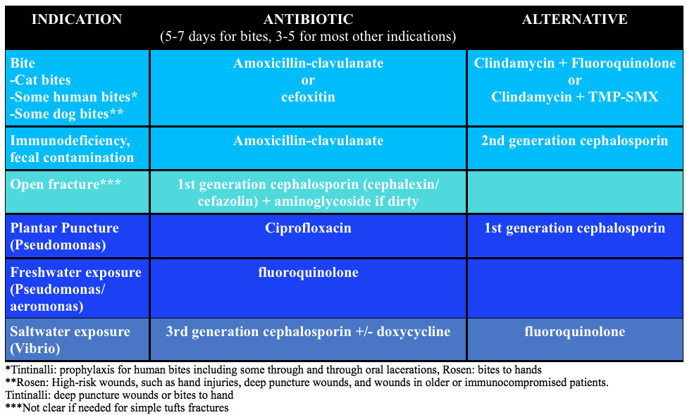
Prophylactic antibiotics:

Screen Shot 2015-11-15 at 5.34.59 PM

Generously Donated Rosh Review Questions 

Question 1.  An 18-year-old woman presents with a laceration to her face from a dog bite that occurred 24 hours ago. The patient owns the dog. Examination reveals a 4 cm laceration to the left cheek with no signs of infection. [polldaddy poll=9180209]

Question 2A 30-year-old man presents with a 2 cm linear laceration through his right eyebrow that he sustained after hitting his head on the kitchen cabinet. You determine that the wound will require repair with sutures. [polldaddy poll=9180210]

Answers

  1. Mammal bites to any part of the body should be copiously irrigated and explored followed by an assessment for primary closure. In this patient, primary closure is recommended as the laceration is on the face. Canine bites often involve laceration as well as crush injury to tissue depending on the size of dog. The presence of a crush injury may make primary wound repair difficult. Additionally, devascularization of the tissue may make primary closure contraindicated as the risk of infection increases. Classically, it was taught that lacerations sustained from dog bites should be irrigated, given antibiotics and not primarily repaired because of these risks. However, more recent literature has shown that the risk of infection was no different for primary closure versus healing by secondary intention. Additionally, if the laceration is to a cosmetic area like the face, primary repair should be attempted. As with any laceration, tetanus status should be updated. Copious irrigation and wound exploration is central to good wound care. Exploration should pay particular attention to the presence of foreign bodies especially teeth, which may break off during the bite. Antibiotics (A & C) are not routinely needed for dog bites despite classic teaching. Antibiotics should be reserved for patients with signs of infection, multiple comorbidities or large wounds with gross contamination. If antibiotics are given, they should primarily cover Staphylococcus and Streptococcus species, as these are the predominant organisms in the canine oral cavity. Eikenella and Pasturella are less commonly responsible for infections. Irrigation and antibiotics alone (A) would be indicated for dog bites that are grossly infected or have large defects that cannot be primarily closed. Wound closure and antibiotics without irrigation (D) is also contraindicated as copious irrigation is central to proper wound management.
  2. A pair of clean, non-sterile gloves can be worn by the physician (and any assistants) during laceration repair. The use of sterile gloves has not been proven to be associated with lower infection rates and is not required. Wounds must be prepped prior to closure. This generally involves cleaning and draping the wound, providing local or regional anesthesia, copious irrigation and exploring the wound to evaluate the integrity underlying structures and identify any foreign bodies. The skin surrounding a wound should be cleansed with either 10% povidone-iodine (C) or chlorhexidine gluconate solution. In general, these commercially available antiseptics should not be used for wound irrigation, as they can be toxic to the tissues. Irrigation should then follow with copious amounts of tap water or saline (at least 250 mL). This is best achieved with a large volume syringe attached to an 18-gauge needle or another commercially available irrigation device that achieves adequate pressure for irrigation. Alternatively, patients can irrigate at the sink if the laceration is in area that allows for this. Shaving of hair been shown to increase the risk of infection and should generally be avoided. It is best to apply a small amount of petroleum- or water-based lubricant to the hair to keep it out of the wound. Alternatively, hair can be clipped with scissors when necessary. Eyebrows (B) in particular should not be shaved as they provide anatomic landmarks that aid in wound approximation and removal results in poor short- and long-term cosmetic effect. In general, non-complex facial wounds are closed with nonabsorbable suture material, such as nylon or polypropylene. Most commonly this will be done with 6-0 suture, as it provides the best cosmetic effect. The use of 3-0 (D) and 4-0 suture is reserved for repair of fascia or wounds that are under high stress, such as those that overly major joints or involve the scalp.

References:

  1. Perelman VS, Francis GJ, Rutledge T, et al. Sterile versus nonsterile gloves for repair of uncomplicated lacerations in the emergency department: a randomized controlled trial. Annals of emergency medicine. 43(3):362-70. 2004
  2. Fernandez R, Griffiths R. Water for wound cleansing. The Cochrane database of systematic reviews. 2:CD003861. 2012.
  3. Weiss EA, Oldham G, Lin M, Foster T, Quinn JV. Water is a safe and effective alternative to sterile normal saline for wound irrigation prior to suturing: a prospective, double-blind, randomised, controlled clinical trial. BMJ open. 3(1):. 2013.
  4. American College of Emergency Physicians: Clinical policy for the initial approach to patients presenting with penetrating extremity trauma. Annals of emergency medicine. 33(5):612-36. 1999. [pubmed] **A past policy, no current clinical policy
  5. Zehtabchi S, Tan A, Yadav K, Badawy A, Lucchesi M. The impact of wound age on the infection rate of simple lacerations repaired in the emergency department. Injury. 43(11):1793-8. 2012.
  6. Quinn JV, Polevoi SK, Kohn MA. Traumatic lacerations: what are the risks for infection and has the ‘golden period’ of laceration care disappeared? Emergency medicine journal : EMJ. 31(2):96-100. 2014.

FOAMcastini – ACEP15 Day 3

(ITUNES OR LISTEN HERE)

FOAMcast brings you pearls from conferences we attend and presently it’s the American College of Emergency Physicians annual meeting, ACEP15 in Boston.

On this episode we cover the following topics:

Foodborne Illnesses – Dr. David Pigott

Wound Management – Dr. Benjamin Lawner

  • Irrigation – Need 50-100 cc per centimeter of wound. To get adequate PSI, take an 18g angiocath on a 30 cc syringe = 4-15 PSI
  • Some predictors of poor healing and infection
    • Location – head and neck more vascular so less likely to get infected
    • Length – >5 cm more likely to have problems
    • Diabetics – microvascular damange makes healing more problematic

Tips for Presentations – Dr. Haney Mallemat (@CriticalCareNow)

  • Avoid bullet points
  • Be brief
  • Use a sans serif font (and only one font throughout)
  • Limit animations (they can be distracting)


FOAMcastini – ACEP15 Day 2

(ITUNES OR LISTEN HERE)

FOAMcast brings you pearls from conferences we attend and presently it’s the American College of Emergency Physicians annual meeting, ACEP15 in Boston. On this episode we cover the following topics:

Extracorporeal Membrane Oxygenation (ECMO) – Dr. Haney Mallemat (@CriticalCareNow)

ECMO is promising in certain devestating disease processes – essentially heart or lung failure. For example, in the CHEER trial, the investigators had a 54% rate of neuro-intact survival after cardiac arrest with ECMO []. Yet, ECMO can be confusing. Dr. Mallemat simplified this for the emergency physician (see this site for more complete explanations)

Screen Shot 2015-10-27 at 5.55.44 PM

Stop the Madness: Diagnostic Imaging in Nephrolithiasis – Workshop with Drs. Eddy Lang, Rebecca Smith-Bindman, Grant Innes, and Lauren Westafer

Debunking Trauma Myths – Billy Mallon

FOAMcastini – ACEP15 Day 1

(ITUNES OR LISTEN HERE)

FOAMcast brings you pearls from conferences we attend and presently it’s the American College of Emergency Physicians annual meeting, ACEP15 in Boston.

Jeremy Hoffman and Rick Bukata summarize recent, relevant literature each year (recently known as #hofkata).  This year, our top three favorite

A top 5 list for emergency medicine: a pilot project to improve the value of emergency care.  Schuur et al. JAMA Internal Medicine. 2014.

  • Just say NO to:
    • CTs for trauma patients who are negative using the NEXUS and Canadian tools for cervical spine injuries.
    • CT for ruling out PE without some sort of risk stratification.
    • MRI for Low back pain w/o red flags.
    • Head CT in with mild TBI who do not meet criteria using the New Orleans OR Canadian Head CT tools.
    • Routine coagulation profile (unless they have a known coagulopathy or are hemorrhaging)

Emergency Physician Perceptions of Medically http://www.mindanews.com/buy-valtrex/ Unnecessary Advanced Diagnostic Imaging. 

The effect of malpractice reform on emergency department care.  Moskow et al.   N Engl J Med. 2015 Jan 8;372(2):192.

Dr. Jeff Kline (@klinelab) spoke on pulmonary embolism.

  • High Risk PE? Consider lysis (this is controversial, we are simply reporting Dr. Kline’s talk)
    • Size and location. Massive and proximal= bad
    • SBP <90 for more than 15 min OR 40mmHg drop from baseline
    • Signs of RV strain – echocardiography showing RV dilation OR hypokinesis?
    • Elevated troponin or BNP
    • ECG findings suggestive of cardiac strain: sinus tachycardia, incomplete right bundle branch block, complete right bundle branch block, T-wave inversion in leads V1 – V4.
  • Kline also participated in a knowledge translation workshop where he argued that sub-segmental PEs, without DVT on ultrasound, are NOT a real thing.  This is controversial but he also argued that treating these is associated with harm [Carrier et al]


Episode 36 – Rib and Sternal Fractures

(ITUNES OR Listen Here)

The Free Open Access Medical Education (FOAM)

This week we cover a joint piece between the Ultrasound Podcast and SonoIn5 on diagnosis of rib and sternal fractures with ultrasound.

Technique:  Linear probe, in line with the long axis of the bone (vertical for sternum, horizontal-ish for ribs).

Diagnosis: Cortical disruption (step off). Excellent sensitivity for sternal fractures [1-3]

Sternal Fracture
Sternal Fracture
  • Caution with sternal fractures as the sternomanubrial joint can mimic fracture, but looks more “bumpy” (see below)

Core Content – Rib and Sternal Fractures

Tintinalli (7e) Chapters 258, 259; Rosen (8e) Chapter 45 

Rib Fractures

Diagnosis:

  • Chest x-ray initial test of choice – may miss 50% of fractures, unclear if this is clinically significant [6]
  • Ultrasound has found to have excellent sensitivity [7]
  • Rib films are NOT recommended [4-6].

Complications: Traumatic rib fractures may be associated with other traumatic injuries such as pneumothorax, hemothorax, or in the case of lower rib fractures, intra-abdominal injury. However, rib fractures themselves have been associated with mortality, most often as sequelae of pulmonary embarrassment including pneumonia, intubation, and death. Mortality in elderly patients with rib fractures is significantly higher than the younger counterparts at 22% and 10% respectively [8,9].

  • Mortality is between 3-13%
  • Risk stratification (see this post): Battle and colleagues developed a prognostic scoring system, not externally validated and unclear if it would change practice, that highlights common sense predictors of poorer outcomes:
    • Age (>65)
    • Higher number of rib fractures
    • Chronic lung disease
    • Hypoxia (<90%)
    • Pre-injury anticoagulant use [11]

Treatment

  • Analgesia:
    • Often includes NSAIDS (ibuprofen), acetaminophen, and narcotics +/- gabapentin (ibuprofen and gabapentin depending on renal function)
    • Epidural analgesia – highly recommended in the EAST guidelines [14].
    • Paracostal analgesia (ex: ON-Q pump) – not sufficient evidence for EAST recommendation (2005) [14]
  • Pulmonary Hygiene (formerly pulmonary toilet):  involved incentive spirometry, coughing, mobilization (up, out of bed), and possibly chest physical therapy
  • ORIF, “rib fixation” or “rib plating,” is increasingly common in the US and studies have found improvements in ICU LOS and ventilator days [15]

Disposition

  • Many rib fracture patients will need to be admitted to the hospital for pain control, observation, and pulmonary hygiene.
    • Some rib fracture patients may benefit from care at trauma centers.  Lee et al  wrote that 3+ rib fractures exists as an indication for transfer to a level 1 trauma center and many places ascribe to this, it depends on the hospital and physicians.
    • While patients in the ED may look good, patients may benefit from high intensity floors (ie stepdown units) and many patients get observed in ICUs, again, depending on local practice patterns. Some protocols risk stratify patients (i.e. to the ICU vs floor) by incentive spirometry.
  • Patients with adequate pain control who are low risk (younger, <3 rib fractures, good effort on incentive spirometry) may be discharged from the ED with analgesia and education on importance of pulmonary hygiene

Sternal Fractures – more common with ubiquity of airbags and seatbelts.

Diagnosis:  Classically the “gold standard” has been lateral x-ray. However, CT technology has improved since those studies. Ample literature suggests that ultrasound has excellent sensitivity [1-3].

Complications: Historically, sternal fractures were associated with injuries of the great vessels, high mortality, and blunt cardiac injury (BCI) [16-18].  The most recent iteration of the EAST guidelines states, “the presence of a sternal fracture alone does not predict the presence of BCI and thus should not prompt monitoring in the setting of normal ECG result and troponin I level” (Level 2) [18].

Treatment: Analgesia. Most patients with isolated sternal fractures (no pneumothorax, hemothorax, BCI, or hemodynamic instability) that have adequate pain control can be discharged from the ED [1-2].

Blunt Cardiac Injury

A broad category including a range of injuries from clinically silent dysrhythmias to cardiac wall rupture or vasospasm. BCI often results from high impact injury and should be considered in patients with significant thoracic trauma including rib fractures, sternal fracture, pneumothorax, hemothorax, and pulmonary contusion.

Diagnosis: There is no gold standard test.  One can rule out BCI with a normal ECG and a single normal troponin I [18].

Management: If an ECG or troponin is abnormal, admit to telemetry for monitoring and echo.

Generously Donated Rosh Review Questions 

Question 1.  A 23-year-old man presents with chest pain after a motor vehicle collision. The patient’s chest struck the steering wheel. He has no other complaints or injuries. Chest X-ray is unremarkable. ECG shows sinus tachycardia with anterior ST depressions. A troponin is sent and is positive at 3.50 mg/dl. [polldaddy poll=9134639]

Question 2A 20-year-old man presents with left rib pain after falling while playing soccer and striking his chest. Vital signs are normal. On physical examination, the patient has tenderness to palpation over the 4th rib in the midaxillary line. [polldaddy poll=9134640]

Question 3.  A 32-year-old woman was the restrained driver involved in a head-on motor vehicle collision (MVC) 2 days prior to presentation. She is complaining of chest pain and bruising to her chest. Her blood pressure is 118/78 mm Hg, pulse is 88 beats/minute, respirations are 18 breaths/minute and oxygen saturation is 96% on room air. You note bony tenderness and ecchymosis to her sternum. You order a chest X-ray and diagnose a non-displaced sternal fracture. [polldaddy poll=9134643]

Answers

  1. This patient presents with a myocardial contusion and should have an echocardiogram performed to look for any cardiac damage. Myocardial contusion describes a nebulous condition. It can occur through several mechanisms including a direct blow to the chest and compressive force over a prolonged period of time. Histologically, the disorder has similar findings to those seen after acute myocardial infarction. The majority of contusions heal spontaneously but small pericardial effusions may develop. Delayed rupture after resorption of hematoma is feared but rare complication. Patients with myocardial contusion will present after trauma with external signs of trauma and typically have other concomitant thoracic lesions (pulmonary contusion, pneumothorax, hemothorax). Patients will typically have tachycardia (up to 70%). ECG may show dysrrhythmia or ST changes but may also be normal. Although it is not effective to admit all patients for workup for myocardial contusion and the disease has a very low rate of cardiac complications, in the presence of ECG changes and elevated biomarkers, observation and echocardiography are a reasonable approach. Echocardiogram can be used to diagnose pericardial effusion, thrombi formation and valvular disruption.Cardiac catheterization (A) is not necessary after a myocardial contusion as coronary artery obstruction is not part of the pathophsyiology. The patient should not be discharged home (B)without an echocardiogram. Pericardiocentesis (D) is only necessary in the presence of a large pericardial effusion or one causing cardiac tamponade.
  2. This patient presents with signs and symptoms consistent with a rib facture. A chest X-ray should be performed to rule out any other pathology including pneumothorax and pulmonary contusion. Rib fractures are a common injury after thoracic trauma and the incidence increases with increasing age. They may be associated with a number of potential complications including pulmonary contusions, hemothorax, penumothorax and post-traumatic pneumonia. Fractures are most common at the posterior angle, which represents the weakest area. The ribs most commonly fractured are the 4th – 9th ribs. The 9th – 11th ribs are mobile, which reduces the risk of fracture. However, fractures of these ribs are more likely to be associated with intraabdominal injuries. Rib fractures should be suspected based on history and clinical evaluation. Patients will present with chest pain and tenderness over the area. Imaging should be obtained to rule out the more serious associated complications of pneumothorax, hemothorax and pulmonary contusion. Chest X-ray is the appropriate modality for this but often will not demonstrate the presence of a single rib fracture when it is in fact present. This is particularly true of non-displaced fractures. Rib belts (B) are discouraged as they may decrease the depth of respiration and lead to atelectasis and pneumonia. CT scan of the chest (D) is not routinely required for management of a simple rib fracture. Analgesia and discharge home (A) is likley to occur once more serious pathology is ruled out with a chest X-ray. Patients with rib fractures should also receive an incentive spirometer to help reduce the complication of pneumonia.
  3. Isolated, non-displaced sternal fractures are associated with low overall mortality rates. Fractures and dislocations of the sternum are caused primarily by anterior blunt chest wall trauma during a head-on MVC. Isolated fractures of the sternum most commonly occur when the chest wall is thrust against a diagonal seatbelt strap during rapid deceleration in a frontal impact MVC. They are more common in older individuals and women. Most fractures are transverse and non-displaced and can be diagnosed on a lateral chest radiograph. Although a fracture of the sternum can be seen following major thoracic trauma, its presence alone does not indicate severe underlying thoracic injury. However, if other significant underlying thoracic injuries are suspected, a CT-scan of the thorax should be performed

References:

  1. You JS, Chung YE, Kim D, Park S, Chung SP. Role of sonography in the emergency room to diagnose sternal fractures. Journal of clinical ultrasound : JCU. 38(3):135-7. 2010. [pubmed]
  2. Engin G, Yekeler E, Güloğlu R, Acunaş B, Acunaş G. US versus conventional radiography in the diagnosis of sternal fractures. Acta radiologica (Stockholm, Sweden : 1987). 41(3):296-9. 2000. [pubmed]
  3. Jin W, Yang DM, Kim HC, Ryu KN. Diagnostic values of sonography for assessment of sternal fractures compared with conventional radiography and bone scans. J Ultrasound Med. 2006 Oct. 25(10):1263-8; quiz 1269-70.
  4. ”Pulmonary Trauma” Tintinalli’s Emergency Medicine: A Comprehensive Study Guide.  7th ed. Ch 258.
  5.  “Thoracic Trauma” Rosen’s Emergency Medicine. 8th ed. Chapter 45.
  6. Henry TS, Kirsch J. ACR Appropriateness Criteria® rib fractures. Journal of thoracic imaging. 29(6):364-6. 2014. [pubmed]
  7. Chan SS. Emergency bedside ultrasound for the diagnosis of rib fractures. The American journal of emergency medicine. 27(5):617-20. 2009. [pubmed]
  8.  Ziegler DW, Agarwal NN. The morbidity and mortality of rib fractures. J. Trauma. 1994;37(6):975–9.
  9. Bulger EM, Arneson M a, Mock CN, Jurkovich GJ. Rib fractures in the elderly. J. Trauma. 2000;48(6):1040–6
  10. Flagel BT, Luchette F a, Reed RL, et al. Half-a-dozen ribs: the breakpoint for mortality. Surgery. 2005;138(4):717–23; discussion 723–5.
  11. Battle CE, Hutchings H, Evans P. Risk factors that predict mortality in patients with blunt chest wall trauma: a systematic review and meta-analysis. Injury. 2012;43(1):8–17.
  12. Livingston DH, Shogan B, John P, Lavery RF. CT diagnosis of Rib fractures and the prediction of acute respiratory failure. The Journal of trauma. 64(4):905-11. 2008. [pubmed]
  13. Battle CE, Hutchings H, Lovett S.  Predicting outcomes after blunt chest wall trauma: development and external validation of a new prognostic model Critical Care 2014, 18:R98
  14. Pain Management in Blunt Thoracic Trauma (BTT)J Trauma. 59(5):1256-1267, November 2005.
  15. Doben AR, Eriksson EA, Denlinger CE. Surgical rib fixation for flail chest deformity improves liberation from mechanical ventilation. Journal of critical care. 29(1):139-43. 2014. [pubmed]
  16. Screening for Blunt Cardiac Injury. J Trauma. 73(5):S301-S306, November 2012
  17. Karangelis D, Koufakis T, Spiliopoulos K, Tsilimingas N, Bouliaris K, Desimonas N. Management of isolated sternal fractures using a practical algorithm. J Emerg Trauma Shock. 7(3):170-. 2014. [article]
  18. Dua A, McMaster J, Desai PJ et al. The Association between Blunt Cardiac Injury and Isolated Sternal Fracture. Cardiology Research and Practice. 2014:1-3. 2014. [article]

FOAMcastini – Core Content Journal Club

(ITUNES OR Listen Here)

This is an exciting week of primary literature, particularly as many large critical care trials were published in major journals despite being “negative studies.”  We are excited by this as, too often, we see “negative studies” discarded.  Further, these studies examined things some practices we seem to believe in: balanced crystalloids, apneic oxygenation (see Dr. Scott Weingart’s podcast on the FELLOW study), treating fever. We love that the Free Open Access Medical Education (FOAM) community and study authors are examining beloved practice and open to questioning the very things we believe in. Well done.

We cover two core content papers out by Dr. Paul Young (@dogICUma) in JAMA and NEJM this week. His trials are as clever as his Twitter handle.

SPLIT – The FOAM world has sung the praises of balanced fluids given they have more physiologic composition. The thought, as detailed in this post, is that 0.9% NaCl contains an ABnormally large amount of chloride which may cause a hyperchloremic metabolic acidosis. Prior literature suggests an increased incidence of kidney injury with saline compared with balanced solutions. Thus, Dr. Paul Young and colleagues sought to study this with the best trial, to date, on this topic.

  • Multicenter, blinded,  cluster-randomized, double-crossover trial of adult ICU patients receiving crystalloids randomizing patients to 0.9%NaCl or Plasma-lyte (balanced solution).
    • Sites used one fluid for seven weeks and then crossed over to the other fluid (labeled Fluids A and B).
  • Primary outcome: AKI according to the RIFLE criteria  within 90 days – no difference between groups.
    • 9.6% in Plasma-lyte group vs 9.2% in the saline group (absolute difference 0.4% [95% CI, −2.1%-2.9%]; RR, 1.04 [95% CI, 0.80-1.36]; P = .77)
  • Secondary outcomes: No difference in renal replacement therapy, ICU days, mechanical ventilation, or mortality
  • A few things to keep in mind:
    • 70% of patients were admitted to the ICU from the OR (mostly cardiac surgery) and only ~15% from the ED
    • Patients got a median of 2L (1L -3.5L) of the study fluid, that’s it. These were not large volume resuscitations.
    • 90% of patients received fluids prior to enrollment, 60% got balanced crystalloid and only 30% 0.9% NaCl.

HEAT – We can’t help treating fever. We like the numbers euboxic, elevated temp? It must be bad! Doctors, parents, nurses treat fever reflexively.  Yet, there’s a thought that fever may be evolutionary and could potentially be protective. Treating pain or discomfort? That’s one thing, but here the authors sought to determine if there was a clinically important benefit to treating the number in ICU patients.

  • Randomised controlled, double blinded study of n=690 ICU patients with T>38F + suspected infection randomized to receive either 1 g paracetamol (acetaminophen/APAP) or placebo every 6 hours.
  • Primary outcome: median ICU-free days to day 28 – no difference 23 (IQR 13-25) in paracetamol group vs 22 in placebo group (IQR 12-25); P=0.07
  • No difference in secondary outcomes of mortality at 28 and 90 days
  • Limitations: ~30% of patients in both arms received open label APAP after the course of the study drug

Bottom Line Pearls:

  • It appears we may be SPLITting hairs over fluid choices. Giving a couple of liters? Fluid choice may not matter. SPLIT does not provide literature for larger volume resuscitations.
  • Treating fever in ICU patients with suspected infection doesn’t have an effect on ICU free days. Treat discomfort and pain with APAP but don’t expect to save lives or ICU beds by doing so.
  • “Negative studies” are important. So is examining our practice.

Episode 35 – The Trachea

(ITUNES OR Listen Here)

The Free Open Access Medical Education (FOAM)

Jane Brody wrote an article, “What Comes After the Heimlich Maneuver” that ran in the NY Times and stirred up a ruckus on Twitter. This is a reasonable article on choking and details the limitations of the Heimlich maneuver.  Unfortunately, the article ends  instructing the layperson do to a cricothyrotomy (cric) with a sharp knife and “something like a straw or casing of a ballpoint pen (first remove the ink cartridge).  “

Dr. Seth Trueger (@MDaware) wrote a post, Bad Idea Jeans, discouraging this practice saying that deciding which patient needs a cric is one of the more difficult but more important parts of this procedure.

On another note, our friend Dr. Andy Neill has found that medical students are able to perform crics with Papermate pens on cadavers [1]. However, it appears that most pens may not be suitable [2]. Further, while medical students are nearly lay people, we do not think this the cric should be within the domain of lay people (especially without patients already declared dead and preserved).

Cricothyrotomies – In reality, this is a bloody procedures that should only be done by those with proper training when the airway cannot be otherwise secured. The actual procedure has been detailed by those far smarter and with more experience than the FOAMcast crew. We recommend checking out Dr. Scott Weingart’s compilation of resources here.

The anatomy of a cricothyrotomy by Dr. Andy Neill

Core Content – Tracheostomy Emergencies and Neck Infections

Tintinalli (7e) Chapters 242, 119

Tracheostomy (trach) (we refer to tracheostomy and tracheotomy interchangeably although there are some technical differences)

Anatomy of the trach tube (may vary)

Screen Shot 2015-10-07 at 8.40.17 AM

Things we have to know about trach emergencies

Screen Shot 2015-10-07 at 7.43.30 AM

Pediatric Trachea (Stridor) Pearls

Screen Shot 2015-10-07 at 8.20.11 AM

Generously Donated Rosh Review Questions 

Question 1. A 72-year-old man who is 1 week out from an ischemic stroke presents with respiratory distress. He had a tracheostomy placed 6 days ago for sudden respiratory failure. The patient is hypoxic and tachypneic on presentation with minimal breath sounds bilaterally. There is no subcutaneous air around the stoma.[polldaddy poll=9110296]

Question 2A 3-year-old boy presents in severe respiratory distress. His mother informs you that he has been ill for the last 5 days, initially with a low-grade fever and “barky cough.”  He was seen at an urgent care facility 4 days ago and given a  “breathing treatment” and discharged on steroids. He has become progressively worse despite compliance with the steroid regimen, which prompted his mother to call an ambulance this morning. He is otherwise healthy and up-to-date on his immunizations. On examination, the child is toxic in appearance and febrile. His oropharynx is clear. You hear both inspiratory and expiratory stridor.  [polldaddy poll=9110301]

Answers

1.The patient’s presentation is concerning for airway obstruction and the first step in management is suctioning of the tracheostomy tube. Tracheostomy tubes are placed for long-term mechanical ventilation in patients with anticipated prolonged or permanent respiratory failure. The two most common complications are obstruction and dislodgement. Sudden onset of respiratory failure often indicates mucous plugging or equipment failure. Suctioning of the tracheostomy is a simple procedure that may quickly relieve the patient’s symptoms. 2 to 3 ml of normal saline should be instilled into the tube followed by suctioning. Patients with slower decline in respiratory status may have a worsening of their underlying pulmonary pathology or may have developed a pulmonary infection.  A cricothyrotomy (A) will not lead to effective oxygenation or ventilation as the cricothyroid membrane is above the tracheostomy site. The tracheostomy tube should not be removed and replaced with either an endotracheal tube (C) or a new tracheostomy tube (B) at this time because the tracheostomy tract has not matured at 6 days (this usually occurs at 15-30 days). If equipment failure in the form of a tracheostomy tube malfunction is suspected, the tube should be replaced with fiberoptic visualization to ensure that a false lumen isn’t created.

2.The patient is suffering from acute bacterial tracheitis. Bacterial tracheitis is the result of severe inflammation of the epithelial lining of the trachea leading to thick mucopurulent secretion production. This clinically manifests as viral prodrome with fever, URI symptoms, barky cough and stridor that intensifies and progresses to include a toxic appearing child with signs of airway obstruction, inspiratory and expiratory stridor, cyanosis, and severe respiratory distress. Another clue is that the child has been treated with medications (aerosolized epinephrine and steroids) for croup and has not improved clinically. Bacterial tracheitis is most common in children between the ages of 3 to 5 years. Most patients require orotracheal intubation for respiratory distress and ICU admission. The patient should be started on broad-spectrum intravenous antibiotics. Croup (B) is the most common cause of upper airway distress and obstruction in children between 6 months to 6 years of age with peak incidence at 2 years of age. Croup begins as a prodrome of low-grade fever and URI symptoms and is characterized by a barky cough, inspiratory stridor, and hoarse voice. Children are less toxic in appearance and rarely develop respiratory failure. The mainstays of treatment are steroids and aerosolized epinephrine. Epiglottitis (C) is characterized by abrupt onset of fever and sore throat and children classically present with difficulty in breathing, anxiety, stridor and drooling. This is less common in vaccinated children, such as the patient above and typically occurs in slightly older children. There is generally not a prodrome associated with epiglottitis. Peritonsillar abscess (D) occurs more commonly during adolescence and presents with trismus, unilateral sore throat, fever, tonsillar asymmetry, and uvula deviation away from the affected tonsil. The age of this patient and normal oropharynx examination make this diagnosis very unlikely.

References:

  1. Neill A, Anderson P. Observational cadaveric study of emergency bystander cricothyroidotomy with a ballpoint pen by untrained junior doctors and medical students. Emergency medicine journal : EMJ. 30(4):308-11. 2013. [pubmed]
  2. Owens D, Greenwood B, Galley A, Tomkinson A, Woolley S. Airflow efficacy of ballpoint pen tubes: a consideration for use in bystander cricothyrotomy. Emergency medicine journal : EMJ. 27(4):317-20. 2010. [pubmed]

FOAMcastini – The “Left Shift”

(ITUNES OR Listen Here)

The Free Open Access Medical Education (FOAM)

The blog Brown Coat Nation (University of Illinois, Chicago) has a new series entitled “Inconceivable.” The idea is to expose medical terminology that we tend to use incorrectly. The first installment is focused on the misuse of the term “left shift,” and it’s the focus of this FOAMcastini.

The Core Content

The correct use of the term “left shift” refers to the presence of banded (immature) neutrophils in the blood. It does not refer to an elevated white blood cell count with a high percentage of neutrophils. An elevated white blood cell count with an abnormally high percentage of neutrophils should be called “neutrophillic leukocytosis.” Only the presence of immature neutrophils in the periphery (including bands) can accurately be called a “left shift.”

The term “left shift” is derived from the diagrams of the six stages of neutrophil development in the bone marrow. On the far left, you see the most basic precursor: the myeloblast. On the far right of the diagram one finds the mature segmented neutrophil (also known as the “polymorphonuclear leukocyte, or PMN). But just to the left of that is the “banded” neutrophil (the 5th stage of neutrophil development in which the large band of nuclear material has not yet “disbanded” into segments).

When an infection runs rampant, sometimes the bone marrow runs out of mature neutrophils to send to the periphery. So, the marrow panics and releases immature banded neutrophils that normally would not be considered “ready for prime time.”

Here’s some relevant spaced repetition on SIRS: Along with leukocytosis (>12,000) and leukopenia (<4,000), Bandemia >10% is one of the SIRS criteria (Systemic Inflammatory Response Syndrome). Temperature (>38C or <36C), Heart rate >90 (“sirs-ycardia”), tachypnea (RR>20) or PCO2 <32) are the other SIRS criteria. The presence of at least two these categories constitutes “positive SIRS”. In pediatrics, the temperature, heart rate, and respiratory rates should be age-adjusted.
Check out our previous episode on appendicitis for a reminder on whether leukocytosis (with or without neutrophillic predominance) is useful in risk stratification.

Episode 34 – Tachyarrhythmias

(ITUNES OR Listen Here)

The Free Open Access Medical Education (FOAM)

The St. Emlyn’s team ran a post on the REVERT trial, which added a new spin to the traditional (and traditionally ineffective) Valsalva maneuver for stable supraventricular tachycardia (SVT). In this post, Dr. Rick Body goes through the trial covering not only the results but also drops pearls on methodology.

  • Randomized 433 patients with SVT to one of the following:
    • “Modified” Valsalva maneuver: patient sitting up performs Valsalva using a syringe to maintain 40 mm Hg for 15 sec and then placed supine with passive leg raise immediately after procedure (see video)
    • “Standard” Valsalva maneuver: patient sitting up performs Valsalva using a syringe to maintain 40 mm Hg for 15 sec while maintaining upright position
  • 43% of the patients in modified Valsalva group versus 17% in the standard technique achieved sinus rhythm at one minute yielding an absolute risk reduction of 26.2% (p<0.001) with a number needed to treat of about 4 (3.8).

Core Content – Supraventricular Tachycardia (SVT) and Ventricular Tachycardia (VT)

Tintinalli (7e) Chapter 22;  Rosen’s Emergency Medicine (8e) Chapter 79

SVT 

Broad term referring to tachycardias originating above the ventricles, including the regular rhythms of sinus tachycardia, AV nodal reentrant tachycardia, AV reentrant tachycardia, and the irregular rhythms of multifocal atrial tachycardia, atrial fibrillation, and some forms of atrial flutter.

Diagnosis: JACC algorithm

Treatment:

  • Unstable patients – electrical cardioversion at 0.5-1 J/kg (100J for an adult) + ABCs!
  • Stable patients-
    • Valsalva maneuver – we like this method of having a patient blow on a syringe. Unfortunately, prior to the REVERT trial, the valsalva maneuver success rate has been documented ~19% [3].
    • Adenosine (0.1mg/kg or 6 mg in adult; 2nd dose 0.2 mg/kg or 12 mg in adult, with occasional dose adjustments) – administration can be tricky because of the drug’s short half life, necessitating proximal administration, elevation of the arm, and a quick saline flush afterwards. You can combine the adenosine IN the flush as detailed in this post, meaning no stopcock.
    • Calcium channel blockers or beta-blockers (verapamil, diltiazem or even metoprolol, esmolol) – Recently the calcium channel blockers have increased in popularity in the FOAM world and these are Rosenalli approved [4,5].

VT

Diagnosis: Typically wide QRS complex (95% with QRS >120 ms) and fast (150-200 beats per minute).

  • SVT with abberency can have a wide complex but this should be treated as VT [4,5] (see this video)
  • Monomorphic – complexes have same morphology
  • Polymorphic – complexes of various morphologies, associated with poor prognosis [4,5]

Treatment:

  • Unstable patients – electrical cardioversion at 0.5-1 J/kg (100J for an adult) + ABCs!
  • Stable patients with monomorphic VT
    • Electrical cardioversion
    • Procainamide – Level B recommendation for first line treatment of monomorphic VT[6].
    • Amiodarone – common in the US but per the AHA guidelines “reasonable in patients with sustained monomorphic VT that is hemodynamically unstable, refractory to conversion with countershock, or recurrent despite procainamide or other agents. (Level of Evidence: C)” [6].
      • Note: Dangerous if prolonged QT [6]
    • Lidocaine – “may be reasonable” [6]
  • Stable patients with polymorphic VT
    • Electrical cardioversion
    • Beta-blockers (particularly if ischemic)
    • Amiodarone
    • Cardiac catheterization if potentially ischemic cause [6]
  • Torsades de Pointes – withdraw offending agent, magnesium sulfate IV if “a few episodes” per the AHA

Generously Donated Rosh Review Questions 

Question 1. A 26-year-old woman presents with dizziness and palpitations. She reports episodes of these symptoms beginning about 1 week ago, which initially only lasted a few minutes. However, for the past two days, she has had about 4 episodes a day which last about 20 minutes each. Her social history is significant for heavy caffeine intake. Her pulse is 166 bpm and her blood pressure is 140/70. Her rhythm strip is seen below. [polldaddy poll=9061960]

Screen Shot 2015-09-05 at 9.52.22 AM

Question 2. A 33-year-old woman with chronic persistent asthma presents with palpitations. Her vital signs are HR 210, BP 118/73, and pulse oxygenation of 97% on room air. An ECG is shown below. [polldaddy poll=9061966]

Screen Shot 2015-09-05 at 9.53.15 AM

References:

  1. Whinnett ZI, Sohaib SM, Davies DW. Diagnosis and management of supraventricular tachycardia. BMJ (Clinical research ed.). 345:e7769. 2012
  2. Link MS. Clinical practice. Evaluation and initial treatment of supraventricular tachycardia. The New England journal of medicine. 367(15):1438-48. 2012.
  3. Smith et al. Effectiveness of the Valsalva Manoeuvre for reversion of supraventricular tachycardia. Cochrane Database Syst Rev. 2013 Mar 28;3:CD009502. doi: 10.1002/14651858.CD009502.pub2
  4. ”Cardiac Rhythm Disturbances.” Tintinalli’s Emergency Medicine: A Comprehensive Study Guide.  7th ed. pp 136-146.
  5. “Tachyarrhythmias.” Rosen’s Emergency Medicine. 8th ed. Chapter 79.
  6. Zipes DP, Camm AJ, Borggrefe M et al. ACC/AHA/ESC 2006 Guidelines for Management of Patients With Ventricular Arrhythmias and the Prevention of Sudden Cardiac Death. Journal of the American College of Cardiology. 48(5):e247-e346. 2006