Episode 25 – Skin and Skin Structure Infections

(ITUNES OR LISTEN HERE)

The Free Open Access Medical Education (FOAM)

We review the post by Bryan Hayes, PharmD, FAACT on Academic Life in Emergency Medicine,  Sulfamethoxazole-Trimethoprim for Skin and Soft Tissue Infections: 1 or 2 Tablets BID?

The Take Home: Most abscesses do not need antibiotics after incision and drainage.  If the patient has systemic signs (fever, tachycardia), co-morbidities, or a concurrent cellulitis, they may need antibiotics.  Sulfamethoxazole-trimethoprim (SMX-TMP / Bactrim) is one of the most commonly used agents in this case, as it covers MRSA.

Dosing: Two double strength (DS) tablets twice daily is a commonly prescribed regimen; yet,1 DS tablet twice daily is sufficient in most cases with exceptions for patients >100 kg, immunocompromised, or in trauma [1]. ; however, this increases the likelihood of adverse events (nausea, hyperkalemia) without notable substantial positive return.

The Bread and Butter

We cover cellulitis and abscesses, necrotizing infections, and Erythema Multiforme/Stevens-Johnson Syndrome/Toxic Epidermal necrolysis using Tintinalli (7e) Chapter; Rosen’s (8e) Chapter 137 as well as the IDSA guidelines.  But, don’t just take our word for it.  Go enrich your fundamental understanding yourself.
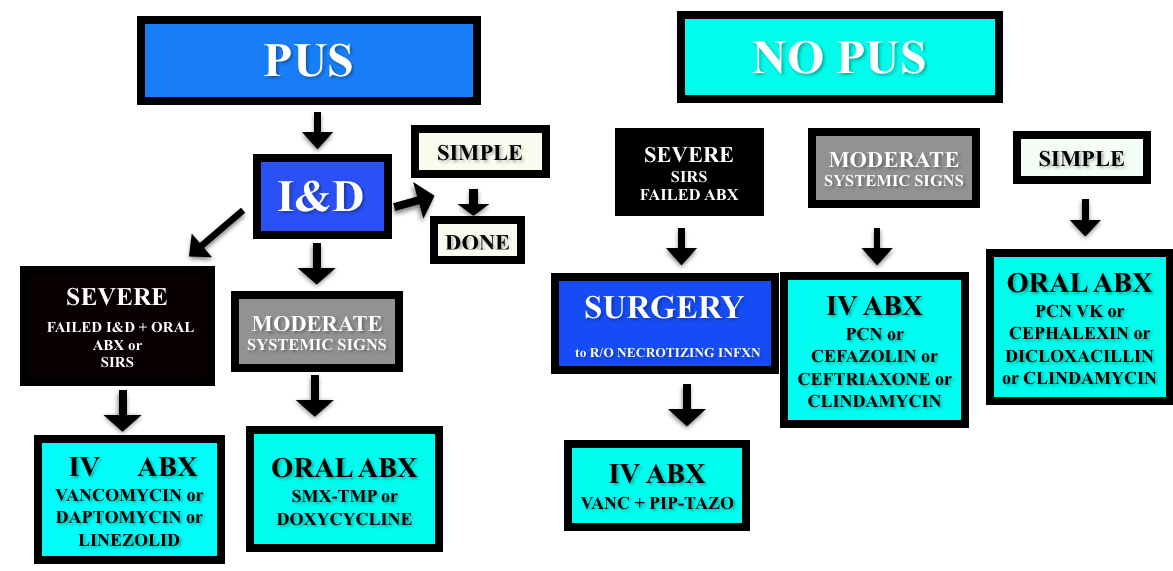
Cellulitis/Abscess

Non-purulent – cover for strep (penicillin, cephalexin/cefazolin).  Even in areas with high incidences of community MRSA, the recommendations for non-purulent skin infections is strep coverage. Five days of treatment is probably enough, and IDSA and Rosenalli approved (see this post)

Purulent – Incise & Drain (I&D).  Cover for MRSA in patients that failed initial I&D or those with systemic signs.

Adapted from the 2014 IDSA Guidelines

Necrotizing Infections

Presentation – pain out of proportion to exam findings, abnormal tachycardia (particularly out of proportion to degree of fever). Crepitus is not reliable (present in 13-30% of patients).

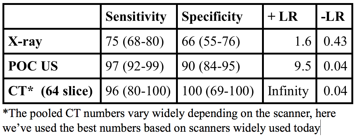
Diagnosis – clinical. X-rays, CT, and MRI have all been used but both x-rays and CT lack sensitivity and it’s probably not a good idea to send a sick patient to MRI.  The gold standard diagnosis is operative findings.

  • The LRINEC score was derived to aid in diagnosis using lab values (sodium, creatinine, white blood cell count, hemoglobin, glucose, CRP) but has not been sufficiently predictive in validation attempts [10].

Classification

  • Type I – polymicrobial. Common in diabetics, immunocompromised.
  • Type II – monomicrobial (often Group A strep or clostridia).
  • Type III? – vibrio, but apparently this is controversial.

Treatment

  • Intravenous fluid and general resuscitation, surgical consult, and antibiotics.

Erythema Multiforme/Stevens-Johnson/Toxic Epidermal Necrolysis (for more, see this Hippo EM podcast)

Courtesy of Rosh Review

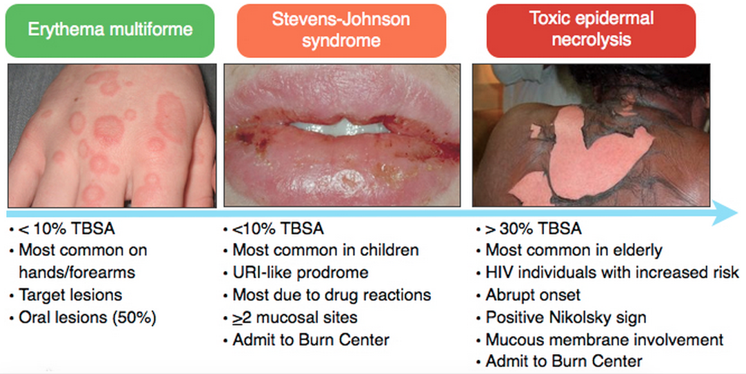
Medications most associated with SJS/TEN:  Antibiotics (sulfa), Anti-epileptics, Nonsteroidals

Treatment of SJS/TEN:  Fluid resuscitation, Burn Center/ICU care, and IVIG

Generously Donated Rosh Review Questions 

Question 1. A 22-year-old woman presents to the emergency department with a painful rash. She has had several days of malaise, arthralgias, and low-grade fever, and today developed diffuse painful erythema across her body which is beginning to blister. She takes no medications, but reports completing a course of trimethroprim-sulfamethoxazole one week ago for a urinary tract infection. Examination reveals diffuse tender erythema over her trunk and extremities with multiple ill-defined large bullae, some of which have ruptured, leaving large areas of denuded skin behind. Oral ulcerations are also noted. [polldaddy poll=8689023]

Question 2.  A 54-year-old man with diabetes presents with severe leg pain. The pain has worsened over the last two days with increased swelling of the calf. He has no chest pain or shortness of breath. Vital signs are: T 101.8°F, BP 98/62, HR 118, RR 18. Physical examination is notable for erythema of the calf, mild tenderness, and crepitus. You initiate IV fluids and broad-spectrum antibiotics. [polldaddy poll=8689082]

Answers

1. This patient has toxic epidermal necrolysis (TEN). TEN is an acute inflammatory process characterized by tender erythema, painful bullae formation, and subsequent exfoliation. It often begins with prodromal symptoms such as fever, malaise, and myalgias. TEN is considered a dermatologic emergency and patients may appear toxic on presentation. Medications (within the first few months of administration) are the most common cause, with sulfa and penicillin antibiotics, anticonvulsants, and oxicam non-steroidal anti-inflammatory drugs commonly implicated. Management of TEN involves admission to a burn unit, fluid resuscitation, and prevention of secondary infection. Steroids are not an indicated treatment. Staphylococcal scalded skin syndrome (C) occurs primarily in infants and young children. Infection with exotoxin-producing Staphyloccus aureus leads to diffuse erythroderma and subsequent exfoliation. Mucous membranes are not usually involved. Treatment is fluid resuscitation and antibiotics. Infection with HSV-1 and HSV-2 results in localized skin infection, though in patients with underlying immunosuppression or malignancy it may lead to disseminated herpes simplex virus infection (A), characterized by diffuse vesicles and ulcerations and multisystem involvement. Photosensitive drug reactions (B) are characterized by confluent erythema, macules, papules, or sometimes vesicles in sun-exposed areas such as the face, neck, and arms, occurring within 1-3 weeks of the patient taking an offending agent.  Medications commonly associated with photosensitivity include sulfonamides, thiazides, furosemide, and fluoroquinolones

2. Necrotizing fasciitis is an infection of the subcutaneous tissue that spreads rapidly across the fascial planes and is often fatal even with aggressive treatment. Risk factors for necrotizing infections include diabetes, vascular insufficiency, and immunosuppression. Classically patients have pain out of proportion to examination. Later findings include diffuse swelling, erythema, induration and crepitus. The gold standard for diagnosis is direct visualization in the operating room by a surgeon. Surgeons may elect to perform a bedside biopsy prior to full exploration. Management includes aggressive IV hydration, broad-spectrum antibiotics, and surgical debridement. CT scan with intravenous contrast of the lower extremity (A) may demonstrate findings suggestive of a necrotizing infection including subcutaneous gas, stranding along the fascial planes or fluid collection. However, the negative predictive value of CT scan has not been quantified and is not yet considered the gold standard. Doppler ultrasound of the lower extremity (B) may be helpful in identifying venous thrombosis as a cause of edema and fullness of the leg. Additionally, sonography may visualize an area of deep fluid collection and may demonstrate artifact from a significant amount of subcutaneous air if present. Measurement of serum lactate and CPK (C) is helpful when positive but not sensitive enough to rule out the diagnosis of necrotizing fasciitis. The Laboratory Risk Indicator for Necrotizing Fasciitis (LRINEC) incorporates other laboratory markers (CRP, WBC, Hemoglobin, Sodium, Creatinine, Glucose) into a decision-rule however lacks sufficient sensitivity in larger studies.

References

1.Stevens DL, Bisno a. L, Chambers HF, et al. Practice Guidelines for the Diagnosis and Management of Skin and Soft Tissue Infections: 2014 Update by the Infectious Diseases Society of America. Clin Infect Dis. 2014.

2.  Shehab N1, Patel PR, Srinivasan A, Budnitz DS. Emergency department visits for antibiotic-associated adverse events. Clin Infect Dis. 2008 Sep 15;47(6):735-43.

3.  Bourgois FT, Mandl KD, Valim C et al.  Pediatric Adverse Drug Events in the Outpatient Setting: An 11-Year National Analysis. Pediatrics. Oct 2009; 124(4): e744–e750.
4. Goldman JL, Jackson MA, Herigon JC et al.  Trends in Adverse Reactions to Trimethoprim-Sulfamethoxazole.  Pediatrics. Jan 2013; 131(1): e103–e108.

5. Khow KS, Yong TY. Hyponatraemia associated with trimethoprim use. Curr Drug Saf. 2014;9:(1)79-82. [pubmed]

6.Antoniou T, Hollands S, Macdonald EM, et al. Trimethoprim-sulfamethoxazole and risk of sudden death among patients taking spironolactone. CMAJ. 2015.

7. Hepburn MJ, et al. Comparison of short-course (5 days) and standard (10 days) treatment for uncomplicated cellulitis.  Arch Intern med 2004; 164, 1669-1674

8. Hurley HJ, Knepper BC, Price CS, et al. Avoidable antibiotic exposure for uncomplicated skin and soft tissue infections in the ambulatory care setting. Am J Med. 2013;126(12):1099–106.

9. Pallin DJ, Binder WD, Allen MB et al.  Clinical Trial: Comparative Effectiveness of Cephalexin Plus Trimethoprim-Sulfamethoxazole Versus Cephalexin Alone for Treatment of Uncomplicated Cellulitis: A Randomized Controlled Trial.  Clin Infect Dis. (2013) 56 (12): 1754-1762.

10.Liao CI, Lee YK, Su YC et al. Validation of the laboratory risk indicator for necrotizing fasciitis (LRINEC) score for early diagnosis of necrotizing fasciitis. Tzu Chi Medical Journal (2012) 24(2):73-76

Episode 23 – SBO and Mesenteric Ischemia

(ITUNES OR LISTEN HERE)

The Free Open Access Medical Education (FOAM)

This week we’re covering Dr. Jacob Avila’s post on ultrasound for small bowel obstruction (SBO)  located at Ultrasound of the week.  He has an accompanying video on 5minSono.

Point of care ultrasound has good operating characteristics for diagnosis of SBO with a LR+ 9.5, LR- 0.04, far better than abdominal x-ray [1].

Screen Shot 2015-01-25 at 10.20.44 AM

What to look for:

  • Dilated loops of bowel > 2.5 cm in diameter
  • Additional clues:  “To and fro” peristalsis
  • The piano key sign, Tanga sign
Piano Key Sign
Piano Key Sign

Problems with abdominal x-ray:

  • Rosen’s: Abdominal x-rays are “diagnostic in approximately 50 to 60% of cases of SBO, equivocal in 20 to 30%, and normal, nonspecific, or misleading in 10 to 20%” [2].
  • American College of Radiology: they can “prolong the evaluation period … while often not obviating the need for additional examinations, particularly CT” [3].

Limitations:

  • While ultrasound can diagnose SBO, there is little evidence to suggest that we can identify transition points or strangulation/necrosis.  As such, there can still be a role for CT scan, particularly in first time SBO to identify a transition point.
  • The EAST guidelines acknowledge the utility of ultrasound yet this practice is far from accepted in the surgical community.  Surgical colleagues will likely still want concrete imaging such as an x-ray or CT; however, ultrasound performed concurrent with the history and physical may speed up patient’s disposition to definitive care/imaging.
Possible algorithm for use of US in SBO

More FOAM on the topic:

The Bread and Butter

We cover key points on SBO and Acute Mesenteric Ischemia from Rosenalli, that’s Tintinalli (7e) Chapter 86; Rosen’s (8e) Chapter 92.  But, don’t just take our word for it.  Go enrich your fundamental understanding yourself.

Small Bowel Obstruction

Etiology of intestinal obstruction: “HANG IV.” Hernia, Adhesions (most common cause), Neoplasm, Gallstone ileus, Intussusception, Volvulus

Treatment

  • Intravenous fluids – resuscitate the patient!
  • Antiemetics.  If a patient is compromising their airway, an aspiration risk, or vomiting despite antiemetics, consider the use of a nasogastric tube.  Shockingly, “use of nasogastric decompression is considered dogma by many emergency physicians and surgeons, its effect in decreasing the duration of SBO has scant support in the medical literature” [2, 5].  This point demonstrates that SBO is not a monolithic disease entity but a spectrum of pathology with variable treatments depending on patient’s sickness.
  • Antibiotics that cover gram-negative and anaerobic organisms
  • Admit. Most of these patients will likely go to the surgical service; however,

Acute Mesenteric Ischemia

Screen Shot 2015-01-24 at 7.18.02 PM

 Generously Donated Rosh Review Questions (scroll for answers)

Question 1. A 73-year-old man presents with vomiting and abdominal pain for 2 days. The patient has a remote history of cholecystectomy and appendectomy. Examination reveals a markedly distended abdomen and absent bowel sounds. Lab studies show an elevated WBC count and a lactate of 4.3 mmol/L. An abdominal radiograph is obtained that is shown below. [polldaddy poll=8607202]

Screen Shot 2015-01-26 at 8.40.19 AM

Question 2. An 87-year-old woman presents with worsening abdominal pain over the last 24 hours. She has minimal tenderness on examination but an elevated lactic acid. An abdominal CT Scan demonstrates mesenteric ischemia. [polldaddy poll=8607198]

References:

1. Taylor MR, Lalani N. Adult small bowel obstruction. Acad Emerg Med. 2013;20(6):528–44.

2. Roline CE, Reardon RF.  “Disorders of the Small Intestine.  Rosen’s Emergency Medicine.  8th ed. pp 1216-1224.e2.

3. Ros PR, Huprich JE. ACR Appropriateness Criteria on suspected small-bowel obstruction. J Am Coll Radiol. 2006;3:(11)838-41.

4. Vicario SJ, Price TG.  “Bowel Obstruction and Volvulus.” Tintinalli’s Emergency Medicine: A Comprehensive Study Guide.  7th ed. pp 581-583.

5.  Fonseca AL, Schuster KM, Maung AA, Kaplan LJ, Davis KA. Routine nasogastric decompression in small bowel obstruction: is it really necessary? Am Surg. 2013;79:(4)422-8.

Answers.

1. D. This patient presents with a high-grade small bowel obstruction (SBO) with evidence of bowel ischemia (elevated lactate). Mortality has fallen in the last century with aggressive surgical treatment (from 60% to 5%). The abdominal radiograph above shows multiple air-fluid levels consistent with an SBO. Radiographs are abnormal in 50-60% of cases and are more likely to demonstrate abnormality when the obstruction is high-grade versus partial. Two views (upright and supine or supine and decubitus) should be obtained. Mechanical obstruction refers to the presence of a physical barrier to the flow of intestinal contents. In a simple obstruction, the intestinal lumen is partially or completely obstructed causing intestinal distension proximally but does not cause compromise of the vascular supply. In a closed-loop obstruction, a segment of bowel is obstructed at two sequential sites usually by twisting on a hernia opening or adhesive band leading to compromise of blood flow eventually resulting in bowel ischemia. Ischemia may only be seen on CT scan or occasionally, on laparoscopy or laparotomy. However, an elevated lactate in the setting of an SBO is highly suggestive of intestinal ischemia. The presence of blood in stool (either gross blood or guaiac positive stools) also suggests the presence of ischemia or infarction. When compromise of the vascular supply is suspected, the patient should have an emergent surgical consultation for operative management. Immediate management should also include placement of a nasogastric tube for decompression of the proximal parts of the intestines, intravascular volume resuscitation and intravenous antibiotics when vascular compromise is suspected or confirmed. CT scan of the abdomen and pelvis (A) is considered complimentary to plain films and is more sensitive and specific. Additionally, CT scan can reveal the site and cause of obstruction. However, surgical evaluation of a high-grade SBO should not be delayed for advanced imaging. Colonoscopy (B) is not indicated in small bowel obstruction. There is an increased risk of perforation. An enema and polyethylene glycol (C) is the treatment for constipation, and may worsen the outcome in patients with high-grade bowel obstruction.

2. Arterial emboli account for more than 50% of cases of mesenteric ischemia. The classic presentation of mesenteric ischemia is abdominal pain out of proportion to examination. Most commonly, thrombi develop in the left ventricle or atrium and embolize into the aorta. From the aorta, the emboli pass into one of the branches supplying the circulation to the gut. Thesuperior mesenteric artery is the most common site of embolization because of its large diameter and narrow angle of takeoff from the aorta. Mesenteric ischemia usually involves the small intestine and sometimes the right colon. The large intestine has significantly more collateral flow and is not as susceptible to ischemia. Aortic dissection (A) may lead to mesenteric ischemia depending on the location of the dissection. It is also possible to have a primary dissection of the mesenteric blood supply (e.g. SMA).Primary arterial thrombosis (C) of the mesentery is much less common and arises from progression of underlying atherosclerotic disease. Patients will often have a history of intestinal “angina” or chronic mesenteric ischemia during which symptoms occur after eating when the gut requires additional blood supply which is limited by the atherosclerotic changes. Venous thrombosis (D) is the least common etiology of mesenteric ischemia and most commonly affects the superior mesenteric vein.

FOAMcastini – ACEP tPA Clinical Policy Update

(ITUNES OR LISTEN HERE)

The Free Open Access Medical Education (FOAM)

Comment on the 2015 ACEP tPA Clinical Policy Draft here

ACEP tPA Clinical Policy 2012 – This policy has been entrenched in controversy since it was published.  Why?  Well, largely because the evidence was given a stronger level recommendation than the data supported, conflicts of interest abounded, and the data (mostly from NINDS (The SGEM review), ECASS, and IST-3) were problematic.  This has been well covered by these reviews of the clinical policy:

What changed in the 2015 draft?

Screen Shot 2015-01-13 at 10.22.04 AM

Episode 22 – The Knee

(ITUNES OR LISTEN HERE)

The Free Open Access Medical Education (FOAM)

This week we’re covering a post from the incredible pediatric resource, Don’t Forget the Bubbles, “Knee X-ray Interpretation” by Dr. Tessa Davis.  We use a systematic approach to assessing chest x-rays, so why not knee x-rays?

  •  Know the anatomy
  •  Look at:
    • Effusion
    • Main bones
    • Tibiofemoral alignment
    • Tibial plateaus
    • Intercondylar eminence
    • Patellar tendon disruption
    • Patellar fracture

The Bread and Butter

We summarize some key topics from Rosenalli, that’s Tintinalli (7e) Chapter s271, 281; Rosen’s (8e) Chapters 57, 136.  But, don’t just take our word for it.  Go enrich your fundamental understanding yourself.

Knee Dislocation

  • Anterior is most common (40%), posterior (33%)
  • Approximately 50% of knee dislocations may be relocated upon presentation to the hospital (this does not reduce risk of badness)
  • Most worrisome sequelae = popliteal artery disruption.  Of patients with popliteal disruption, the amputation rate rises to 90% 8 hours after the injury without surgical intervention.
  • Workup may depend on your institution (ex: angiogram vs. CT angio vs. ultrasound) but all patients will need an ABI + 24 hour of pulse checks per current standards.
Screen Shot 2015-01-08 at 1.10.48 PM
Algorithm (adopted from Rosen’s)

Septic Arthritis

  • Most Common Organisms: S. aureus, N. gonorrhea
  • Hematogenous spread
  • Most Common Location: knee, hip

Risk factors such as immunocompromised hosts and use of steroids are risk factors for septic arthritis but the ones with the highest likelihood ratio (LR+ >10 is ideal):

  • Skin infection overlying prosthetic joint (LR+ 15)
  • Joint surgery within the preceding 3 months (LR+ 6.9)
  • Age > 80 (LR+ 3.5)

Diagnosis:  In the red, hot, swollen, painful joint, think septic arthritis.  Clinical and laboratory indicators aren’t great. Synovial fluid analysis, particularly the culture exists as the gold standard.  Arthrocentesis Trick of the Trade from ALiEM. Here are the operating characteristics from Margaretten et al:

  • Fever: Sensitivity 57%
  • Lab tests: White Blood Cell count (WBC), sedimentation rate (ESR), and c-reactive protein don’t perform well
    • WBC LR+  1.4 (1.1-1.8); LR- 0.28 (0.07-1.10)
    • Erythrocyte sedimentation rate 1.3 (1.1-1.8); LR- 0.17 (0.20-1.30)
    • C-reactive protein  1.6 (1.1-2.5); LR- 0.44 (0.24-0.82)
  • Synovial fluid gram stain and culture is the “gold standard.”

Treatment: Intravenous antibiotics and washout of the joint by orthopedics in the operating room

 Generously Donated Rosh Review Questions 

Question 1. A 67-year-old man with a history of gout presents with atraumatic left knee pain. Physical examination reveals an effusion with overlying warmth and erythema. There is pain with passive range of motion. He reports a history of gout in this joint in the past. [polldaddy poll=8568492]

Question 2.  A 27-year-old woman presents with severe left knee pain after an MVC where she was the front passenger. She states her knee hit the dashboard. An X-ray of the patient’s knee is shown below. After reduction, the physical examination reveals swelling of the knee and an Ankle-Brachial Index (ABI) of 0.8. [polldaddy poll=8569540]

Screen Shot 2015-01-12 at 5.13.06 PM

Answers.

1. D. Septic arthritis is a bacterial or fungal infection of a joint typically spread hematogenously unless there is direct bacterial http://www.mindanews.com/buy-imitrex/ contamination. The synovium is highly vascular and lacks a basement membrane making it susceptible to bacterial seeding. Certain conditions predispose individuals to septic arthritis including diabetes, sickle cell disease, immunocompromise, alcoholism or pre-existing joint disease like rheumatoid arthritis or gout. Fever is present in less than half of cases of septic arthritis so with clinical suspicion an arthrocentesis is indicated. The knee is the most common joint affected and patients have pain (especially on passive range of motion) and decreased range of motion often accompanied by warmth, erythema and fever. This patient may have an acute gouty flare, but the clinician must exclude an infection. On joint fluid analysis, the white blood cell count of a septic joint is typically > 50,000. Indomethacin (B) is a non-steroidal anti-inflammatory agent commonly used in the treatment of acute gout. Gout is an arthritis caused by deposition of monosodium urate monohydrate crystals in the joint space. Acute flares involve a monoarticular arthritis with a red, hot, swollen and tender joint. Acute episodes of gout result from overproduction or decreased secretion of uric acid. However, measurement of serum uric acid (C) does not correlate with the presence of absence of an acute flare. A radiograph of the knee (D) may show chronic degenerative changes associated with gout but will not help to differentiate a gouty arthritis versus septic arthritis.

2. C. Obtain Angiography. This patient presents with a knee dislocation and signs of a popliteal artery injury requiring angiography for diagnosis. A knee dislocation refers to a dislocation of the tibia in relation to the femur and not a patellofemoral dislocation. A tibiofemoral dislocation is a limb-threatening emergency due to the high rate of popliteal artery injury. The neurovascular bundle (popliteal artery, popliteal vein and common peroneal nerve) runs posteriorly in the popliteal fossa. The popliteal artery is tethered to the femur and tibia by a fibrous tunnel and is inherently immobile making it susceptible to injury during dislocation. Knee dislocations typically occur in major trauma. An MVC where the knee strikes the dashboard is a common scenario. The dislocation is usually clinically obvious and should be emergently reduced regardless of the presence of confirmatory X-rays. The leg should rapidly be assessed for any “hard” signs of vascular injury including an absence of pulse, limb ischemia, rapidly expanding hematoma, the presence of a bruit or thrill and pulsatile bleeding. Neurologic status should also be assessed prior to and after reduction. After reduction, all patients should have ankle-brachial index (ABI) performed. A normal ABI is > 0.9. Any patient with an ABI less than this should be further investigated for a popliteal injury with angiography. Splint and elevation (D) may be appropriate once a vascular injury is ruled out. The patient should not be discharged home (A) with an abnormal ABI. Observation and repeat ABI (B) is indicated if the initial ABI is normal.

Episode 20 – Anticoagulation

(ITUNES OR LISTEN HERE)

The Free Open Access Medical Education (FOAM)

We review Dr. Rory Spiegel’s, A Case of Identity Part Two, post on EMNerd which is essentially a take down of dual antiplatelet therapy (DAT) in acute coronary syndrome (ACS).  The bottom line?  There’s no demonstrable and clinically significant benefit from DAT as demonstrated in the trials below.

CURE trial – composite endpoints of questionable clinical significance and an enormous sample size.

  • 2.1% absolute decrease in cardiovascular death and myocardial infarction (MI), completely powered by the 1.5% absolute difference in MIs. Almost all of these MIs were Type IV and peri-procedural. Mortality between groups was identical at 30 days and end of follow up (1.0% vs 1.1% and 2.3% vs 2.4%, respectively).

ACCOAST – RCT of prasugrel or placebo prior to angiography

  • No difference in cardiovascular death, myocardial infarct, stroke, urgent revascularization or glycoprotein IIb/IIIa rescue therapy (10.8% vs 10.8%)
  • Approximately 1% increase in major bleeding

CREDO – RCT with placebo or clopidogrel 3-24 hours prior to urgent cardiac catheterization

  • No statistical difference  in the rates of death, stroke or MI at 28 days
  • Statistical significance of a secondary endpoint of the 1-year outcomes with a 2% absolute reduction in the rate of death, MI, and stroke, largely the result of a 1.9% reduction of MIs.
  • 1% increase in major bleeding events

Thienopyridine Meta-Analysis

  • In patients with non-ST elevation ACS, pretreatment with thienopyridines is not associated with reduced mortality but comes at a cost of a significant excess of major bleeding.

Composite endpoints are problematic (see this post, “Would You Rather“) and statistical significance claimed in these trials is largely a product of composite outcomes rather than patient oriented measures.

The Bread and Butter

We summarize some key topics from the following readings, Goldfrank (10th ed) Chapter 60, EMPractice October 2013 (there’s almost nothing in Rosenalli on this topic) but, the point isn’t to just take our word for it.  Go enrich your fundamental understanding yourself!

Aspirin

  • Irreversibly inhibits platelets (for the duration of platelet’s life)

Ibuprofen

  • Reversibly inhibits platelets

Novel Oral Anticoagulants (NOACs)

NOACs have gained increased popularity and are slowly supplanting warfarin for common anticoagulation indications such as non-valvular atrial fibrillation (NVAF) as well as treatment of venous thromboembolisms such as pulmonary embolism (PE) and deep venous thrombosis (DVT).

Direct thrombin inhibitor – dabigatran (Pradaxa).  This drug was the first to supplant warfarin for NVAF in the United States, billed as more patient friendly given the lack of purported need for routine monitoring.  Recent investigations by Cohen et al, however, demonstrate that monitoring may, in fact, be safer.  Further, in a real world, retrospective cohort of Medicare beneficiaries given either dabigatran or warfarin for atrial fibrillation, major bleeding of the dabigatran cohort was higher than in the warfarin cohort 9.0% (95% CI 7.8 – 10.2) versus 5.9% (95% CI 5.1 – 6.6) after propensity matching [Hernandez].  For more on the problems with dabigatran, check out Emergency Medicine Literature of Note.

  • Predominantly renal excretion
    • Caution with impaired renal function (can cause dabigatran to stick around longer)
    • Hemodialysis an option in acute overdose; however, most people would probably not want to put a dialysis catheter in a coagulopathic patient.
  • Half-life ~ 15 hours
  • Can elevate the PTT. If the PTT is normal, likely not coagulopathic secondary to dabigatran [Dager et al].
  • No reversal agent

Factor XA inhibitors – these have XA in the name….rivaroXAban, apiXAban, edoXAban.

Rivaroxaban – approved for NVAF and treatment of DVT/PE.  Half life approximately 6-9 hours.

Apixaban – approved for NVAF and treatment of DVT/PE. Half life about 12 hours.

Edoxaban – approved for NVAF. Half life about 10-14 hours.

Bleeding Duration from ACCP
  • Cleared by liver and kidneys.
  • Can elevate the prothrombin time (PT), but not reliably. Specific assays exist but are not widely available and are expensive.
  • No specific reversal agent although andexanet alfa is in the pipeline.  It’s a Factor Xa decoy (Andexanet Alfa) that binds up the F10A inhibitors like a sponge. Read more here.
  • In the setting of major bleeding, guidelines recommend 4 factor PCCs.  A recent study demonstrates reduction in bleeding using 4 factor PCCs on healthy patients given edoxaban [Zahir et al, EMLitofNote].  The benefit of 4 factor PCCs is predominantly based on improvement in numbers, not patient oriented benefit and is discussed in these posts by Dr. Spiegel The Sign of Four, The Sign of Four Part 2.

More FOAM on Anticoagulation Reversal

Generously Donated Rosh Review Questions 

Question 1. A 65-year-old man with a metal aortic valve presents with hematemesis. His vitals are BP 95/50 and HR 118. The patient is on warfarin and has an INR of 7.3. [polldaddy poll=8501352]

Question 2. A 66-year-old woman with atrial fibrillation on warfarin presents with dark stools for 2 days. Her vitals are T 37.7°C, HR 136, BP 81/43, RR 24, and oxygen saturation 94%. Her labs reveal a hematocrit of 19.4% (baseline 33.1%) and an INR of 6.1. [polldaddy poll=8505511]


Answers.

1. D.  The patient presents with life-threatening bleeding and an elevated INR from warfarin use requiring immediate anticoagulant reversal regardless of the indication for anticoagulation. Warfarin acts by inhibiting vitamin K recycling thus limiting the effectiveness of vitamin K dependant clotting factors (factors II, VII, IX and X). The effect of warfarin can be measured using the prothrombin time or the INR. Warfarin is indicated for anticoagulation for a number of disorders including the presence of a metal valve. Patients with metal valves are at a higher 1-year risk of clot formation around the valve and subsequent embolic stroke. The therapeutic goal of warfarin in a patient with a metallic valve is usually between 2.5 –  3.5 or 3.0 – 4.0. Despite the increased stroke risk, patients with life-threatening bleeding should always have their warfarin reversed by administration of vitamin K and fresh frozen plasma (FFP). Alternatively, prothrombin complex concentrates can be given instead of FFP.

Warfarin is not amenable to hemodialysis (A) for removal or reversal. Although patients with a mechanical valve are at an increased stroke risk (increased 1 year risk) reversal should not be delayed (B), as the patient is more likely to die in the immediate situation from their gastrointestinal bleed. Platelet transfusion (C) will not help, as warfarin does not inhibit platelet function.

2. C.  The patient has a life-threatening gastrointestinal bleed in the setting of anticoagulation with warfarin, a vitamin K antagonist. Warfarin acts by inhibiting the synthesis of vitamin K-dependant factors in the coagulation cascade (II, VII, IX, X, protein C, and protein S). The anticoagulant effect of warfarin should be reversed as part of the patient’s emergent treatment. Fresh frozen plasma (FFP) contains all factors in the coagulation cascade and should be given in patients with major bleeding and elevated INR. Vitamin K should be given IV in critically ill patients with elevated INR because it shortens the time to effect.

Vitamin K should not be given intramuscularly (B) because absorption via this route is highly variable. Vitamin K should also not be given orally (D) in critically ill patients because the onset of action will be delayed. Additionally, absorption in patients with gastrointestinal bleeding may be variable. Vitamin K should be given along with FFP (A) because the factors inhibited rely on vitamin K for function.

Episode 18 – Falls and Geriatrics

(ITUNES OR LISTEN HERE)

The Free Open Access Medical Education (FOAM)

We review Dr. Ken Milne’s podcast, The Skeptic’s Guide to Emergency Medicine Episode #89,  special episode on falls in the geriatric.  This episode is the first in the HOP (Hot Off the Press) series in which Dr. Milne has paired with Academic Emergency Medicine and the Canadian Journal of Emergency Medicine to review a paper, with the author, the same week the paper is published.

Why HOP is special:

  • Reducing the knowledge gap by disseminating hot-off- the press
  • Concurrent peer review from global audience.  Peer review is a flawed process and in this way, Dr. Milne takes his skeptical perspective to the paper and the author.
  • Key comments from social media will then be published in these journals, reaching the traditional academic readership.

Pearls from the Carpenter et al systematic review

Fall Statistics:

  • Fall Rates – > 65 y/o – 1 in 3 people fall per year; > 80 years old – 1 in 2 people fall per year
  • Elderly patients who fall and are admitted have a 1 year mortality of ~33% [1,3]. So, geriatric falls are bad, it seems logical to wish to predict who is going to fall.

Predictors of Falls:

  • The best negative likelihood ratio (-LR) was if the patient could cut their own toenails –LR 0.57 (95% CI 0.38-0.86) (remember, the target for a -LR is 0.1). This outperformed traditional assessments like the “get up and go test.”
  • Previous history of falls is a big predictor of falls.  Of elderly patients who present to the ED with a fall, the incidence of another fall by 6 months later is 31%.  Of those patients who present with a fall as a secondary problem,14% had another fall within 6 months.
  • The Carpenter instrument has a promising -LR of 0.11 (95% CI = 0.06-0.20) but has not been validated
    • Carpenter instrument: Nonhealing foot sores, self-reported depression, not clipping one’s own toenails, and previous falls

The Bread and Butter

We summarize some key topics from the following readings, Tintinalli (6e) Chapter 307 (This chapter was removed from the seventh edition) ; Rosen’s 8(e) Chapter 182 – but, the point isn’t to just take our word for it.  Go enrich your fundamental understanding yourself!

Abdominal Pain Abdominal pain in the elderly is much higher risk than the younger cohort. This is complicated by vague presentations.  Abdominal pain in the elderly often causes one to raise an eyebrow and ponder chest pathology such as an atypical presentation of ACS.  However, the converse can also be true.  Chest discomfort may really reflect intra-abdominal pathology.  Bottom line – presentations are vague and badness is common.

Geriatric abdominal pain stats:

  • Fever and WBC unreliable.  Per Rosen’s “Elders with potentially catastrophic intra-abdominal processes may not present with a fever or an elevated white blood cell count.”
  • Much higher risk than younger patients – 2/3 patients admitted and 1/5 go directly to the operating room.
  • Most common serious pathologies:  Biliary pathology (cholecystitis), small bowel obstruction, appendicitis.
  • Vascular pathologies such as abdominal aortic aneurysm (AAA) and mesenteric ischemia also have an important place in the differential given increased incidence in the elderly.

FOAM resources:

Polypharmacy  Elderly patients are often on a host of medications but have physiologic alterations that make them susceptible to increased adverse events.

  • 12-30% of admitted elderly patients have adverse drug reactions or interactions as a primary or major contributing factor to their admission and 25% of these drug reactions or interactions are serious or life-threaten
  • Garfinkel et al demonstrated that reducing medications in elderly nursing home patients may actually be better for their health.
  • Some of the highest risk medications, in general, for our elderly patients: diuretics, nonopioid analgesics, hypoglycemics, and anticoagulants.  A patient’s presentation (syncope, fall) may be a manifestation of a medication side effect.
  • There are many high risk pharmaceuticals in the ED, but be very cautious of: narcotics, nonsteroidal anti-inflammatory agents, sedative-hypnotics, muscle relaxants, and antihistamines.
    • NSAIDS – patients may have reduced renal function and due to loss of lean muscle mass, creatine may not be accurate and NSAIDs may tip the patient into renal insufficiency. These drugs may also worsen hypertension and congestive heart failure as a result of salt retention.  NSAIDs are also associated with gastrointestinal bleeding.  Be cautious – acetaminophen is the safer bet.
    • Narcotics – may predispose patients to falls (which are bad in the elderly).  These drugs may also constipate patients, which can cause abdominal pain.  Give guidance and make sure the patient has a solid bowel regimen.
  • Start low and go slow.  It’s much easier to add doses of medications than clearing excess medications.
  • Cautiously start new medications. Furthermore, as drugs may be responsible for the patient’s symptoms that brought them to the ED, review the medication list. If possible, consider discussing discontinuation of medications with the patient’s PCP.

FOAM resources:

Delirium – Delirium in the elderly ED patients is associated with a 12-month mortality rate of 10% to 26% [5].  Be wary of chalking up alterations in mental status to dementia or sundowning.


Generously Donated Rosh Review Questions (Scroll for Answers)

Question 1. [polldaddy poll=8443367]

Question 1. An 87-year-old woman presents to the ED after her caregiver witnessed the patient having difficulty swallowing over the past 2 days. The patient is having difficulty with both solids and liquids. She requires multiple swallowing attempts and occasionally has a mild choking episode. She has no other complaints. Your exam is unremarkable. [polldaddy poll=8443380]

Bonus Question: What proportion of elderly patients with proven bacterial infections lack a fever?

References:

1.Carpenter CR, Avidan MS, Wildes T, et al. Predicting Geriatric Falls Following an Episode of Emergency Department Care: A Systematic Review. Acad Emerg Med. 2014 Oct;21(10):1069-1082.

2. “The Elder Patient.” Chapter 182.  Rosen’s Emergency Medicine, 8e.

3.  “The Elderly Patient.” Chapter 307.  Tintinalli’s Emergency Medicine: A Comprehensive Review, 6e.

4. Garfinkel D1, Zur-Gil S, Ben-Israel J. The war against polypharmacy: a new cost-effective geriatric-palliative approach for improving drug therapy in disabled elderly people.  Isr Med Assoc J. 2007 Jun;9(6):430-4.

5. Gower LE, Gatewood MO, Kang CS. Emergency Department Management of Delirium in the Elderly. West J Emerg Med. May 2012; 13(2): 194–201.

Answers:

1.D.  Physiologic changes of aging affect virtually every organ system and have many effects on the health and functional status of the elderly. Compared to healthy adults, elderly patients have a decreased thirst response that puts them atincreased risk for dehydration and electrolyte abnormalities. Cell-mediated immunity (A) is decreased, which increases susceptibility to neoplasms and a tendency to reactivate latent diseases. Peripheral vascular resistance (B) is increased contributing to development of hypertension. Sweat glands (C) are decreased in the elderly, which puts them at risk for hyperthermia.

2.B.  Dysphagia can be divided into two categories: transfer and transport. Transfer dysphagia occurs early in swallowing and is often described by the patient as difficulty with initiation of swallowing. Transport dysphagia occurs due to impaired movement of the bolus down the esophagus and through the lower sphincter. This patient is experiencing a transfer dysphagia. This condition is most commonly due to neuromuscular disorders that result in misdirection of the food bolus and requires repeated swallowing attempts. A cerebrovascular accident (stroke) that causes muscleweakness of the oropharyngeal muscles is frequently the underlying cause.
Achalasia (A) is the most common motility disorder producing dysphagia. It is typically seen in patients between 20 and 40 years of age and is associated with esophageal spasm, chest pain, and odynophagia. Esophageal neoplasm (C)usually leads to dysphagia over a period of months and progresses from symptoms with solids to liquids. It is also associated with weight loss and bleeding. Foreign bodies (D) such as a food bolus can lead to dysphagia, but patients are typically unable to tolerate secretions and are often observed drooling. These patients do not have difficulty in initiating swallowing.

Bonus. Up to one half.

FOAMcastini – ACEP Wednesday

(ITUNES OR LISTEN HERE)

FOAMcast is bringing you pearls from conferences we attend and, first up, the American College of Emergency Physicians annual meeting, ACEP14.  Weekend review, Monday review, Tuesday review

Scientific Assembly Wednesday Pearls

(there’s too much to choose from, so follow #ACEP14)

Debating Clinical Policies: Implications for tPA and Beyond – Drs. David Newman, David Seaberg, and Edward Sloan

  • The ACEP clinical policy on TPA is hotly debated, as it gives Level A evidence to TPA in acute ischemic stroke.  This policy is being reconsidered and big props to ACEP for doing this, most professional organizations aren’t that responsive.
  • TPA has a NNT of 8 and a NNH of 16.  The TPA supporters typically reference NINDS, ignoring the other RCTs.  They also reference large sets of registry data.
TPA in Stroke courtesy of Dr. Andy Neill
  • Check out his SMART EM podcast on the topic

Evidence-Based Approach to the “Other” Stroke – Dr. Jon Edlow

  • Prothrombin complex concentrate (PCCs) are all the rage, particularly since the 4 factor PCC was approved last year in the United States.  It improves patients numbers of coagulopathy, but not necessarily patient outcomes (Dr. Rory Spiegel on the topic).
  • Fresh frozen plasma, dosing is based on INR and the patient’s weight, it’s not an empiric “2 units.”
  • Blood pressure control may be safe in Intracerebral hemorrhage, but the studies such as INTERACT don’t show that it benefits patients (The SGEM).

Chest Pain in the ED: Is One Troponin Enough? – Dr. David Newman

  • The miss rate for MI is often quoted as 2%, but it’s more like 0.2% per the Pope et al study.  So, we’re pretty good at this.
  • ACEP has a policy stating that a single troponin after 8 hours of chest pain is sufficient
  • States that have tort reform have shown fewer lawsuits and less money without compromising patient outcomes.
  • See the SMART EM talk on this

Clinical Pearls From the Recent Medical Literature – Drs. Jerome Hoffman and Richard Bukata (#hofkata)

  • Topical analgesia for corneal abrasions – the FOAM world has been buzzing with the notion of using tetracaine for corneal abrasions (Rebel EM, The SGEM).  Hofkata reviewed this paper by Waldman et al that showed no difference in visual analog scores for normal saline compared with tetracaine for corneal abrasions.  Tetracaine was perceived more effective so there may be a role for dilute proparacaine but we’ll need some more studies.
  • Cough medicines don’t work, as demonstrated by Smith et al but honey might per Cohen et al (The SGEM)
EMRA award!
EMRA award! Thanks, y’all!

FOAMcastini – ACEP Tuesday

(ITUNES OR LISTEN HERE)

FOAMcast is bringing you pearls from conferences we attend and, first up, the American College of Emergency Physicians annual meeting, ACEP14.  Weekend review, Day 1 review.

Scientific Assembly Day 2 Pearls

(there’s too much to choose from, so follow #ACEP14)

Simple Complaints in Patients with HIV – Dr. John Perkins

  • HIV is a risk factor for coronary artery disease (CAD) and these patients are prone to thrombotic complications [Boccara et al]
  • Dr. Amal Mattu has really championed this point, as in this videocast

Resuscitation Pearls – Dr. Scott Weingart 

  • REBOA and ECMO are exciting and coming…but most of us don’t have them.  Watch the literature.
  • “Normal” vital signs shouldn’t reassure us in trauma. Don’t wait for patients to become hypotensive (this is a danger of euboxia)
  • The Shock Index (Heart Rate/Systolic Blood Pressure) is one way to help detect badness amongst “normal” vital signs in these patients (See this post)
  • ACLS algorithms, they’re helpful for people who don’t specialize in resuscitation.  Think about the individual patient and target interventions accordingly.  Oh, and do good CPR.
    • The AHA supports this, for example, they recommend against the routine use of calcium and sodium bicarbonate [2010 Guidelines].

End of Life/Palliative Care – Dr. James Adams

  • Hospice and palliative care are INTENSIVE. Listen to Dr. Ashley Shreves on the EMCrit podcast if you’re not convinced of this (actually, listen regardless, it’s worth it).
  • A Do Not Resuscitate (DNR) order only speaks to whether or not a patient wants CPR if they die.  No more, no less.  But, for more on this, check out this blog post.
  • In general, physicians don’t broach end-of-life topics with patients. Dr. Adams quoted a statistic “Approximately 50% of doctors don’t know their patient’s resuscitation wishes.”  The consensus in the room was that it really doesn’t take that much time to initiate these conversations but brief questions asking about a patient’s wishes, checking in to see if they have sufficient resources, or.  (Lauren’s take on the topic).

ACEP’s New Additions to Choosing Wisely

The cliff notes, courtesy of Dr. Seth Trueger

Screen Shot 2014-10-28 at 3.48.17 PM

Also, the first 5 from 2013:

Screen Shot 2014-10-28 at 3.48.55 PM

FOAMcastini – ACEP Round Up 1

(ITUNES OR LISTEN HERE)

FOAMcast is bringing you pearls from conferences we attend and, first up, the American College of Emergency Physicians annual meeting, ACEP14.  However, Jeremy and I both worked overnights so we got into town a little late.  Our friends and ACEP luminaries, Drs. Justin Hensley, Howie Mell, and Todd Slesinger.

For updates, follow #ACEP14

A Few Council Pearls:

Opiates are a huge problem in the United States.  A Town Council met and discussed this issue and the role of emergency physicians in this “epidemic.”  There were a lot of opinions about how emergency departments may contribute to this problem and can possibly play a role in the solution. Further reading on this topic below and, look out for the December ALiEM Journal Club on this paper Lack of Association Between Press Ganey Emergency Department Patient Satisfaction Scores and Emergency Department Administration of Analgesic Medications.

Naloxone (Narcan) – The council approved resolutions in support of naloxone for everyone.  There was also a resolution on developing a clinical policy for emergency physicians prescriptions of naloxone. Watch out for it.

Medical Marijuana – Apparently http://www.mindanews.com/buy-inderal/ every year brings some bickering about medical marijuana….and every year, the council defeats the resolutions.  This year was no different…no support for medical marijuana from ACEP.

 Emergency Department Pharmacists – These folks are indispensable in the ED (and in the FOAM world), and ACEP recognized this with the passing of Resolution 44 (what this means, clinically, not sure).  And if you haven’t work with them – you’re missing out.  We’re huge fans of EMPharmD and, naturally, Bryan Hayes (@PharmERToxGuy)

There’s also a lot of politics that goes into these bills, for that part, we got Dr. Kevin Klauer, Council Speaker on FOAMcast to explain.

But the real news… Dr. Kevin Klauer’s haircut.

Dr. Kevin Klauer
Dr. Kevin Klauer’s former look


Also, congratulations to the new ACEP president-elect, Dr. Jay Kaplan and all others elected to new ACEP board positions.

And, of course, the conference is fun (and, it turns out, Dr. Seth Trueger (@MDaware) is actually the nice one).

Episode 17 – The Spleen!

(ITUNES OR LISTEN HERE)

The Free Open Access Medical Education (FOAM)

We review Dr. Scott Weingart’s episode 133 on pre-hospital REBOA (resuscitative endovascular balloon occlusion of the aorta). Weingart interviews Dr. Gareth Davies about the encounter, underscoring the increasing use of REBOA.

For a quick REBOA refresher, check out Episode 121.

REBOA (Review of REBOA) – First described in 1954 in the Korean War, this is a form of hemorrhage control below the level of the chest without having to do a thoracotomy with aortic cross clamping, which has sparse mortality benefit and can be dangerous to providers.  Most of the REBOA literature is from swine models and case-series, although there are currently larger trials underway.

  • Outline of procedure – Obtain arterial access through the common femoral artery, pass a vascular sheath, float a balloon catheter to the appropriate section of the aorta, and inflate the balloon to occlude blood flow. The aorta is divided into three zones so that balloon occlusion is performed in Zone 1 for abdominal injuries or Zone 3 for pelvic injuries, while Zone 2 is a proposed no-occlusion zone.

The Bread and Butter

Rosen’s Chapter 46, 134 and Tintinalli Chapter 260.

What DOES the spleen do?

Answer: More than most appreciate. The spleen filters the blood, removing abnormal or old red blood cells (RBCs), debris, and antibody coated bacteria from the bloodstream.  It also serves as a reservoir for RBCs and platelets and synthesizes antibodies, opsonins, etc.

Splenic Trauma – EAST Guidelines (their podcast)

Diagnosis – suspect spleen trauma clinically, with hypotension, left upper quadrant abdominal pain, or even minimal trauma, especially after infectious mononucleosis.

  • CT with IV contrast (hemodynamically stable patients).  While FAST and DPL can detect peritoneal free fluid, they can’t detect subcapsular bleeds.
  • Unstable patients – operating room versus interventional radiology

Grading – 1 is 1 (<1 cm laceration depth), 3 is 3 (>3 cm laceration depth). Everything else is somewhere in between.   Higher grades typically result in more aggressive interventions. Historically these get operative intervention

  • Grade 1: < 1 cm laceration depth or<10% subcapsular hematoma
  • Grade 2: 1-3 cm laceration or 10-50% subcapsular
  • Grade 3: > 3 cm laceration depth or >50% subcapsular hematoma
    • Grade 3 or higher should be considered for angiography with embolization (Level II, III – EAST)
  • Grade 4: partially devascularized spleen or contrast blush
  • Grade 5:  a very battered, devascularized spleen

There’s slightly more to spleen grading, check out this post from Dr. McGonigal

Trivia:  The punctate extravasation sometimes seen on angiogram after blunt trauma?

Answer: The Seurat Spleen (pubmed), named after the pointillist painter.

Treatment –

  • Unstable patients: Operating room or Angiographic Intervention (IR) (Level II – EAST)
    • Note: Board exam? Send the patient to the OR.  In reality, there is some practice variation. Many would still argue the patient belongs in the OR, some places have combined OR/IR suites, and some opt for IR.
    • Post splenectomy patients will need immunizations for the encapsulated bacteria
  • Stable patients: Nonoperative management, which often comprises in-hospital monitoring, serial abdominal exams and hematocrits, etc is becoming increasingly common as first line for higher grade splenic injuries.  The key here is that the team must be able to take the patient to the OR or IR should the situation change.  Angioembolization has also gained momentum as management

Post-Splenectomy Sepsis (Review)– Most common in the first years after splenectomy and in children.

Presentation – Patients may present with a vague flu-like illness or gastroenteritis but may go on to develop septic shock, DIC, and multiorgan dysfunction. In addition, meningitis without overwhelming infection or shock is a common presentation of pneumococcal infection in asplenic patients.

Etiology – encapsulated bacteria (Streptococcus pneumoniae, Haemophilus influenza, and Neisseria meningitidis), capnocytophaga canimorsus (dog bites), and parasites such as babesiosis (endemic in New England), malaria, and ehrlichiosis.

Management – Labs, blood cultures, antibiotics (typically ceftriaxone)

Splenic Sequestration – Second most common cause of death in kids with Sickle Cell Disease <5 years of age.

Classic presentation – LLL: LUQ, lethargy, lightness (pallor).

Labs: 3 point drop in hemoglobin, increased reticulocyte count, and thrombocytopenia.

Generously Donated Rosh Review Questions (Scroll for Answers)

Question 1.  [polldaddy poll=8376275]

Question 2.  A 23-year-old man presents with a stab wound to the abdomen. His vital signs are HR 132, BP 88/45. He has a positive FAST.[polldaddy poll=8376283]


References:

Chapter 46, 134. Rosen’s Emergency Medicine, 8e.

Chapter 260. Tintinalli’s Emergency Medicine: A Comprehensive Study Guide, 7e. New York, NY: McGraw-Hill; 2011


Answers.

1.  C.  Splenic artery aneurysms are the most common type of visceral arterial aneurysms, accounting for up to 60% of cases. Etiologies include arterial fibrodysplasia, portal hypertension, and increased splenic AV shunting in pregnancy. Clinical presentation is vague with left upper quadrant pain with radiation to the left shoulder or subscapular area. Most of the aneurysms are <2 cm in diameter; only 2% result in life-threatening rupture. Treatment is surgical resection if the patient is symptomatic. Otherwise, asymptomatic patients can undergo transcatheter embolization. Of those aneurysms that rupture, up to 95% occur in young pregnant women.

Hepatic artery aneurysms (A) represents 20% of visceral artery aneurysms and are caused by atherosclerosis, infection, and abdominal trauma. Clinical presentation can mimic cholecystitis. Inferior mesenteric artery aneurysms(B) are uncommon. Superior mesenteric artery aneurysms (D) are the 3rd-most common visceral aneurysms. IV drug abusers are at increased risk.

2. D. This patient presents with hemorrhagic shock from a penetrating abdominal trauma and should be immediately transferred to the operating room for an exploratory laparotomy. Stab wounds are the most common form of penetrating trauma. About 70% of anterior stab wounds penetrate the peritoneum. It is difficult to predict the specific organ injured based on the external location of the wound. Initial management should focus on securing the airway, assessing and supporting the patients breathing and circulation. IV access and supplemental oxygen should be provided. In hypotensive trauma patients, early blood transfusion should be initiated and consideration should be made for massive transfusion protocol. Concomitant with the primary and secondary survey, a Focused Assessment with Sonography for Trauma (FAST) exam should be performed. The speed and accuracy of the FAST has almost completely replaced the need for diagnostic peritoneal lavage. In a FAST exam, images are obtained of the splenorenal space, hepatorenal space (Morrison’s pouch), heart and bladder (pouch of Douglas). A FAST exam has high sensitivity in detecting as little as 100 ml of fluid. A FAST examination showing free fluid in any of the abdominal views in the presence of hypotension should lead to the patient being transported to the operating room for exploratory laparotomy according to the Advanced Trauma Life Support (ATLS) algorithm. Angiographic embolization (A) is useful in patients with pelvic fractures and bleeding from pelvic vessels. CT scan of the abdomen and pelvis (B) can be performed in trauma patients who are stable to further assess for injuries.Diagnostic peritoneal lavage (C) does not play a role in management of penetrating trauma patients with hypotension and positive FAST examination.