Episode 16 – Headaches

(ITUNES OR LISTEN HERE)

The Free Open Access Medical Education (FOAM)

This week we review a post from Dr. Rob Orman’s ERCast, Is it really a sinus headache?

POUND- 4 criteria is very indicative of migraine (+LR 24), 3 criteria also likely (+LR 3), although most of this comes from the outpatient literature [1].

  • Pounding headache
  • hOurs: headache lasts 4-72 h without medication
  • Unilateral headaches
  • Nausea
  • Disabling: disrupts daily activities

The Bread and Butter

We summarize some key topics from the following readings, Tintinalli (7e) Chapter 159 ; Rosen’s 8(e) Chapter 20, 103 – but, the point isn’t to just take our word for it.  Go enrich your fundamental understanding yourself!

In Emergency Medicine, our job is to investigate and think about the life and limb threatening causes, even to mundane problems.   Things such as intracranial bleeds, meningitis, masses – these are huge deals and are covered well and hammered into our heads.  For FOAM core content on this, check out the St. Emlyn’s podcast.  On this episode, we’re running a mini-ophthalmology headache special and focusing on headaches that treatment may render “sight saving.”

Temporal Arteritis – often in patients older than 50 years of age and more common in those with a history of polymyalgia rheumatica. May be accompanied by visual changes including the “classic” amaurosis fugax or “curtain” of unilateral vision loss.  If not treated, these patient can lose vision permanently.

  • Unilateral or localized headache, often in the temporal or retro-orbital area
  • Jaw claudication (pain with chewing) – most specific sign
  • Decreased pulse in temporal artery or tenderness
  • Sedimentation Rate (ESR) >50

Treatment

  • Prednisone 40-60 mg if thinking about diagnosis
  • Temporal artery biopsy within 48 hrs

Acute Angle Closure Glaucoma – Classically, these patients present with unilateral mid-dilated pupils and severe nausea, vomiting, and headaches.  The history can, naturally, be less classic and more vague.  Also, if not treated, this can lead to vision loss.

  • Elevated intraocular pressure (>20 mmHg)
  • Decreased visual acuity
  • Fixed irregular semidilated (midposition) pupil
  • Slit lamp — shallow AC (closed angle), injected conjunctiva; corneal microcystic edema (cloudy)

Treatment –

  • Ophthalmology consult stat
    • They may want topical b-blocker, cholinergic, alpha-2 agonist, eye drops or administration of acetazolamide

Idiopathic Intracranial Hypertension (Pseudotumor Cerebri) – Common in young, overweight women or those on oral contraceptives.  Untreated, they can suffer vision loss.

  • Elevated opening pressure (>20-25 cm H20) on lumbar puncture

Treatment

  • Neuro follow up
  • Acetazolamide +/- furosemide
  • Therapeutic lumbar punctures

Cerebral Venous Sinus Thrombosis – may present as atypical headache with stroke like symptoms in patients without known vascular risk factors.  The neurological findings may be transient.  Often associated with post-partum patients, patients with hypercoaguable states (Factor V mutations, protein C or S deficiency, antithrombin III deficiency, etc), patients on OCPs.

Diagnosis – CTV or MRV (magnetic resonance venography) after CT scan, which may be normal.

Treatment – Anticoagulation, although this is somewhat controversial

Generously Donated Rosh Review Questions (Scroll for Answers)
Question 1. A 73-year-old woman with a history of hypertension presents with a unilateral headache for 3 weeks. She states that she has a throbbing pain at her right temple and has pain in her jaw with opening and closing. The vision in her right eye has worsened over the previous day. Her blood pressure is 173/100.
 [polldaddy poll=8340282]
Question 2. A 71-year-old woman presents to the ED with daily headaches for 2 months. She describes the headache as a dull pain that is most intense in the morning and resolves by the afternoon. On exam you note 4/5 motor weakness of the left upper and lower extremity.
[polldaddy poll=8340292]
References:
1. Detsky ME,McDonald DR, Baerlocher MO, Tomlinson GA,McCrory DC, Booth CM. Does this patient with headache have a migraine or need neuroimaging? JAMA. 2006 Sep 13;296(10):1274-83.

2. Chapter 20, 103.  Rosen’s Emergency Medicine, 8e.

3.Chapter 159.  Tintinalli’s Emergency Medicine: A Comprehensive Study Guide, 7e. New York, NY: McGraw-Hill; 2011

Answers
1. D. This patient presents with a unilateral, subacute headache with associated jaw claudication and vision change; symptoms consistent with temporal arteritis. Temporal arteritis or giant cell arteritis is a systemic inflammatory process of small and medium-size arteries. The most commonly involved vessels are the ophthalmic vessels and the extracranial branches of the aortic arch. The disease typically affects patients over 70 years of age and is more common in women than in men. Patients present with a subacute headache that is throbbing in nature and may be present for weeks to months. Often, patients will have symptoms for more than 2 months. Patients may also report jaw claudication secondary to vascular insufficiency of the masseter and temporalis muscles. Physical examination may reveal tenderness over the temporal artery. Systemic symptoms may also be present including fever, joint pains, and weight loss. Diagnostic testing in the Emergency Department generally begins with an erythrocyte sedimentation rate (ESR) with a cutoff of 50 mm/hour although the level may be >100 mm/hour. However, the ESR will be normal in 10-25% of patients. The gold standard diagnostic test is a temporal artery biopsy. In patients with a high-clinical likelihood of temporal arteritis, treatment should be initiated regardless of initial diagnostic testing as delay can lead to permanent visual loss. Prednisone should be started at 60 – 120 mg/day.

Carbamazepine (A) is the treatment of choice for trigeminal neuralgia, not temporal arteritis. The patient does not present with symptoms consistent with hypertensive emergency requiring emergent antihypertensive treatment withlabetalol (B). A non-contrast head CT scan (C) is not helpful in temporal arteritis as the disease does not involve the intracranial contents.

2.  B  More than half of patients diagnosed with a brain tumor complain of headache. However, the headache associated with brain tumor is highly variable. Patients may describe it as continuous or intermittent, unilateral or bilateral, sharp or dull. It is associated with neurologic deficits less than 10% of the time. However, in the setting of aneurologic deficit and chronic headache (as in this scenario with motor weakness), a mass lesion should be strongly considered as the cause. Patients may also complain of nausea, vomiting, visual change, and gait disturbance. Headaches due to brain tumors are classically associated with pain that is worse in the morning (as in this case). However, this is rare.

Central venous thrombosis (A) results from hypercoagulable states and is associated with acute to subacute headaches with vomiting and sometimes seizures. Risk factors include the use of oral contraceptives, postpartum or postoperative states, and any hypercoagulable state such as factor V Leiden mutation, antithrombin III deficiency, protein S or C deficiency, or polycythemia. The diagnosis is usually made by MRI venogram. Migraine headache (C) is classified as a primary headache and can be quite variable in presentation. These headaches can be associated with nausea, vomiting, photophobia, and phonophobia. The headache may also be preceded or accompanied by an aura that develops gradually over minutes, usually lasts 60 minutes, and is reversible. Auras may include neurologic symptom but commonly include scintillating scotomas (dark spots) or flashing lights. Temporal arteritis (D) occurs almost exclusively in patients older than 50 years and is much more common in women. Headache is the most common symptom of temporal arteritis and usually occurs over the frontotemporal region. It is strongly associated with a history of polymyalgia rheumatic. It is not associated with focal neurologic deficits, but it can lead to vision loss due to ischemic optic neuritis.

FOAMcastini 3 – All About the Pearls

(ITUNES OR LISTEN HERE)

In this tiny episode, Jeremy reviews his favorite pearl from each episode, loooking at episodes 4-14.  Why the repetition?  Well, it turns out we learn better when information is spaced in small aliquots over time (spaced repetition).

Episode 4 – Transfusions and Ingested Foreign Bodies.

  • Most common infection from blood transfusion – Parvovirus B19

Episode 5 – Psychiatry and Increased ICP.

  • If re-initiating medications in the emergency department, some need to be titrated up slowly, such as quetiapine (Seroquel) and risperidone.
  • Figure out why patients stopped taking their medications.

Episode 6 – Hepatic Emergencies.

  • INR peaks after about 24-36 h in acute liver failure
  • AST>3000 suggests toxic or ischemic injury
  • ALT OR AST>300: Alk phos is useful.

Episode 7 – Heart Failure.

  • Right Ventricle MI: ST elevation in lead III greater than lead II – you know what to do (cath lab).  ST elevation in II greater than III – think pericarditee (pericarditis).
  • Right sided leads: elevation in V4R most specific but elevation in V3R-6R are indicative of RVMI.

Episode 8 – Acid-Base and Hyponatremia.

  • Strong Ion Difference (SID), which is the difference between the sums of concentrations of the strong cations and strong ions (typically Sodium minus Chloride). Small SID = acidic (example SID of 0.9% NaCl = 0)

Episode 9 – Pregnancy Emergencies.

  • The discriminatory zone is out.  Get ultrasounds in pregnant patients, regardless of the quantitative beta-hCG. A certain beta-hCG level can not be used to rule in or rule out ectopic pregnancy or viable intrauterine pregnancy (IUP), get the ultrasound and ensure you identify the uterus.

Episode 10 – Pediatric GI Emergencies.

  • Jaundice in the first 24 hours is NOT normal and is bad.

Episode 11 – Ebola and Transmission Precautions.

  • Airborne precautions recommended for: Measles (rubeola), Varicella, Tuberculosis.  Ebola is not airborne, but often we treat it as such.

Episode 12 – Anaphylaxis and Angioedema.

  • Biphasic anaphylaxis is extremely rare and prolonged ED observation does not really help as these reactions can occur up to 6 days later.  Rosenalli recommend observation 2-4 hours.

Episode 13  Tricyclic Antidepressants and Sodium Channel Blockade.

  • Tricyclic antidepressants can cause a rightward axis, in addition to prolonged QRS on an ECG.  This can resolve with treatment with sodium bicarbonate.

Episode 14 – Hand.

  • Flexor TenosynovitisKanavel’s Cardinal Signs (look for words that start with “F”).
    • Pain on passive extension -happens early
    • Finger held in flexion – see above point..extension hurts!
    • Fusiform (uniform) swelling of finger – most common
    • Tenderness along flexor tendon sheath – happens later

Episode 15 – Atrial Fibrillation/Flutter

(iTunes or listen here)

The Free Open Access Medical Education (FOAM)

This week we review a post from Academic Life in Emergency Medicine, written by Brent Reed on selecting rate control agents in the management of atrial fibrillation.  This is a follow up post to Bryan Hayes’ summary of emergency department (ED) management of acute atrial fibrillation.  Both are high yield.

Acute Management:

  • No clear cut winner in the beta-blocker vs. calcium channel blocker battle

Long Term Atrial Fibrillation Management in General:

Avoid beta-blockers in:

  • Obstructive lung disease (asthma/COPD)
  • Peripheral vascular disease
  • Diabetics
  • Severe congestive heart failure (CHF)
  • Erectile dysfunction

Avoid calcium-channel blockers in:

  • Severe CHF and acute decompensated heart failure (ADHF)

Of note, in patients

The Maryland Critical Care Project has a great post with many of Dr. Amal Mattu’s key FOAM talks embedded on Tachydysrhythmias You Gotta Know.

The Bread and Butter

We summarize some key topics from the following readings, Tintinalli (7e) Chapter 280, 295 ; Rosen’s 8(e) Chapter  50 – a well written chapter, but, the point isn’t to just take our word for it.  Go enrich your fundamental understanding yourself!

Atrial Fibrillation

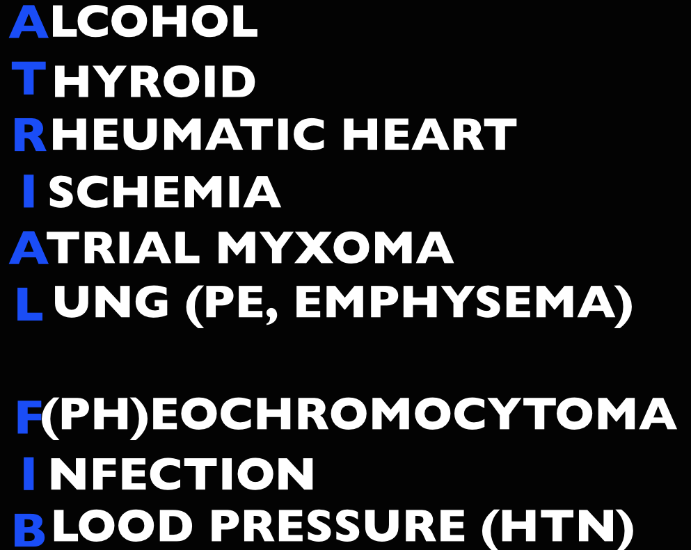
Etiology

Screen Shot 2014-09-16 at 3.57.07 PM

Management

Unstable (hypotension, pulmonary edema, altered mental status, chest pain) – cardioversion.

  • Pads in either an anterior-lateral (AL) or anterior-posterior (AP) position followed by synchronized cardioversion at 100-200 J biphasic.  Current literature shows no significant difference in pad placement [1]
  • If cardioversion fails, try amiodarone load or diltiazem. Check out EMCrit on Crashing A Fib

Stable

  • Rate control. A target of <120 beats per minute is acceptable in the ED [2-3].  First line agents are nodal blocking agents such as diltiazem and metoprolol
    • Diltiazem 0.25 mg/kg IV over 2 minutes with a peak effect in 2-7 minutes. Can repeat at 0.35 mg/kg IV over 2 minutes.
    • Metoprolol 5-10 mg IV.
  • Rhythm control with cardioversion.  While there’s no proven benefit to rhythm control, many patients would prefer to be in sinus rhythm and ED cardioversion of stable new-onset atrial fibrillation is appropriate in a select population, notably, when the onset is <48 hours (or <72 hours per Rosen).  The pooled literature suggests a thromboembolism rate <0.8% [4].
    • Note: A recent article in JAMA by Nuotio et al found a higher rate of embolic events in patients who were electively cardioverted after >12 hours in atrial fibrillation.The 30 day risk of thromboembolism when cardioverted between 12-48 hours was 1.1%, compared to the ~2% risk if cardioverted after 48 hours. While the risk is still small, it is higher than the ~0.3% risk of thromboembolism with anticoagulation on board.
  • Treat the underlying cause (ex: sepsis, pulmonary embolism, hyperthyroidism, etc)
  • May also consider Amiodarone, Digoxin (mean >11 hours to rate control) [3]

In atrial fibrillation with pre-excitation (WPW), an often wide and irregular rhythm with different/changing morphologies to the QRS do NOT treat with an AV Nodal blocking agent as this may result in death (Adenosine, Beta-blocker, Calcium-channel blocker, etc). Treat with procainamide or shock

Disposition – Admit patients that present unstable, with underlying co-morbidities, or those that are not rate controlled.  Depending on the patient’s follow up and local practice patterns, the

Atrial Flutter

How to Avoid Misdiagnosing Atrial Flutter – Dr. Amal Mattu

Management – same as atrial fibrillation

  • More sensitive to electrical cardioversion, less sensitive to chemical cardioversion

Multifocal Atrial Tachycardia

Irregular narrow complex tachycardia with p waves of at least 3 morphologies (this can be difficult to see, so look in multiple leads, particularly V2)

 Etiology -often seen in advanced pulmonary disease

Management  – Treat the underlying cause, do NOT cardiovert MAT

 Learn from the master ECG educator, Dr. Amal Mattu

Generously Donated Rosh Review Questions (Scroll for Answers)

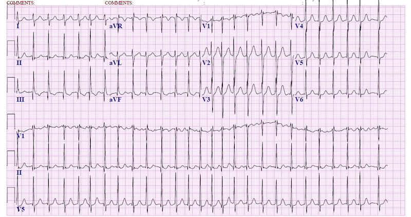
Question 1.  A 72-year-old man with a history of hypertension, diabetes, and congestive heart failure presents to the ED with heart palpitations for the past 4 days. He denies any chest pain, shortness of breath, abdominal pain, or history of similar palpitations. In the ED, his vital signs are BP 135/75, HR 138, RR 14, and oxygen saturation 98% on room air. His ECG is seen below. Which of the following is the most appropriate next step in management?

Rosh Review
Rosh Review

A. Chemical cardioversion

B. Rate Control

C. Synchronized cardioversion

D. Warfarin

Question 2.  When do you worry about giving calcium channel blockers, beta-blockers, or digoxin in a patient with atrial fibrillation?

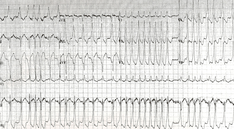
Question 3. An 18-year-old woman presents with palpitations and near syncope. Her vitals are T 98.7F, HR 199, BP 113/66, RR 32, and oxygen saturation 94%. Her ECG is shown below. What treatment is indicated?

Rosh Review
Rosh Review

A. Administer adenosine 6 mg IV

B. Administer diltiazem 10 mg IV

C. Administer lopressor 10 mg IV

D. Administer procainamide 100mg IV

References

1.  Kirkland S, Stiell I, AlShawabkeh T, Campbell S, Dickinson G, Rowe BH. The Efficacy of Pad Placement for Electrical Cardioversion of Atrial Fibrillation/Flutter: A Systematic Review. Acad Emerg Med. 2014;21(7):717–726.

2. Chapter.  Rosen’s Emergency Medicine, 8e.

3.Chapter.  Tintinalli’s Emergency Medicine: A Comprehensive Study Guide, 7e. New York, NY: McGraw-Hill; 2011

4. Cohn BG, Keim SM, Yealy DM. Is Emergency Department Cardioversion of Recent-onset Atrial Fibrillation Safe and Effective? J Emerg Med. 2013;45(1):117–27.

Answers

1.  B. Atrial fibrillation is caused by chaotic, disorderly firing from a second focus within the atria, resulting in uncoordinated atrial contractions. Patients with atrial fibrillation may present with palpitations, chest pain, shortness of breath, or they may be asymptomatic. Atrial fibrillation can be classified as chronic or paroxysmal, with paroxysms lasting minutes to days. On ECG, there are irregularly irregular narrow QRS complexes. In addition, no discernible p-waves are noted, rather fibrillatory waves are seen. Unless the patient is hemodynamically unstable, the mainstay of therapy is rate control. This is achieved through medications that act on the AV node such as calcium channel blockers (eg diltiazem or verapamil), beta-blockers, or digoxin. Due to digoxin’s slow onset of action and side effects, it is considered a second line medication.

If atrial fibrillation has been present for >48 hours, there is an increased risk of atrial thrombus formation. An echocardiogram should be obtained in these patients to exclude thrombus formation prior to rhythm control. Patients with chronic atrial fibrillation usually are placed on warfarin (D) or a similar anticoagulant to prevent thromboembolism.Chemical cardioversion (A) (amiorodone, procainamide or flecainide) can be attempted in patients with paroxysmal atrial fibrillation for less than 48 hours. Synchronized cardioversion (C) is used in patients who are hemodynamically unstable. This can be achieved by administering 50 – 100 J of electricity in synchronization mode.

2.  If a patient has an accessory pathway, such as Wolff-Parkinson-White Syndrome.

3. D. This patient presents with near syncope in the setting of atrial fibrillation with abberant conduction most likely secondary to Wolff-Parkinson-White (WPW) syndrome and should be chemically or electrically cardioverted. WPW syndrome refers to the presence of an accessory pathway between the right atrium and right ventricle. This accessory pathway has a shortened refractory period and can bypass normal conduction down the AV node. Because of the shortened refractory time, the accessory pathway in WPW can conduct atrial impulses much faster than the AV node can allowing for a ventricular rate between 150 and 300 beats per minute. Any tachycardia greater than 200 beats per minute in an adult should raise suspicion for an accessory pathway.

Patients with WPW can be asymptomatic or may present with severe tachydysrhythmias. The most common presenting dysrhythmia is reentrant tachycardia (70-80%) and second is atrial fibrillation (10-30%). In these tachydysrhythmias, the patient can conduct orthodromically (down the AV node and back up the accessory pathway), antidromically (down the accessory pathway and up the AV node) or in both directions. Patients who have any antidromic conduction will present with wide complex tachycardias. In patients with irregularly irregular wide-complex tachycardias, atrial fibrillation with WPW is the most common diagnosis. If the patient is unstable, electrical cardioversion should be pursued immediately as these patients run the risk of degrading into ventricular tachycardia and ventricular fibrillation. If the patient is stable, procainamide can be administered for chemical cardioversion. Procainamide is a class Ia anitdysrhythmic agent. The dose of procainamide (D) is 18-20 mg/kg administered at a rate of 20-30 mg/min.

In patients with WPW, antidysrhythmic agents that block the AV node are contraindicated. Blocking the AV node causes unopposed electrical conduction down the accessory pathway. This can lead to ventricular dysrhythmias. Additionally, the accessory pathway in WPW responds paradoxically to AV nodal blocking agents by further decreasing its refractory time. Adenosine (A), beta-blockers (C), calcium-channel blockers (B) and digoxin all block the AV node.

Episode 14 – Hand

(iTunes or listen here)

The Free Open Access Medical Education (FOAM)

This week we review EM in 5’s episode on the Neuro Exam of the Hand.

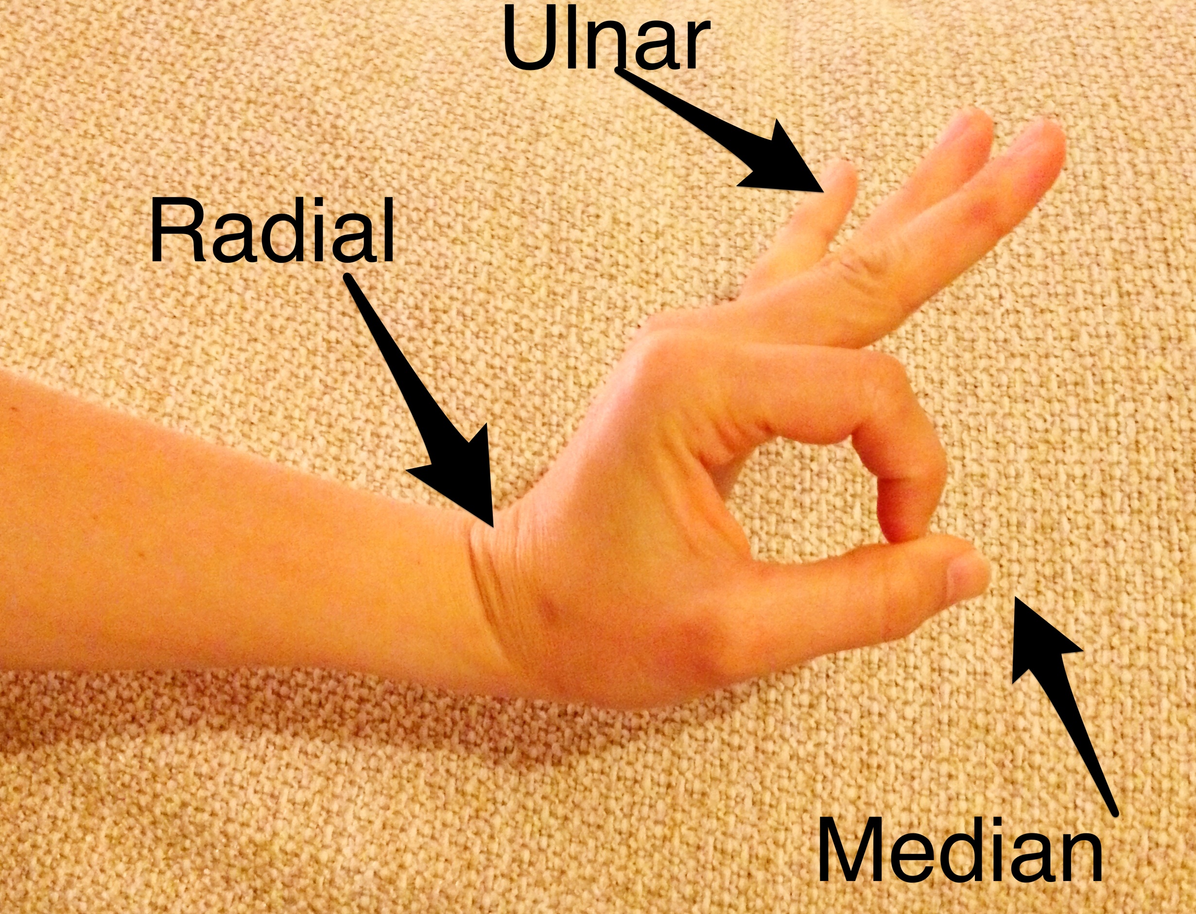
All 3 motor nerves (median, ulnar, radial) in one motion:

  • Make the “ok sign” and try to break the ring formed by the thumb and index finger – median
    • Memory aid: The median is “ok.”
  • Spread 3-5 fingers apart and adduct against resistance – ulnar
    • Memory aid: The intertriginous areas between abducted fingers make a “U” for ulnar
  • Dorsiflex wrist – radial
    • Memory aid…if you look really hard, the the dorsiflexed hand and arm appear to be a sideways, lower case “r”

…all in one

Motor Exam of the Hand

The Bread and Butter

We summarize some key topics from the following readings, Tintinalli (7e) Chapter 280, 295 ; Rosen’s 8(e) Chapter  50 – a well written chapter, but, the point isn’t to just take our word for it.  Go enrich your fundamental understanding yourself!

Flexor Tenosynovitis

Kanavel’s Cardinal Signs (look for words that start with “F”).

  • Pain on passive extension -happens early
  • Finger held in flexion – see above point..extension hurts!
  • Fusiform (uniform) swelling of finger – most common
  • Tenderness along flexor tendon sheath – happens later [3]

Treatment – serious surgical emergency and failure to treat adequately can result in necrosis, proximal spread, and loss of use of hand.

  • IV Antibiotics – penicillinase resistant penicillin (ampicillin-sulbactam, piperacillin-tazobactam) + vancomycin (if suspect MRSA)
  • Consult hand surgery
  • Admit

Hand Pearls

Mallet Finger:  Disruption of the distal extensor tendons, often caused by a jamming injury, hyperextension, or crush.  Look for bruising at the distal interphalangeal joint. Closed injury –  immobilization. Open- extensor tendon repair.

High pressure injection injuries (paint gun, on the job stuff): Worse than they look.  Tetanus, IV antibiotics, consult hand.

Digit amputation storage: Cover the stump with saline gauze and wrap amputated digit in saline soaked gauze.  Put the saline soaked gauze covered digit in airtight plastic bag and place that bag in a bag on ice.

Amputations have best success of reimplantation if they are:

  • Clean cut (saw, etc) versus a crush injury
  • Distal (distal finger > proximal finger > hand)

Learn to Splint Like a Pro (via ERCast) and document a thorough neurovascular exam pre and post-splinting.

Bonus Rosenalli Myth Buster:  Common teaching prevails that epinephrine should not be used with anesthesia of the fingers, nose, toes, penis, etc.  FOAM has debunked this (SMART EM podcast), as well as peer reviewed literature: Waterbrook et alTruth, and epinephrine, at our fingertips: unveiling the pseudoaxioms.

  • Use of epinephrine in finger, nose, etc IS Rosenalli approved [Ch 40, Tintinalli 7e; Ch 3 Rosen’s 8e].

Generously Donated Rosh Review Questions (Scroll for Answers)

Question 1.  Which of the following is classically seen in flexor tenosynovitis?

A. Extended position of the involved digit

B. Fusiform swelling of the digit

C. Tenderness over the extensor sheath

D. Vesicular eruption over the flexor surface

Question 2. A 42-year-old man presents to the ED with an amputation of his left thumb just proximal to the interphalangeal joint. The injury occurred 1 hour ago at a rural construction site while the patient was operating a power miter saw. The thumb is brought in, in a sandwich bag, along with the patient. Which of the following is true regarding predictors of successful replantation?

A. Crush injuries have a high success rate of replantation

B. Digits have better tolerance for ischemia than limbs have

C. The amputated part should be kept cold and dry

D. The patient’s social history adds little value to the success rate of replantation

References

1. Chapters 3, 50.  Rosen’s Emergency Medicine, 8e.

2.Chapters 40, 280, 295.  Tintinalli’s Emergency Medicine: A Comprehensive Study Guide, 7e. New York, NY: McGraw-Hill; 2011

3. Draeger RW, Bynum DK Jr. Flexor tendon sheath infections of the hand. J Am Acad Orthop Surg. 2012 Jun;20(6):373-82. doi: 10.5435/JAAOS-20-06-373.

Answers.

1. B.  The flexor tendons of the fingers are covered by a double layer of synovium to promote gliding of the tendon underneath. Infections in the synovial spaces in the hand tend to spread along the course of the flexor tendon sheaths and may extend proximally to the hand. Infections are usually due to penetrating trauma involving the sheath but occasionally from hematogenous spread. Four cardinal signs of acute flexor tenosynovitis are usually present to help distinguish tenosynovitis from other hand infections. These criteria are referred to as the Kanavel’s signs. Flexor tenosynovitis is a surgical emergency. Consultation with a hand surgeon is warranted along with intravenous antibiotics. The affected digit is held in a flexed (A), not extended, posture. The tenderness is over the flexor (C) sheath, not extensor. Vesicles (D) are not commonly associated with flexor tenosynovitis. A localized herpes simplex infection may cause vesicles to form on a digit

2. B.  Ischemia time is one of the most important predictors of successful replantation. Digits have less muscle mass to oxygenate and tolerate ischemia better than amputations more proximally along the limb. Replantation of limbs must be completed within 4–6 hours, but digits can tolerate an ischemic time of up to 8 hours, given proper preservation.  Crush injuries (A) have a low success rate for replantation due to the significant destruction of neurovascular structures. The amputated body part should be irrigated with normal saline to remove gross contamination, wrapped insterile gauze moistened with saline (C), and placed in a sterile, watertight container. This container should be placed in ice water, but the digit itself should not be submerged. Research has repeatedly shown that tobacco use (D), especially smoking after surgery, will worsen the chance of successful replantation. Obtaining a social history is important in these cases.

Episode 13 – Tricyclic Antidepressants and Sodium Channel Blockade

(iTunes or listen here)

The Free Open Access Medical Education (FOAM)

We review Dr. Amal Mattu’s episode, Why You Should Care When Things are Totally RAD, on his fabulous weekly ECG video series. This week’s episode reviews Rightward Axis Deviation (RAD) and the two “can’t miss” causes in the Emergency Department (ED): Sodium channel blocker toxicity (tricyclic antidepressants and cocaine) and pulmonary hypertension (pulmonary embolism).

Causes of Rightward Axis Deviation – Lead misplacement, Hyperkalemia (can do anything on the ECG)
Ventricular ectopy (VT), Lateral MI (Q-waves in lead I), Left posterior fascicular block, Right ventricular hypertrophy, Dextrocardia

Sodium Channel Blocker Toxicity ECG clues:

  • Tachycardia (most common)
  • Right Axis Deviation
  • Tall R wave in aVR
  • Tall R in V1
  • Prolonged QRS
  • Prolonged QTc

EMCrit Episode 98 – Dr. Scott Weingart reviews the treatment of tricyclic antidepressant (TCA) toxicity. Top pearls

  • When using sodium bicarbonate (NaHCO3) to treat TCA toxicity, the pH doesn’t seem to rise above 7.5 (but you still have to check it). Ventilate these patients appropriately (given breakdown of NaHCO3 to CO2).
  • When infusing NaHCO3, check a VBG every hour to follow the pH and the potassium.
  • Calcium can be critically low in these patients, replete and check accordingly.

The Bread and Butter

We summarize some key topics from the following readings, Tintinalli (7e) Chapter 171 ; Rosen’s 8(e) Chapter 151, Goldfrank’s Toxicology Chapter 73 but, the point isn’t to just take our word for it. Go enrich your fundamental understanding yourself!

Tricyclic Antidepressants

Includes drugs like amitriptyline, nortriptyline and work on a variety of receptors. Other drugs, such as carbemazepine and cyclobenzaprine demonstrate TCA-like properties, albeit with different toxicologic properties (ALiEM post) Properties include anticholinergic, antihistamine, and alpha-1 adrenergic blockade in addition to sodium channel blockade and inhibition of the reuptake of norepinephrine and serotonin.

Clinical Presentation

  • Anticholinergic Symptoms predominate early on – tachycardia, flushed/dry skin, mydriasis, altered mental status, urinary retention (Mad as a Hatter, Hot as a Hare, Dry as a Bone, Blind as a Bat)
  • Mumbling speech
  • Seizures and ventricular dysrhythmias typically occur within the first few hours after ingestion.

Diagnosis – serum TCA levels and urine drug screens have no value in acute treatment (that’s per Rosen) [2]. In addition to history and physical examination, the ECG may be helpful:

  • QRS >100 ms*
  • Tall R wave in lead aVR (>3mm), R/S(avr) > 0.7
  • T40-ms axis between 120° and 270° (difficult to measure)

*It is widely taught, included in Rosen’s, Tintinalli, and Goldfrank, that seizures occur with a QRS >100 ms and ventricular dysrhythmias occur with a QRS >160 ms [1-3]. A 2004 meta-analysis demonstrated operating characteristics that were less than favorable, with the following characteristics:

  • QRS duration >100 ms to predict seizure: Sensitivity 69% (CI 57-78), +LR 3.18, -LR 0.38
  • QRS duration to predict ventricular dysrhythmia: Sensitivity 79% (CI 58-91), +LR 1.77, – LR 0.39
    • Note: It appears the studies in this analysis used a QRS of 100 ms whereas traditional teaching references 160 ms as the threshold for most ventricular dysrhythmias
  • Most predictive of ventricular dysrhythmia: R/S waves ratio (+LR 15.67) but this was evaluated in only one study (Sensitivity 47%)

[4]

Treatment

In acute overdose with an ingestion < 1 hour prior, activated charcoal may be given if no contraindications exist.

Symptomatic treatment and observation for at least six hours.

Hypotension – crystalloid intravenous fluids, vasopressors if needed (norepinephrine)

Seizure – Most seizures are self-limiting and spontaneously terminate within 3 minutes but benzodiazepines and standard seizure treatment can be used .

Prolonged QRS (>100 ms) or Dysrhythmia

Sodium Bicarbonate – 1-2 mEq/kg bolus. Each amp of NaHCO3 has 50 mEq so the average 70 kg patient can get 1.5-2.5 amps. An infusion of NaHCO3 may be used (3 amps of NaHCO3 in 1 L of D5W).

  • Caution: must follow pH (can titrate as high as 7.5-7.55) as well as potassium.

If the NaHCO3 pushes the patient’s pH to the 7.5-7.55 limit and the QRS remains wide, 200 mL of hypertonic saline (3%) can be used.

Lidocaine, a class IB sodium channel blocker/antiarrhythmic can also be used although it may seem counterintuitive.

Magnesium may also be used

Disposition:

Asymptomatic patients six hours after ingestion (no sinus tachycardia) can be medically cleared.

Generously Donated Rosh Review Questions (Scroll for Answers)

Question 1. A 24-year-old man presents to the ED via the police because of altered mental status and violent behavior. The patient reportedly had been on a drug binge, using amphetamines and cocaine for the last 2 days. On arrival, he is afebrile, his heart rate is 140 beats per minute, blood pressure is 180/110 mm Hg, and he is respiring at 22 breaths per minute. He is agitated and combative, but there is no evidence of traumatic injury. His pupils are dilated, and he is diaphoretic. Which of the following statements is true regarding the treatment of amphetamine and cocaine toxicity?

A. Antipsychotics are the preferred initial therapy for agitation and psychosis

B. Beta-adrenergic blockers are safe for managing tachycardia and dysrhythmias

C. Body packers with leaking packets should receive whole-bowel irrigation

D. Vasodilators are first-line therapy for treating amphetamine-induced hypertension

E. Wide-complex tachydysrhythmias should be treated initially with sodium bicarbonate

Question 2. A 27-year-old man is brought to the ED by EMS after being found wandering in the street. His BP is 155/70, HR 115, T 37.5°C, RR 16, pulse ox 99% on room air, and finger stick glucose 98. On exam, the patient is confused with mumbling speech. His pupils are 7 mm and reactive. His face is flushed. Mucous membranes and skin are dry. Which of the following toxidromes is this patient exhibiting?

A. Anticholinergic

B. Cholinergic

C. Sedative-hypnotic

D. Sympathomimetic

More FOAM:

Life in the Fast Lane TCA case

Life in the Fast Lane TCA ECG

References:

1. Chapter 151. Rosen’s Emergency Medicine, 8e.

2.Chapter 171. Tintinalli’s Emergency Medicine: A Comprehensive Study Guide, 7e. New York, NY: McGraw-Hill; 2011

3.Chapter 73. Goldfrank’s Toxicologic Emergencies, 9e, 2010

4. Bailey B, Sc M. A Meta-Analysis of Prognostic Indicators to Predict Seizures , Arrhythmias or Death After Tricyclic Antidepressant Overdose . 2004;42(6):877–888. doi:10.1081/CLT-200035286.

Episode 12 – Back to Biphasics

(iTunes or listen here)

The Free Open Access Medical education (FOAM)

We review Episode 1 of Dr. Salim Rezaie’s REBEL Cast covering exposing the dogma behind biphasic anaphylaxis reactions.

Traditionally, we’re taught to observe patients in the Emergency Department (ED) for 4-6 hours to watch out for biphasic reactions, as the rate of biphasic reactions can approach 20%.

Grunau BE et al. Incidence of Clinically Important Biphasic Reactions in Emergency Department Patients with Allergic Reactions or Anaphylaxis. Ann of EM 2014; 63(6): 736 – 44.

  • Retrospective chart review of 430,000 visits
  • Anaphylaxis n=496 (2 had biphasic reactions, Allergic reactions n=2323 (3 had biphasic reactions)
  • No deaths, biphasic reactions occurred anywhere from 16 minutes into the ED stay to 6 days later

Rohacek M et al. Biphasic Anaphylactic Reactions: Occurrence and Mortality. Eur J All Clin Imm 2014; 69(6): 791 – 7.

  • Retrospective study in Europe of presentations to one hospital from 2001-2013 of n=1334 allergic reactions, n=524 anaphylaxis reactions
  • 2.3% (n=12) met criteria for clinically important biphasic reactions

Additional FOAM on the topic: The SGEM

The Bread and Butter

We summarize some key topics from the following readings, Tintinalli (7e) Chapter 27 ; Rosen’s 8(e) Chapter 119 but, the point isn’t to just take our word for it. Go enrich your fundamental understanding yourself!

Anaphylaxis

Diagnosis

Two or more systems involved after likely allergen exposure (within hours):

  • Skin – generalized hives, itch-flush, swollen lips-tongue-uvula
  • Pulmonary – dyspnea, wheezing, bronchospasm, stridor
  • Cardiovascular -hypotension or associated symptoms (syncope, incontinence)
  • Gastrointestinal symptoms- crampy abdominal pain, vomiting

Hypotension after exposure to known allergen for that patient (minutes to several hours)also qualifies

Differential Diagnosis -vasovagal (most common mimicker), myocardial ischemia, status asthmaticus, epiglottitis, angioedema, foreign body, carcinoid, vocal cord dysfunction, drug reactions, psychogenic

Treatment

Epinephrine. Commit the dose to memory and look up if, needed as this is a huge source of medication errors [Gaeta et al, Benklefat ].

  • Adults: 0.3mg, Pediatrics 0.01mg/kg of 1:1000 epinephrine intramuscular (IM) to lateral thigh (pediatric patients >30kg get the adult dose).
  • If repeated doses of IM epinephrine required and patient continues to remain hypotensive, start intravenous epinephrine. ALiEM’s post on making the dirty epi drip.

Adjuncts:

  • IV fluids for blood pressure/shock
  • Corticosteroids may help prevent recurrence although they take 4-6 hours to work, so are unhelpful in acute attacks [Choo et al]. Rosen recommends either prednisone 0.5-1mg/kg orally or methylprednisolone 80-125 mg IV. (You don’t have to give IV in all cases)
  • Histamine blockers (H1 and H2 such as diphenhydramine and famotidine, respectively) may help with the dermatologic symptoms and pruritis.
  • Glucagon in patients that aren’t responding or are on beta-blockers. ALiEM post.
  • Give patients that are going home a prescription for an EpiPen (for pediatric patients, have one parent go fill the script during the observation period) and show them how to use the autoinjector. These things are expensive and do expire, and there are some coupons out there to help out.

Disposition – Clearly patients with ongoing symptoms and/or shock should stay in the hospital. However, most patients can be discharged home once they are improved and the effects of the epinephrine have worn off. Tintinalli recommends about 4 hours, referencing a study by Brady et al from 2007. Interestingly, in this study there were 2 biphasic reactions that occurred at 20 hours and 46 hours after the initial ED visit. So, not sure how they came up with 4 hours.

FOAM Pearls Supported by the Literature and Rosenalli –

Iodine Allergy is not a thing.

Shellfish allergy does NOT put a patient at increased risk of contrast allergy more than any other allergen [Kaufman et al].

Cross-reactivity between penicillin and cephalosporins is often quoted at 10-20% but, in reality, is far less and a review demonstrates cross reactivity of 1% in patients reporting a penicillin allergy [Campagna et al]. Rosen’s agrees with this assessment and states that the overall cross reactivity is minimal. ALiEM post on this myth

ACE-Inhibitor Induced Angioedema

Cause: The vasodilation and non-pitting edema of the mucosal, dermal, and subcutaneous tissues thought to be mediated by the build up of bradykinin and substance P. Non-allergic, often asymmetric.

Presentation: Swelling of the lips, tongue, airway most often although it can also involve the genitals and viscera.

Treatment:

  • Stop the ACE-Inhibitor.
  • Active, anticipatory airway management. Perhaps, even an awake airway (The EMCrit Way, The Strayer Way).
  • Epinephrine, corticosteroids, and histamine blockers do not work. While fresh frozen plasma may work for hereditary angioedema, but it doesn’t really work in ACE-inhibitor angioedema and there’s no proven therapy [Winters et al].
  • Investigations underway for icatibant (bradykinin 2 receptor antagonist) Bas et al, Schmidt et al and Ecallantide (reversible kallikrein inhibitor)

Question 1. A 55-year old man who is taking several antihypertensive medications presents to the ED with nausea, vomiting, shortness of breath, and a rash after eating a home-cooked Thai meal at a friend’s house about 1 hour ago. The symptoms began within seconds of the first bite of his meal. Despite the patient being administered 2 doses of intramuscular epinephrine, diphenhydramine, dexamethasone, and crystalloid fluids, his blood pressure remains at 75/38 mm Hg.Which other medication should be considered in this patient?

  • A. Cimetidine
  • B. Glucagon
  • C. Norepinephrine
  • D. Octreotide

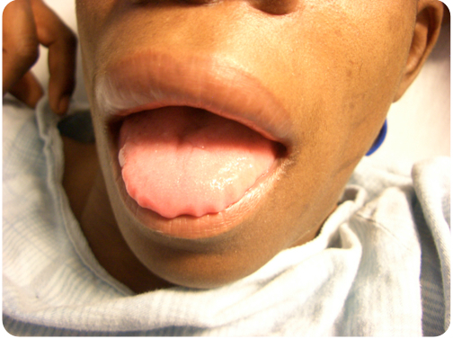
Question 2. A 55-year-old woman presents to the ED for swelling of her tongue and lips.

Photo: Rosh Review
Photo: Rosh Review

She recently started a new antihypertensive medication. Which of the following is the direct mediator for her condition?

  • A. Angiotensin
  • B. Bradykinin
  • C. C1-esterase inhibitor
  • D. Histamine

Answers.

1. B. The patient is experiencing an acute anaphylactic reaction, most likely to peanuts that are commonly found in Thai cooking. Although uncommon, patients taking beta-blocking agents for hypertension may exhibit refractory hypotension despite being administered fluids and epinephrine. This is because epinephrine acts by binding to adrenergic receptors, which includes beta-receptors. To circumvent the beta-receptor, glucagon can be administered, which will bypass the beta-adrenergic second messenger system, potentiate the circulating epinephrine, and help restore vasomotor tone.

Cimetidine (A) is an antihistamine. Although it may help in mild allergic reactions, it will not treat hypotension in severe anaphylaxis. In addition, cimetidine prolongs the metabolism of beta-blockers. Octreotide (D) may be used in management of esophageal variceal bleeding control, treatment of carcinoid syndrome, and refractory hypoglycemia after sulfonylurea-induced hypoglycemia. There is no role in anaphylaxis. Norepinephrine (C) also binds adrenergic receptors that may be inhibited in patients who take beta-blocking medications.
2. B. Angioedema is the clinical manifestation of transient, localized, nonpitting swelling of the subcutaneous layer of the skin or submucosal layer of the respiratory or gastrointestinal tracts. There are many cases of angioedema, but the condition is usually divided into hereditary, acquired, and drug-induced causes. Hereditary angioedema (HAE) is caused by deficiency or dysfunction of C1-esterase inhibitor and is usually precipitated by stress or trauma. Acquired angioedema is also due to deficiency or dysfunction of C1-esterase inhibitor, but is not due to a genetic cause; rather, it appears later in life. The exact etiology is unknown, but the condition is exceedingly rare. The most common cause of drug-induced angioedema is due to an adverse reaction from ACE inhibitors. When ACE is inhibited by medications, angiotensin I is not converted to angiotensin II, and bradykinin is not metabolized. It is thought that the increased level of bradykinin is responsible for angioedema induced by ACE inhibitors. Angioedema can result in severe airway compromise or, less commonly, compromise in the GI tract that is associated with abdominal pain. Evaluation should focus on ruling out laryngeal edema and airway compromise. Although direct visualization is best, asking the patient to phonate a high-pitched “E” is one quick way of assessing for laryngeal edema. If the patient is able to phonate a high-pitched “E,” then the presence of laryngeal edema is unlikely. Treatment is mainly supportive with special attention to airway protection. Angioedema caused by deficiency or dysfunction of C1-esterase inhibitor can be treated by replacing C1-esterase inhibitor with fresh frozen plasma or other recombinant agents.

Angiotensin (A) is a peptide hormone that causes vasoconstriction and a subsequent increase in blood pressure. It is part of the renin-angiotensin system, which is a major target for drugs (ACE inhibitors) that lower blood pressure. An elevated level of angiotensin is not responsible for angioedema. C1-esterase inhibitor (C) serves as the main regulator of the kallikrein-kinin system. As a result of decreased amounts of functional C1-INH, when the kallikrein-kinin system is activated, it is not kept in check. This leads to increased formation of bradykinin and the resultant increased vascular permeability and edema formation and is the cause of hereditary angioedema, not ACE-inhibitor induced angioedema. Histamine (D) has many roles in the body, but its primary role is within the immune system. Mast cells release histamine through a process known as degranulation when they have been sensitized with IgE antibodies and then come in contact with an appropriate antigen leading to the development of urticaria and pruritus.


Episode 11 – Ebola and Transmission Precaution Pearls

Episode 11 (iTunes or listen here)

The Free Open Access Medical education (FOAM)

We review Mount Sinai Emergency Medicine Residency’s blog post on Ebola. The Pearls:

  • Signs and symptoms of ebola: Fever (>101.5F, 41C), severe HA, myalgias, vomiting, abdominal pain, unexplained hemorrhage, hypotension plus an epidemiologic risk factor in the past 3 weeks.
  • Risk factors:  contact with blood or other body fluids of a patient known or suspected to have ebola, residence or travel to endemic areas, and direct handling of bats, rodents, or primates from disease endemic areas.
  • CDC recommends screening in those with :
    • percutaneous/mucous membrane exposure or direct skin contact with body fluids of a person with a confirmed or suspected case of ebola without appropriate personal protective equipment
    • laboratory processing of body fluids of suspected or confirmed ebola cases without appropriate PPE or standard biosafety precautions
    • participation in funeral rites or other direct exposure to human remains in the geographic area where the outbreak is occurring without appropriate personal protective equipment.
  • Personal protective equipment is key in prevention of ebola spread.  Ebola is not airborne but due to the case mortality rate, fear, and questionable history of aerosol transmission in the past, we treat it like it is.  Recommended protection in the United States:  fluid impermeable gown, N95 respirator, eye shield and in situations with large amounts of fluids -double gloving, disposable shoe covers, and leg coverings (CDC recommendations).

Check out the CDC website on Ebola

EMDocs post on Ebola

EMCRIT Ebola algorithm


The Bread and Butter

We summarize some key topics from the following readings, Tintinalli (7e) Chapters 157, 148  but, the point isn’t to just take our word for it.  Go enrich your fundamental understanding yourself! Airborne Precautions – used for patients known to be or suspected of being infected with organisms transmitted by airborne droplets and small particle residue (<5 micrometers) of evaporated droplets containing microorganisms that can be spread by air currents.

  • Require special, negative pressure rooms with special ventilation and filtration and the N95 respirators.
  • Limited movement of patient within the health care setting and in the ED they need to be in a room with the door closed.
  • Recommended for: Measles, Varicella, Tuberculosis

Droplet Precautions – used for patients known to have or suspected of having serious illnesses transmitted by large particle droplets (>5 micrometers) produced by the patient during talking, sneezing, or coughing or during procedures.

  • Use a mask and wash your hands.  N95 respirator recommended for procedures like bronchoscopy, suctioning, etc.
  • Non Sterile gown if one anticipates substantial contact with the patient or if the patient is incontinent or has wound drainage not contained by dressings.
  • Limit transportation and movement of the patient – they should wear a mask when transported throughout the hospital.
  • Recommended for: Serious infections, Pertussis, Parvovirus B19, Mumps, Rubella

Varicella – Herpes virus that causes chickenpox (primary infection) and a secondary reactivation (herpes zoster/shingles) as the virus may lie latent in dorsal root ganglia.

  • Use Airborne precautions as it’s spread via respiratory secretions but may also spread (although less infectious) from the fluid of the non-crusted vesicles.
  • Symptoms of chickenpox: fever, malaise, headache and a vesicular rash that appears in crops with lesions at varying stages, including papules, vesicles, and crusted lesions, predominantly on the torso and face.
  • Most infections are minor and self-limited but increased sequelae exist in the immunocompromised and elderly.  These subgroups may benefit from antivirals
  • Immunizations now prevalent although individuals can still get mild chickenpox after immunization.
  • Varicella-zoster immune globulin exists but use as postexposure prophylaxis is essentially limited to non-immune pregnant women and the severely immunosuppressed. Healthy non-immunue individuals can be vaccinated after exposure and, if they are high risk and develop symptoms, they can get antivirals.

Generously Donated Rosh Review Questions

Question 1. [polldaddy poll=8241932]

Question 2.  A 3-year-old boy presents to the ED with 3 days of fever, cough, and runny nose. On exam, you note conjunctival injection and an erythematous, nonblanching, nonvesicular, maculopapular rash behind his ears and on his hairline, with a few spots on his chest. [polldaddy poll=8241939]

References:

Safe Management of Patients with Ebola Virus Disease (EVD) in U.S. Hospitals.”  CDC.  August 6, 2014

Cline DM.  “Chapter 157. Occupational Exposures, Infection Control, and Standard Precautions”  Tintinalli’s Emergency Medicine:  A Comprehensive Study Guide (2011).

Answers

1. Chickenpox is a highly contagious but generally benign and self-limited viral disease caused by the varicella-zoster virus (also known as human herpesvirus 3). The disease is characterized by the sudden onset of fever, malaise, and a pustular maculopapular rash that can occur anywhere on the skin or mucus membranes. The lesions then become vesiculated followed by scabbing over the course of 3-4 days before resolving. Skin lesions appear in crops with multiple lesions of various stages appearing on the skin at the same time. Uncomplicated infection is generally treated with supportive measures, including antipyretic, antipruritic, and pain control medications. Antivirals such as acyclovir, valacyclovir, and foscarnet may also be initiated in severe disease or immunosuppressed individuals. Parents should be cautioned to avoid giving their children aspirin or aspirin containing medications due to the risk of developing Reye’s syndrome.

The lesions of chickenpox appear suddenly rather than gradually (A). Smallpox lesions may appear similar to chickenpox lesions, however they are found in the same stage (B) of development. Rubella (German measles) is associated with the sudden onset of a maculopapular rash that first appears on the face then rapidly spreads inferiorly to the neck, trunk, and extremities and fades by the 3rd day (C).

2. Rubeola, or measles, is associated with fever and rash with cough, conjunctivitis, coryza, and Koplik spots. The characteristic rash is erythematous, nonblanching, and maculopapular. It begins on the head, usually behind the ears and around the hairline, with subsequent spread down the face, to the trunk, and extremities (centrifugal spread). The rash may coalesce into salmon-colored patches and typically disappears within 1 week. Koplik spots or pinpoint-sized white lesions on a red background that appear on the buccal mucosa opposite the molars are pathognomonic.

Roseola (A) is a viral infection with the onset of a rash that occurs upon resolution of a high fever. It is common in ages 6–18 months. Rubella (B) is often referred to as “three-day measles.” It is a mild illness, except for congenital infection, which can cause major birth defects. It is associated with fever, rash, and prominent lymphadenopathy, with tender posterior auricular, cervical, and occipital nodes. Varicella (D) (chicken pox) is associated with a flu-like illness and the formation of macules that progress to fluid-filled vesicles in an erythematous base (“dew drops on a rose petal”). Crops of lesions typically appear at the same time with vesicles in various stages of healing.

Episode 10 – Pediatric GI Emergencies

Episode 10 (iTunes or listen here)

The Free Open Access Medical education (FOAM)

We review Dr. Natalie May’s brilliant post on the St. Emlyn’s blog, “When Sick Means Sick: Emesemantics and Vomiting in Kids”  in which she dissects emesis descriptors such as bilious, projectile, and coffee-ground.

The Pearls:

  • Ask for color descriptors or look at the emesis yourself rather rely on typical descriptors of emesis.
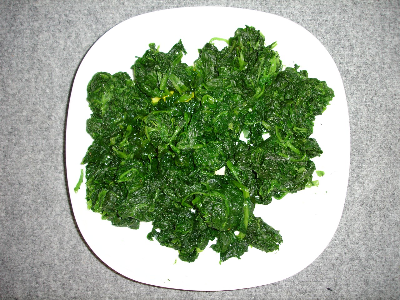
  • Bilious vomiting, medical speaking, means emesis that appears like cooked, green spinach – not the yellow color that parents often mean. While sometimes normal in older children with gastroenteritis, in neonates or anyone sick appearing, this represents a surgical emergency such as volvulus, malrotation, necrotizing enterocolitis etc.
Photo: Laurent Nguyen, Wikimedia Commons
  • Projectile vomiting – Most vomit is projected at least a short distance, so parents may say even reflux is projectile.  Observe a test feed to gauge whether a baby is vomiting or has true projectile vomiting which may represent idiopathic hypertrophic pyloric stenosis.
  • Coffee ground emesis – this is a blackish-brown gritty emesis but parents may mean any brown-ish vomit.  This is typically indicative of upper GI bleed, which is pretty rare in pediatric patients.

The Bread and Butter

We summarize some key topics from the following readings, Tintinalli (7e) Chapters 111,124; Rosen’s 8(e) Chapter 172 but, the point isn’t to just take our word for it.  Go enrich your fundamental understanding yourself!

Neonatal Jaundice

Physiologic Jaundice – Jaundice in healthy, full-term newborns typically develops during the 2nd – 3rd day of life and resolves by the 5th or 6th day.  This occurs a little later in Asian and premature infants.

  • Mean peak total serum bilirubin is 6 mg/dL
  • Use the nomogram in infants >35 weeks gestational age to determine need for phototherapy

Non-physiologic Jaundice – bad.

  • Jaundice in the first 24 hours
  • Bilirubin rising faster than 5 mg/dL in 24 hours •Clinical jaundice >1 week
  • Direct bilirubin >2 mg/dL
  • In healthy term infants total serum bilirubin concentration >15 mg/dL (so remember: average of 6 but more than 15 is bad. But check out the nomogram in non-preemies).

Indirect Neonatal Jaundice – 3 main causes, listed below. Treatment is with phototherapy and mitigation of underlying causes.

  • Increased lysis – this can be due to lysis of red blood cells or sequestration of blood – ABO incompatibility, splenic sequestration, spherocytosis.
  • Decreased hepatic uptake/decreased conjugation – Immature transfer enzymes (babies grow out of this), breastmilk jaundice (lack certain enzymes, idiopathic hypertrophic pyloric stenosis (unclear why), as well as Gilberts, Crigler Najjar Syndrome
  • Increased enterohepatic uptake (i.e. too much reclaimed from the gut) – obstruction or breastfeeding jaundice (dehydrated babies who are breast feeding).

Direct Bilirubinemia

  • Etiology – biliary tree obstruction or biliary atresia, enzyme deficiencies (cystic fibrosis, alpha-1 antitrypsin deficiency, glycogen storage diseases)
  • Conjugated bilirubin is non-toxic so treat the underlying cause

Emergency Department Diagnostics:

  • Total and Fractionated Bilirubin,  Blood Type with Rh factor, Coomb’s test, blood count, Reticulocyte count, consider sepsis work-up .

More FOAM

Intussusception

Presentation: Abdominal pain, vomiting, bloody or guaiac positive stool.  The classic triad is not useful and present in 15-20%.  While intussusception is most common at approximately 1 year of age and, moreover 2 months -6 years, it can present at any time, including the elderly.

Etiology:  The bowel telescopes on itself and

Diagnosis:  Clinical, ultrasound (target sign), or diagnostic and therapeutic air contrast enema. It’s also reasonable to get plain films, if desired.

Treatment:  Air contrast enema in radiology results in approximately 60% success so most recommend a surgery consult in the event there’s a complication or failure in radiology.  Also, give these patients 20cc/kg fluid bolus and treat their pain.

More FOAM:

Generously donated Rosh Review questions (scroll for answers)

Question 1. A 10-month-old previously healthy boy presents with 1 day of bilious vomiting and fever. The patient is ill-appearing. Physical examination reveals a distended and diffusely tender abdomen with guarding and rebound. [polldaddy poll=8224476]

Question 2. A 2-year-old ex-33 week premature girl presents with vomiting, diarrhea and poor feeding. The patient has episodes of fussiness and inconsolable crying followed by periods of lethargy and sleeping. During periods of fussiness, the patient draws her legs up to her chest. [polldaddy poll=8225869]

Also, check out “Ketamine, The Album” – A musical written by and for emergency physicians as a tribute to ketamine


Answers.

1.Correct Answer ( A ) This patient presents with signs and symptoms concerning for an obstruction secondary to a volvulus and requires emergent surgical evaluation. Malrotation is a relatively common occurrence (1 in 500 live births) and about 75% of patients with malrotation will develop volvulus. During embryonic development, rotation of the gut arrests. This allows for the small bowel to twist around the superior mesenteric artery causing an acute obstruction. Patients will present with sudden onset of abdominal distension and bilious emesis. These infants will be ill-appearing and possibly toxic on presentation. Although a number of diagnostic modalities can be employed for definitive diagnosis, the priority in an ill-appearing infant with bilious emesis is emergent surgical consultation. All other interventions risk delaying definitive management. While waiting for the surgical consultation, the patient should have an IV placed, fluid resuscitation begun and a nasogastric tube placed for decompression of the stomach. Additionally, broad spectrum antibiotics should be administered. After consultation, an upper GI series may be obtained for definitive diagnosis.

Stool cultures (B) are useful when there is a suspicion for infectious process such as a parasitic or bacterial infection. Laboratory studies (C) will provide limited data and should not delay definitive management by a surgeon. The patient should receive intravenous hydration, not oral rehydration (D) as there is a high likelihood that this patient will be taken to the operating room. As such, the patient should be kept NPO.

2. D.  This patient presents with symptoms concerning for intussusception and should have an emergent ultrasound performed to make the diagnosis. Intussusception is defined as the telescoping of one segment of the intestine into another. It is the most common cause of obstruction in children younger than 2 years of age. The classic triadof intussusception is abdominal pain, vomiting and bloody stools but all three features are only present in about 33% of patients. Bowel movements may be loose with mucous and blood and appear like “currant jelly.” Often patients will have cycles of severe abdominal pain lasting 10 to 15 minutes during which they are inconsolable. These episodes are followed by periods of painlessness during which the child may be lethargic. Palpation of the abdomen may reveal a sausage-like mass in the right upper quadrant representing the actual intussusception. The lead point for the telescoping may be due to Henoch-Schonlein purpura vasculitis, Meckel’s divericulum, lymphoma or polyps in children over 5 years of age. In younger children, enlarge Peyer’s patches may be the culprit. These occur after viral infections. Ultrasound of the abdomen is the best initial modality for identifying the intussusception. It may reveal the classic findings of a target sign or “pseudokidney” sign. Sensitivity and specificity of ultrasound approach 100%.  Abdominal X-ray (A) may show intussusception but may be negative in up to 20% of patients. CT(B) and MRI (C) of the abdomen and pelvis  are also unreliable in the diagnosis.

Episode 9 – Pregnancy Emergencies

Episode 9 (iTunes or listen here)

The Free Open Access Medical education (FOAM)

We review Dr. Scott Weingart’s Practical Evidence Podcast #3 – ACEP 2012 Management of Early Pregnancy, in which he summaries the ACEP 2012 Clinical Policy on this topic.

  • The discriminatory zone is out. Get ultrasounds in pregnant patients, regardless of the quantitative beta-hCG.
  • A certain beta-hCG level can not be used to rule in or rule out ectopic pregnancy or viable intrauterine pregnancy (IUP), get the ultrasound and ensure you identify the uterus.
  • If an ultrasound (including radiology’s formal ultrasound) is indeterminate for ectopic versus IUP, that patient should have a repeat ultrasound and follow up with OB within 48 hours.

A good FOAM ectopic rule out pathway

The Bread and Butter

We summarize some key topics from the following readings, Tintinalli (7e) Chapters 102-105; Rosen’s 8(e) Chapter 178 but, the point isn’t to just take our word for it. Go enrich your fundamental understanding yourself!

Bleeding after the First Trimester

The two things we worry about the most are placental abruption and placenta previa.

Placental Abruption – premature separation of the placenta from the uterine lining.

  • Classic presentation: painful vaginal bleeding.
  • Actual presentation: 30% do not have vaginal bleeding, uterine pain/tenderness, back pain, hypotension, nausea.
  • Risk factors: hypertension, previous abruption, uterine scar, cocaine, smoking, blunt trauma, older age
  • Diagnosis: Ultrasound, clinical suspicion, MRI. Note – Ultrasound is specific but NOT sensitive.
  • Management: Intravenous access, hemoglobin, type and screen, coagulation panel, resuscitation, ultrasound, likely call OB.

Placenta Previa – implantation of the placenta over the cervical os.

  • Classic presentation: painless vaginal bleeding
  • Diagnosis: Ultrasound
  • Management is the same as for abruption but do NOT do a speculum or cervical exam unless there is no OB service.

Abdominal Pain in Pregnancy

Pregnant women are at risk for the same abdominal emergencies as everyone else – appendicitis, cholecystitis, and pyelonephritis but they’re also at risk for some special pregnancy related issues.

Septic Abortion – evidence of infection with any type of abortion, often due to unsanitary abortions or retained products of conception

  • Presentation: fever, uterine tenderness at under 20 weeks of gestation
  • Treatment: Ampicillin 3g IV, gentamicin 1-2 mg/kg IV, stat OB consult for source control

Chorioamnionitis – infection or inflammation of the placenta and fetal membranes, often after 16 weeks of gestation

  • Presentation: fever, uterine tenderness, fever/maternal tachycardia, sepsis
  • Risk factors: preterm labor
  • Treatment: Ampicillin 3g IV, gentamicin 1-2 mg/kg IV, stat OB consult for source control

HELLP syndrome – hemolysis, elevated liver enzymes, low platelets.

  • Diagnostics: CBC with schistocytes, Platelets <100,000, Elevated transaminases, Normal or elevated BUN/creatinine, abnormal coagulation profile. CT abdomen/pelvis if patient hemodynamically stable.
  • Complications: liver hematoma, splenic or liver rupture.
  • Management: Magnesium, control of blood pressure (labetalol, hydralazine), correction of coagulopathy, delivery of the fetus.

Pre-Eclampsia/Eclampsia

New onset hypertension (BP >140/90) after 20 weeks of gestation in a previously normotensive patient plus one of the following: proteinuria or end organ dysfunction (pulmonary edema, renal dysfunction, visual changes, etc). Eclampsia is a sequelae of pre-eclampsia, like HELLP syndrome, characterized by seizures and coma.

  • Control seizures with magnesium
  • Control BP if DBP >105 mmHg
  • Check for organi injury (CBC/platelet count, transaminases, BUN/Creatinine)

Generously donated Rosh Review questions (scroll for answers)

Question 1. A 27-year-old woman 32 weeks pregnant presents with bright-red vaginal bleeding for 1 day. The patient denies any pain and is not tender on abdominal exam. Her vital signs are BP 115/70, HR 90, and RR 16. [polldaddy poll=8210380]

Question 2. A 26-year-old woman presents with abdominal cramping after a positive home pregnancy test. Her vitals are T 98.7°F, HR 94, BP 110/66, RR 18, oxygen saturation 97%. Her exam is unremarkable. Labs reveal a serum beta HCG of 1000 mIU and she is Rh positive. She states that the pregnancy is wanted. An ultrasound is performed as seen below.[polldaddy poll=8206329]

More FOAM on vaginal bleeding:

Answers.

1. A. Placenta previa is characterized by painless, fresh vaginal bleeding in late pregnancy. Placenta previa occurs in 1% of pregnancies and is defined as a placenta that extends near, partially over, or completely over the cervical os. These patients are at an increased risk for life-threatening hemorrhage. As a result, the first step in management of placental previa is to obtain intravenous access in anticipation of fluid resuscitation and possible transfusion. Obstetrical consultation is also advised.

2. D. This patient presents with abdominal pain and a positive pregnancy test raising the concern for an ectopic pregnancy. Ectopic pregnancy complicates about 1.5 – 2.0% of pregnancies and is potentially life threatening. There are a number of risk factors for ectopic pregnancy including pelvic inflammatory disease, prior tubal surgery and previous ectopic pregnancy. This patient has an early pregnancy based on the low beta hCG. The transvaginal ultrasound shows an early gestational sac without a yolk sac or fetal pole within the uterus. This ultrasound does not rule out the diagnosis of an ectopic pregnancy as an ectopic pregnancy can cause a decidual reaction in the uterus, which appears similar to an early gestational sac. The definitive ultrasound finding for an intrauterine pregnancy would be the presence of a yolk sac or fetal pole. It is expected that above the discriminatory hCG zone of 1500-2500 mIU, a definitive IUP should be identified. Patients with a beta hCG below the discriminatory zone without a definitive IUP can be managed conservatively with repeat hCG level in 48 hours (the level should double every 48 hours) and repeat ultrasound.

Episode 8 – Acid-Base and Hyponatremia

Episode 8 (iTunes or Listen Here)

The Free Open Access Medical education (FOAM)

This week we review Dr. David Story’s talk from SMACC GOLD, “Is Chloride a Poison?”  Dr. Story discusses the Stewart ion approach to acid-base, driven by the independent variable, the Strong Ion Difference (SID), which is the difference between the sums of concentrations of the strong cations and strong ions (typically Sodium and Chloride). He also reviews literature that suggests that there may be morbidity and even mortality associated with large volume infusions of 0.9% NaCl (NS), although more research is required in this arena to determine the patient oriented sequelae. Perhaps we should be using more balanced solutions such as lactated ringers (LR).

Also, SMACC  is awesome, listen to the talks from SMACC GOLD and come meet us in Chicago next June!

Literature on the topic:

Other FOAM Acid-Base Resources:

The Bread and Butter

We summarize some key topics from the following readings, Tintinalli (7e) Chapters 19,21; Rosen’s (3e) Chapters 124, 125 …but, the point isn’t to just take our word for it.  Go enrich your fundamental understanding yourself!

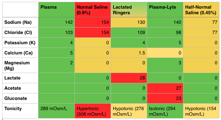
IV Fluids – Know the Composition (PV card from ALiEM)

Red - Too Much, Yellow - Too Little, Green - Just Right
Red – Too Much, Yellow – Too Little, Green – Just Right

Costs – NS (0.9%) is the cheapest, coming in at just over a US dollar per Liter and LR is slightly more expensive (most estimates are approximately $0.50 more per liter).  Plasma-lyte is more expensive, costing several dollars more per liter.

Downsides of Normal Saline (NS, 0.9%)

  • Hypertonic, hypernatremic, hyperchloremic – it has a little too much of everything and is acidic, with a pH of 5.0 [1]
  • The SID (Strong Ion Difference) of NS is 0, far less than the physiologic or normal SID of 38.  This is where the non-anion gap acidosis comes into play [1].

Caution with Lactated Ringers (LR)

  • LR contains calcium and some labs studies have shown that this may cause clotting; thus, major societies say LR is incompatible with blood products.  There are some studies to show that this may not be as big of a deal as previously thought: Albert et alCull et alLorenzo et al.
  • May interfere with Lactate clearance -A study of healthy individuals demonstrated that LR did not affect serum lactate levels [2].  However, often we are doing these large volume resuscitations in the critically ill who may have hepatic insufficiency and, thereby, reduced lactate clearance.  There is worry that LR may increase the serum lactate, making it difficult to gauge resuscitation by lactate clearance.  Paul Marino’s ICU Book states that significant skewing of the lactate is unlikely unless the patient’s ability to hepatically clear lactate is nil and the patient has gotten several liters of LR [1].

FOAM Resources:

Hyponatremia  – Na+ <135 mEq/L

Symptoms: often asymptomatic but may see vague symptoms such as nausea, vomiting, myalgias, lethargy.  Values <120 mEq  more associated with symptoms and <113 mEq, may see seizures/coma.

Causes:

  • Hypovolemic – Extra-renal: dehydration (vomiting, diarrhea, small bowel obstruction, burns), infusion of hypotonic fluids, Renal: thiazide diuretics, Renal Tubular Acidosis, osmotic diuresis, aldosterone/mineralocorticoid deficiency
  • Euvolemic – SIADH (syndrome of inappropriate secretion of antidiuretic hormone), water intoxication, drugs (NSAIDs, APAP, TCAs, sulfonylureas, morphine, carbamazepine, etc), beer potomania
  • Hypervolemic – think organ failure and people who are third spacing fluids. Congestive Heart Failure, Liver Failure, Renal Failure

Other FOAM Hyponatremia Resources:

Generously donated Rosh Review questions (scroll for answers)

Question 1.[polldaddy poll=8191357]

Question 2. A 23-year-old woman presents with seizures. The patient received 2 mg of lorazepam by EMS but continues to seize. Serum lab tests show the following: sodium 118, potassium 3.6, chloride 90, bicarbonate 21, BUN/Cr 10/1.0, glucose 89. [polldaddy poll=8191360]

Additional References:

1.Tizard, H.  Chapter 12: Colloid and Crystalloid Resuscitation.  Marino’s The ICU Book, ed 4.Lippincott Williams & Wilkins, 2007.

2.Didwania A, Miller J, Kassel D, Jet al. Effect of intravenous lactated Ringer’s solution infusion on the circulating lactate concentration: Part 3. Results of a prospective, randomized, double-blind, placebo-controlled trial.Crit Care Med. 1997 Nov;25(11):1851-4.

Answers.

1.C -The syndrome of inappropriate secretion of ADH (SIADH) is defined by the secretion of ADH in the absence of an appropriate physiologic stimulus. Its hallmark is an inappropriately concentrated urine, despite the presence of a low serum osmolality and a normal circulating blood volume. Causes of SIADH include central nervous system disorders, pulmonary disease, drugs, stress, pain, and surgery. Therefore, the above patient, with a known history of lung cancer and hyponatremia, most likely has SIADH and exhibits the following lab findings: serum osmolarity low, urine osmolarity high, urine sodium high. Psychogenic polydipsia (D) is a rare cause of euvolemic hyponatremia and is seen in psychiatric patients who consume large amounts of free water (in excess of 1 L/hr). This large consumption overwhelms the kidney’s ability to excrete free water. Patients will exhibit serum osmolarity low, urine osmolarity low, urine sodium low. Diabetes insipidus (B) results in the loss of large amounts of dilute urine from the loss of concentrating ability in the distal nephron. This may be due to a central cause—such as the lack of ADH secretion from the pituitary—or a nephrogenic cause—such as the lack of responsiveness to circulating ADH. Laboratory workup that invariably shows serum osmolarity high, urine osmolarity high, urine sodium low (A) rarely occurs.

2.A-This patient presents with prolonged seizure activity and hyponatremia and should emergently be treated withhypertonic saline. Hyponatremia is defined as a serum sodium level <135 mEq/L and is the second most common electrolyte abnormality after hypokalemia. The symptoms and signs of hyponatremia depend on the patient’s volume status, the cause and the rapidity of the change in serum sodium. Typically, patients with acute changes will have more severe symptoms including nausea, vomiting, confusion, stupor and seizures. Chronic hyponatremia will typically present with mild neurologic symptoms as well as lower serum sodium levels than acute hyponatremia. In patients without neurologic symptoms, volume status should be assessed and additional labs should be sent off to determine the cause of hyponatremia (urine sodium, osmolarity etc.). Patients with neurologic symptoms should be aggressively treated with 3% hypertonic saline. When correcting serum sodium, it is important to increase the serum sodium by no more than 0.5 mEq/L/hour and by no more than 10 – 12 mEq/day. More rapid changes can lead to central pontine myelinolysis, a crippling neurologic disease.