Episode 22 – The Knee

(ITUNES OR LISTEN HERE)

The Free Open Access Medical Education (FOAM)

This week we’re covering a post from the incredible pediatric resource, Don’t Forget the Bubbles, “Knee X-ray Interpretation” by Dr. Tessa Davis.  We use a systematic approach to assessing chest x-rays, so why not knee x-rays?

  •  Know the anatomy
  •  Look at:
    • Effusion
    • Main bones
    • Tibiofemoral alignment
    • Tibial plateaus
    • Intercondylar eminence
    • Patellar tendon disruption
    • Patellar fracture

The Bread and Butter

We summarize some key topics from Rosenalli, that’s Tintinalli (7e) Chapter s271, 281; Rosen’s (8e) Chapters 57, 136.  But, don’t just take our word for it.  Go enrich your fundamental understanding yourself.

Knee Dislocation

  • Anterior is most common (40%), posterior (33%)
  • Approximately 50% of knee dislocations may be relocated upon presentation to the hospital (this does not reduce risk of badness)
  • Most worrisome sequelae = popliteal artery disruption.  Of patients with popliteal disruption, the amputation rate rises to 90% 8 hours after the injury without surgical intervention.
  • Workup may depend on your institution (ex: angiogram vs. CT angio vs. ultrasound) but all patients will need an ABI + 24 hour of pulse checks per current standards.
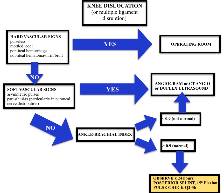
Screen Shot 2015-01-08 at 1.10.48 PM
Algorithm (adopted from Rosen’s)

Septic Arthritis

  • Most Common Organisms: S. aureus, N. gonorrhea
  • Hematogenous spread
  • Most Common Location: knee, hip

Risk factors such as immunocompromised hosts and use of steroids are risk factors for septic arthritis but the ones with the highest likelihood ratio (LR+ >10 is ideal):

  • Skin infection overlying prosthetic joint (LR+ 15)
  • Joint surgery within the preceding 3 months (LR+ 6.9)
  • Age > 80 (LR+ 3.5)

Diagnosis:  In the red, hot, swollen, painful joint, think septic arthritis.  Clinical and laboratory indicators aren’t great. Synovial fluid analysis, particularly the culture exists as the gold standard.  Arthrocentesis Trick of the Trade from ALiEM. Here are the operating characteristics from Margaretten et al:

  • Fever: Sensitivity 57%
  • Lab tests: White Blood Cell count (WBC), sedimentation rate (ESR), and c-reactive protein don’t perform well
    • WBC LR+  1.4 (1.1-1.8); LR- 0.28 (0.07-1.10)
    • Erythrocyte sedimentation rate 1.3 (1.1-1.8); LR- 0.17 (0.20-1.30)
    • C-reactive protein  1.6 (1.1-2.5); LR- 0.44 (0.24-0.82)
  • Synovial fluid gram stain and culture is the “gold standard.”

Treatment: Intravenous antibiotics and washout of the joint by orthopedics in the operating room

 Generously Donated Rosh Review Questions 

Question 1. A 67-year-old man with a history of gout presents with atraumatic left knee pain. Physical examination reveals an effusion with overlying warmth and erythema. There is pain with passive range of motion. He reports a history of gout in this joint in the past. [polldaddy poll=8568492]

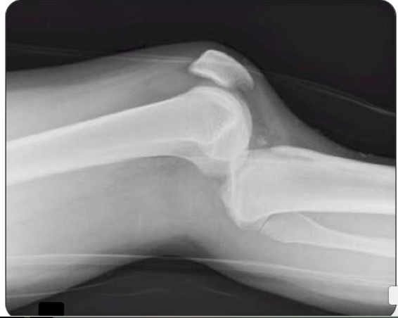
Question 2.  A 27-year-old woman presents with severe left knee pain after an MVC where she was the front passenger. She states her knee hit the dashboard. An X-ray of the patient’s knee is shown below. After reduction, the physical examination reveals swelling of the knee and an Ankle-Brachial Index (ABI) of 0.8. [polldaddy poll=8569540]

Screen Shot 2015-01-12 at 5.13.06 PM

Answers.

1. D. Septic arthritis is a bacterial or fungal infection of a joint typically spread hematogenously unless there is direct bacterial contamination. The synovium is highly vascular and lacks a basement membrane making it susceptible to bacterial seeding. Certain conditions predispose individuals to septic arthritis including diabetes, sickle cell disease, immunocompromise, alcoholism or pre-existing joint disease like rheumatoid arthritis or gout. Fever is present in less than half of cases of septic arthritis so with clinical suspicion an arthrocentesis is indicated. The knee is the most common joint affected and patients have pain (especially on passive range of motion) and decreased range of motion often accompanied by warmth, erythema and fever. This patient may have an acute gouty flare, but the clinician must exclude an infection. On joint fluid analysis, the white blood cell count of a septic joint is typically > 50,000. Indomethacin (B) is a non-steroidal anti-inflammatory agent commonly used in the treatment of acute gout. Gout is an arthritis caused by deposition of monosodium urate monohydrate crystals in the joint space. Acute flares involve a monoarticular arthritis with a red, hot, swollen and tender joint. Acute episodes of gout result from overproduction or decreased secretion of uric acid. However, measurement of serum uric acid (C) does not correlate with the presence of absence of an acute flare. A radiograph of the knee (D) may show chronic degenerative changes associated with gout but will not help to differentiate a gouty arthritis versus septic arthritis.

2. C. Obtain Angiography. This patient presents with a knee dislocation and signs of a popliteal artery injury requiring angiography for diagnosis. A knee dislocation refers to a dislocation of the tibia in relation to the femur and not a patellofemoral dislocation. A tibiofemoral dislocation is a limb-threatening emergency due to the high rate of popliteal artery injury. The neurovascular bundle (popliteal artery, popliteal vein and common peroneal nerve) runs posteriorly in the popliteal fossa. The popliteal artery is tethered to the femur and tibia by a fibrous tunnel and is inherently immobile making it susceptible to injury during dislocation. Knee dislocations typically occur in major trauma. An MVC where the knee strikes the dashboard is a common scenario. The dislocation is usually clinically obvious and should be emergently reduced regardless of the presence of confirmatory X-rays. The leg should rapidly be assessed for any “hard” signs of vascular injury including an absence of pulse, limb ischemia, rapidly expanding hematoma, the presence of a bruit or thrill and pulsatile bleeding. Neurologic status should also be assessed prior to and after reduction. After reduction, all patients should have ankle-brachial index (ABI) performed. A normal ABI is > 0.9. Any patient with an ABI less than this should be further investigated for a popliteal injury with angiography. Splint and elevation (D) may be appropriate once a vascular injury is ruled out. The patient should not be discharged home (A) with an abnormal ABI. Observation and repeat ABI (B) is indicated if the initial ABI is normal.
Episode 21 – Acute Kidney Injury

(ITUNES OR LISTEN HERE)

The Free Open Access Medical Education (FOAM)

Dr. Josh Farkas of the PulmCrit blog has produced a couple of blog posts on the importance of renal protection in sepsis, Renoresuscitation: Sepsis resuscitation designed to avoid long-term complications and Renal microvascular hemodynamics in sepsis: a new paradigm.  Much of this is theoretical and certainly not something that is standard practice, rathery a theory extrapolated from subgroups of several trials.

Suggested renoresuscitation measures:

(1) Avoid renal failure – avoid nephrotoxins (many antibiotics, NSAIDs, ace-inhibitors), avoid hyperchloremic metabolic acidosis.

(2) Avoid volume overload – treating decreased urine output by flooding a patient with fluids is not necessarily the best move.

(3) Protect the glycocalyx of the endothelium – this suggestion proffers more questions than answers. Steroids? Albumin? Certain vasopressors?  Stay tuned, as we’re not really certain what this entails.

The Bread and Butter

We summarize some key topics from Rosenalli, that’s Tintinalli (7e) Chapter 91; Rosen’s (8e) Chapter 97.  But, don’t just take our word for it.  Go enrich your fundamental understanding yourself.

Acute Kidney Injury – typically a creatinine 1.5-2x the patient’s baseline is classified as acute kidney injury.  Urine output can be increased initially but determine whether a patient is making urine and how much, as urine output <0.5 mL/kg/h qualifies as AKI.

RIFLE criteria
RIFLE criteria

Importance – AKI is associated with worse outcomes, although it’s unclear as to whether this is merely a marker of

  • Found in 35-65% of admissions to the intensive care unit, in 5-20% of hospital admissions.  Furthermore, AKI is associated with higher mortality.
  • Renal failure can also cause significant problems for the patient such as electrolyte abnormalities (hyperkalemia the most worriesome, but also hyperphosphatemia) and pulmonary edema.

Etiology – many causes of AKI are reversible or amenable to treatment.

Prerenal – this is one of the most common causes of acute kidney injury and basically is caused by decreased blood flow to the kidney.  Associated with a high BUN/creatinine ratio, increased urine osmolality, a urine sodium concentration less than 20 mEq/L, and FENa less than 1% (this is why getting urine sodium and a concurrent chemistry panel is key).

  • Hypovolemia – volume depleted, hemorrhage, intravascular volume depletion from congestive heart failure or cirrhosis.
  • Hypotension – poor cardiac output (heart failure, valvular problems), shock
  • Decreased flow through the renal artery disease – Nonsteroidal anti-inflammatories: inhibit prostaglandins in the afferent arteriole.  ACE inhibitors prevent the conversion of angiotensin I to angiotensin II, leading to decreased levels of angiotensin II, which when absent decreases the GFR because of dilatation of the efferent arteriole.

Post Renal (Obstructive) – Check out Episode 2 on urologic emergencies.

  • Benign prostatic hypertrophy (BPH) is the most common cause but medications such as anticholinergics and pseudoephedrine. Trauma, stones, strictures, and malignancy can also cause obstruction.

Intrinsic acute renal failure divided into: tubular disease (most common), glomerular disease, vascular disease and interstitial disease.

  • Least common form of AKI in the ED, more common in inpatients.
  • Acute Tubular Necrosis (ATN) most common cause – via nephrotoxins such as aminoglycosides and contrast.
  • Granular “muddy brown” casts – think of necrosis from the “N” in ATN and necrosis tends to be dark.

Indications for emergent dialysis – AEIOU

A- Acidosis

E- Electrolyte emergencies (hyperkalemia!)

I-  Intoxication with dialyzable toxins (ethylene glycol)

O- Overloaded with volume

U- Uremia

 Generously Donated Rosh Review Questions 

Question 1. A 72-year-old man is brought to the ED from a nursing home for evaluation of oliguria. He is found to have an acutely elevated BUN and plasma creatinine from baseline. A Foley catheter is placed; his urine sodium (UNa) is measured below 20 mEq/L and fractional excretion of sodium (FENa) below 1%. [polldaddy poll=8545511]

Question 2.  A 54-year-old man presents to the ED in acute renal failure (ARF). [polldaddy poll=8545512]

Answer 1.  D. This patient’s oliguria with acutely elevated BUN and plasma creatinine suggest that he is in acute renal failure (ARF). His UNa <20 mEq/L and FENa <1% indicate that he has intact reabsorptive function and is able to conserve sodium. This is consistent with prerenal azotemia as the cause for his ARF.

Acute tubular necrosis (ATN) (A), loop diuretics (e.g., furosemide) (B), and osmotic diuresis (e.g., mannitol) (C)all lead to UNa >20 mEq/L and FENa >1% because there is impairment in the ability to concentrate the urine. In such cases, a high-sodium load is excreted.

Answer 2. A.   Acute tubular necrosis (ATN) is a severe form of impairment of tubular epithelial cells caused by ischemia or toxic injury. It is a leading cause of ARF. One of its hallmarks is the presence of brown granular casts on urinalysis. These contain cellular debris rich in cytochrome pigments. In contrast, hyaline casts (B) are usually nonspecific but present after exercise; red cell casts (C) are indicative of glomerular hematuria (e.g., glomerulonephritis); and white cell casts (D) imply renal parenchymal inflammation (e.g., acute interstitial nephritis, pyelonephritis).

Episode 19 – Environment: Mushrooms and Hypothermia

(ITUNES OR LISTEN HERE)

The Free Open Access Medical Education (FOAM)

We review the Tox Talk podcast, Episode 23 – Mushrooms.  Our favorite pearls:

Clitocybe, Inocybe – contain muscarine which stimulates muscarinic receptors (acetylcholine/parasympathetic), causing a cholinergic toxidrome. Think SLUDGE (salivation, lacrimation, urination, defecation, gastric emptying/emesis) and the Killer B’s (bradycardia, bronchorrhea, bronchospasm) or DUMBELLS (diarrhea/diaphoresis, urination, miosis, bradycardia, emesis, lacrimation, lethargy, salivation). Basically, cholinergic toxidrome: SMALL, WET, SLOW.

  • Memory aid: these mushrooms end in -yBE, akin to the “killer B’s” that make cholinergic toxicity deadly.

Gyromitra – (false morel) contains gyromitrin which can cause seizures, in addition to gastrointestinal upset and liver failure.  Treatment: pyridoxine (B6).

  • Memory aid: gyromitra named because they look like the gyri of the brain and, conveniently, make the brain seize through depletion of GABA.

Amanita phalloides – contains amatoxins which cause delayed gastrointestinal symptoms and liver failure., echoing acetaminophen toxicity.

  • Caution: this is different than the amanita muscaria ‘mushroom, which is tricky because that amanita muscaria has neither muscarinic properties nor the toxicity of amanita phalloides.

Bonus pearl: Coprinus species can cause a disulfiram like reaction.

FOAM article on mushrooms by Jo et al

The Bread and Butter

We summarize some key topics from the following readings, Tintinalli (7e) Chapter  ; Rosen’s 8(e) Chapter  – but, the point isn’t to just take our word for it.  Go enrich your fundamental understanding yourself!

Hypothermia starts at 35°C and then is categorized based on severity.

Pearls:

  • Ethanol + hypothermia = bad news.  Ethanol is the most common cause of excessive heat loss in urban areas as people tend to not take warming measures, may be homeless or without heat, and have impaired thermoregulation.  Hypothermia also slows alcohol metabolism, making people drunker for longer.
  • Elderly patients are more susceptible to hypothermia, particularly as they may not sense the cooler temperatures.  Some may also have impaired thermoregulation.
  • Have a low threshold

Diagnostics:

  • Get a temperature, on all patients.  This applies to patient’s “found down” as well as the chronic alcoholic who just seems really drunk.
  • If patients aren’t rewarming 1°C/hr and they’re above 32°C, consider: sepsis, cortisol deficiency, myxedema, ethanol.
  • The J wave or “Osborn” wave is found in many cases of hypothermia, often quoted at ~80%.  However, it is not pathognomonic for hypothermia.
From the Rosh Review

Treatment: Warm the patient.  Don’t call the patient dead until they’re warm and dead, which means their temp is above 30-32°C.

Screen Shot 2014-11-22 at 9.30.13 PM

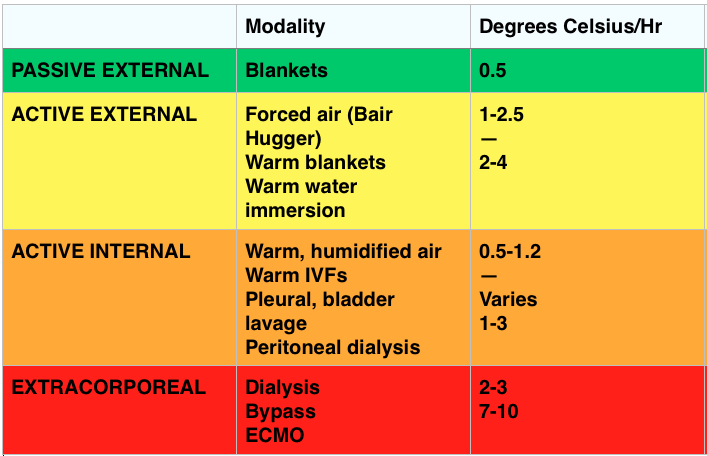
Passive Rewarming – effective when the patient can still shiver (33-35°C).

  • Generates ~1.5°C of heat/hr

Active Rewarming – direct transfer of heat to the patient.

  • Indications: Cardiovascular instability, temp ≤30-32° C, inadequate rate of rewarming or failure to rewarm, endocrine problem, trauma, tox, secondary hypothermia impairing thermoregulation
  • Can be external or internal (which can be minimally invasive like IV fluids or quite invasive with things like bypass or pleural lavage).

Outcomes

  • Unlikely survival with a potassium > 12 mmol/L and recommendations are to terminate resuscitation for potassium >12 mmol/L and consider cessation for potassium between 10-12 mmol/L

FOAM Resources:

EBM Gone Wild on Prognostication

ScanCrit on ECMO in Accidental Hypothermia

EMCrit on Severe Accidental Hypothermia

Generously Donated Rosh Review Questions (Scroll for Answers)

Question 1.  A 40-year-old man with a history of substance abuse is brought in by EMS after being found unconscious outside of a nightclub in the middle of winter. It is unclear how long he was outside. He is unresponsive http://www.mindanews.com/buy-topamax/ with a GCS of 3.[polldaddy poll=8469565]

Question 2.  What is the most common cause of death in hypothermic patients after successful resuscitation?

Question 3.

[polldaddy poll=8473489]

Question 4. What abnormal rhythm is common with temperatures below 32°C?

References:

Danzl DF, Zafren K. Accidental Hypothermia, in Marx JA, Hockberger RS, Walls RM, et al (eds): Rosen’s Emergency Medicine: Concepts and Clinical Practice, ed 7. St. Louis, Mosby, Inc., 2013, (Ch) 140: pp 1883-1885.

Brown D JA, Brugger H, et al. Accidental Hypothermia. N Engl J Med 2012;367:1930-1938.

Mair P, Kornberger E, et al. Prognostic markers in patients with severe accidental hypothermia and cardiocirculatory arrest. Resuscitation 1994;27:47-54.

Answers:

1. D. When the serum potassium is greater than 12 mmol/L resuscitative efforts should be halted as the patient is unlikely to survive and further efforts constitute futile care. Accidental hypothermia is not an uncommon occurrence particularly in colder climates. It may occur in conjunction with substance abuse when an individual becomes impaired and is subsequently exposed to the outdoors. It can also occur as a result of drowning, avalanche and other trauma. Bio-makers other than potassium have been studied including serum lactate (B), pH (C) and clotting time. None have been proven prognostically reliable and therefore should not be used as a guide to determine if resuscitation should be continued. Hypothermic patients that present in cardiac arrest should be warmed to a minimum of 32°C (A) preferably via ECMO or cardiopulmonary bypass. However, if a hypothermic patient is warmed to 32°C and remains in asystole, recovery is unlikely and resuscitative efforts should be terminated. Other indications to cease resuscitative efforts include: obvious signs of irreversible death (e.g. major trauma), valid DNR order, conditions that are unsafe for the rescuer or provider, and an avalanche burial > 35 minutes in which the airway is packed with snow and the patient is asystolic.

2. Pulmonary edema.

2. C. Hypothermia. The ECG demonstrates the presence of J waves or Osborn waves which are seen in hypothermia. One of the first cardiac effects of hypothermia is bradycardia secondary to decreased firing of the cardiac pacemaker cells in cold temperatures. Osborn waves may appear at any temperature below 32°C. The waves are an upward deflection at the terminal portion of the QRS complex. They may represent abnormal ion flux in cold temperatures along with delayed depolarization and early repolarization of the left ventricular wall. As temperatures continue to drop, the ECG will demonstrate prolonged intervals: PR, followed by QRS and then QTc. Both diabetic ketoacidosis (A) and digoxin toxicity (B) may lead to hyperkalemia. In diabetic ketoacidosis, hyperkalemia develops as a result of the acidic pH in the blood and the transport of hydrogen ions intracellularly in exchange for a potassium ion. Digoxin toxicity poisons the cellular Na+/K+ ATPase resulting in elevated extracellular levels of potassium. The ECG manifestations of hyperkalemia begin with peaked T waves. Multiple other findings eventually develop including a shortened QT interval, ST depression, bundle branch blocks, widened QRS, prolonged PR interval, flattened T wave and ultimately a sine wave. Hyperparathyroidism (D) may lead to hypercalcemia. In hypercalcemia, the ECG shows a shortened QT interval, flattened T waves and QRS widening at very high levels.

4.  Atrial fibrillation.

Episode 18 – Falls and Geriatrics

(ITUNES OR LISTEN HERE)

The Free Open Access Medical Education (FOAM)

We review Dr. Ken Milne’s podcast, The Skeptic’s Guide to Emergency Medicine Episode #89,  special episode on falls in the geriatric.  This episode is the first in the HOP (Hot Off the Press) series in which Dr. Milne has paired with Academic Emergency Medicine and the Canadian Journal of Emergency Medicine to review a paper, with the author, the same week the paper is published.

Why HOP is special:

  • Reducing the knowledge gap by disseminating hot-off- the press
  • Concurrent peer review from global audience.  Peer review is a flawed process and in this way, Dr. Milne takes his skeptical perspective to the paper and the author.
  • Key comments from social media will then be published in these journals, reaching the traditional academic readership.

Pearls from the Carpenter et al systematic review

Fall Statistics:

  • Fall Rates – > 65 y/o – 1 in 3 people fall per year; > 80 years old – 1 in 2 people fall per year
  • Elderly patients who fall and are admitted have a 1 year mortality of ~33% [1,3]. So, geriatric falls are bad, it seems logical to wish to predict who is going to fall.

Predictors of Falls:

  • The best negative likelihood ratio (-LR) was if the patient could cut their own toenails –LR 0.57 (95% CI 0.38-0.86) (remember, the target for a -LR is 0.1). This outperformed traditional assessments like the “get up and go test.”
  • Previous history of falls is a big predictor of falls.  Of elderly patients who present to the ED with a fall, the incidence of another fall by 6 months later is 31%.  Of those patients who present with a fall as a secondary problem,14% had another fall within 6 months.
  • The Carpenter instrument has a promising -LR of 0.11 (95% CI = 0.06-0.20) but has not been validated
    • Carpenter instrument: Nonhealing foot sores, self-reported depression, not clipping one’s own toenails, and previous falls

The Bread and Butter

We summarize some key topics from the following readings, Tintinalli (6e) Chapter 307 (This chapter was removed from the seventh edition) ; Rosen’s 8(e) Chapter 182 – but, the point isn’t to just take our word for it.  Go enrich your fundamental understanding yourself!

Abdominal Pain Abdominal pain in the elderly is much higher risk than the younger cohort. This is complicated by vague presentations.  Abdominal pain in the elderly often causes one to raise an eyebrow and ponder chest pathology such as an atypical presentation of ACS.  However, the converse can also be true.  Chest discomfort may really reflect intra-abdominal pathology.  Bottom line – presentations are vague and badness is common.

Geriatric abdominal pain stats:

  • Fever and WBC unreliable.  Per Rosen’s “Elders with potentially catastrophic intra-abdominal processes may not present with a fever or an elevated white blood cell count.”
  • Much higher risk than younger patients – 2/3 patients admitted and 1/5 go directly to the operating room.
  • Most common serious pathologies:  Biliary pathology (cholecystitis), small bowel obstruction, appendicitis.
  • Vascular pathologies such as abdominal aortic aneurysm (AAA) and mesenteric ischemia also have an important place in the differential given increased incidence in the elderly.

FOAM resources:

Polypharmacy  Elderly patients are often on a host of medications but have physiologic alterations that make them susceptible to increased adverse events.

  • 12-30% of admitted elderly patients have adverse drug reactions or interactions as a primary or major contributing factor to their admission and 25% of these drug reactions or interactions are serious or life-threaten
  • Garfinkel et al demonstrated that reducing medications in elderly nursing home patients may actually be better for their health.
  • Some of the highest risk medications, in general, for our elderly patients: diuretics, nonopioid analgesics, hypoglycemics, and anticoagulants.  A patient’s presentation (syncope, fall) may be a manifestation of a medication side effect.
  • There are many high risk pharmaceuticals in the ED, but be very cautious of: narcotics, nonsteroidal anti-inflammatory agents, sedative-hypnotics, muscle relaxants, and antihistamines.
    • NSAIDS – patients may have reduced renal function and due to loss of lean muscle mass, creatine may not be accurate and NSAIDs may tip the patient into renal insufficiency. These drugs may also worsen hypertension and congestive heart failure as a result of salt retention.  NSAIDs are also associated with gastrointestinal bleeding.  Be cautious – acetaminophen is the safer bet.
    • Narcotics – may predispose patients to falls (which are bad in the elderly).  These drugs may also constipate patients, which can cause abdominal pain.  Give guidance and make sure the patient has a solid bowel regimen.
  • Start low and go slow.  It’s much easier to add doses of medications than clearing excess medications.
  • Cautiously start new medications. Furthermore, as drugs may be responsible for the patient’s symptoms that brought them to the ED, review the medication list. If possible, consider discussing discontinuation of medications with the patient’s PCP.

FOAM resources:

Delirium – Delirium in the elderly ED patients is associated with a 12-month mortality rate of 10% to 26% [5].  Be wary of chalking up alterations in mental status to dementia or sundowning.


Generously Donated Rosh Review Questions (Scroll for Answers)

Question 1. [polldaddy poll=8443367]

Question 1. An 87-year-old woman presents to the ED after her caregiver witnessed the patient having difficulty swallowing over the past 2 days. The patient is having difficulty with both solids and liquids. She requires multiple swallowing attempts and occasionally has a mild choking episode. She has no other complaints. Your exam is unremarkable. [polldaddy poll=8443380]

Bonus Question: What proportion of elderly patients with proven bacterial infections lack a fever?

References:

1.Carpenter CR, Avidan MS, Wildes T, et al. Predicting Geriatric Falls Following an Episode of Emergency Department Care: A Systematic Review. Acad Emerg Med. 2014 Oct;21(10):1069-1082.

2. “The Elder Patient.” Chapter 182.  Rosen’s Emergency Medicine, 8e.

3.  “The Elderly Patient.” Chapter 307.  Tintinalli’s Emergency Medicine: A Comprehensive Review, 6e.

4. Garfinkel D1, Zur-Gil S, Ben-Israel J. The war against polypharmacy: a new cost-effective geriatric-palliative approach for improving drug therapy in disabled elderly people.  Isr Med Assoc J. 2007 Jun;9(6):430-4.

5. Gower LE, Gatewood MO, Kang CS. Emergency Department Management of Delirium in the Elderly. West J Emerg Med. May 2012; 13(2): 194–201.

Answers:

1.D.  Physiologic changes of aging affect virtually every organ system and have many effects on the health and functional status of the elderly. Compared to healthy adults, elderly patients have a decreased thirst response that puts them atincreased risk for dehydration and electrolyte abnormalities. Cell-mediated immunity (A) is decreased, which increases susceptibility to neoplasms and a tendency to reactivate latent diseases. Peripheral vascular resistance (B) is increased contributing to development of hypertension. Sweat glands (C) are decreased in the elderly, which puts them at risk for hyperthermia.

2.B.  Dysphagia can be divided into two categories: transfer and transport. Transfer dysphagia occurs early in swallowing and is often described by the patient as difficulty with initiation of swallowing. Transport dysphagia occurs due to impaired movement of the bolus down the esophagus and through the lower sphincter. This patient is experiencing a transfer dysphagia. This condition is most commonly due to neuromuscular disorders that result in misdirection of the food bolus and requires repeated swallowing attempts. A cerebrovascular accident (stroke) that causes muscleweakness of the oropharyngeal muscles is frequently the underlying cause.
Achalasia (A) is the most common motility disorder producing dysphagia. It is typically seen in patients between 20 and 40 years of age and is associated with esophageal spasm, chest pain, and odynophagia. Esophageal neoplasm (C)usually leads to dysphagia over a period of months and progresses from symptoms with solids to liquids. It is also associated with weight loss and bleeding. Foreign bodies (D) such as a food bolus can lead to dysphagia, but patients are typically unable to tolerate secretions and are often observed drooling. These patients do not have difficulty in initiating swallowing.

Bonus. Up to one half.

FOAMcastini – SMACC

(ITUNES OR LISTEN HERE)

FOAMcast will be back shortly with regular core content-cutting edge mash ups.  However, we would be remiss not to take a moment to focus on a conference that inadvertently created FOAMcast…and is coming to Chicago in June 2015.  SMACC – Chicago (#smaccUS) June 23-26, 2015.

The etiology:  FOAMcast was dreamed up whilst milling around the exhibition hall at SMACC, discussing how even core content and “basic” medicine seemed cutting edge and important here.  These projects can be dreamed up via Twitter or e-mail but I think there’s something special engendered by the propinquity of Free Open Access Medical education (FOAM) mixed with the physical conference. Our fate was sealed when Dr. Victoria Brazil happened to stop by to say “hi” while we were hyped up on “long blacks” and blabbering away about what we would call our project.  For better or worse, FOAMcast was born.

  • Note: Please do not blame Dr. Brazil for our off-beat humor or the podcast.  She had no idea what we were up to and does not endorse FOAMcast or Drs. Jeremy Faust and Lauren Westafer.

The talks at SMACC were unparalleled.  The speakers inspiring, the slides clean, the material relevant, and the audience questions thoughtful.  We learned from social workers (Liz Crowe’s hilarious talk), nurses, medics, and doctors from around the globe.  In fact, we became friends, even the pre-med university student.  We learned from them all.

The Core Content – There were a cornucopia of excellent core content talks; for example, Natalie May’s pediatric pearls, Aortic Catastrophes, and the Meaning of Acidosis by Dr. David Story.  There are too many to list and they’re all worth a listen and can be found on iTunes or via the Intensive Care Network.  Even the sonowars were brilliant.  For example, Drs. Matt Dawson and Mike Mallin taught us to visualize cardiac view using humans.  The awkward apical 4 chamber view for cardiac ultrasound:

Apical 4 Chamber View - Cardiac Ultrasound
Awkward (Apical) 4 Chamber View – Cardiac Ultrasound

In this episode, we only had time to hit just a few heavy hitters that haven’t made it onto our other podcasts.

Dr. Haney Mallemat – The Art and Science of Fluid Responsiveness

  • Dr. Mallemat beautifully describes various methods of assessing fluid responsiveness – from IVC ultrasound (used alone, approximately equal to CVP), stroke volume variation, to passive leg raise and more advance ultrasound techniques.
  • Use dynamic markers rather than static numbers, which seemed to be universally lousy.  Trend the patient’s response in order to give them “as much fluid as they need, and not one drop more.”

Dr. Scott Weingart – Sepsis in New York: Our First 15,000 Patients

  • Source control is key in sepsis.  If a patient has an infected gallbladder, obstructing kidney stone, etc – call surgery. Advocate for these patients.
  • ProCESS (and now ARISE) have demonstrated that protocols don’t necessarily have to be followed in order to reduce mortality in sepsis.  We have become increasingly good at identifying and treating sepsis since the original EGDT trial.  In his words – you don’t have to do sh*t, you just have to give a sh*t (Note: you still have to provide basic resuscitation, antibiotics, etc; you just don’t have to do the fancy stuff).
  • He had more pearls about lactate – such as in his collaborative, the number predicted badness but trends mattered less.

Dr. Cliff Reid – Resuscitation Dogmalysis

  • One cannot predict blood pressure based on the presence or absence of a pulse in various anatomic locales (i.e. if there’s a pulse at the radial artery, then their systolic blood pressure is at least >80 mmHg).  This myth was taught for years and still persists in some trauma bays; however, even the evidence and the two most recent iterations of ATLS agree with Dr. Reid [Deakin et al]

Dr. Rob Mac Sweeney – ARDS: An Evidence Based Update

  • The Berlin definition of ARDS [ARDS Definition Task Force]:
    • Acute worsening of respiratory failure (< 1 week)
    • Edema not solely due to hydrostatic pulmonary edema (i.e. should not be due to heart failure or fluid overload)
    • Bilateral infiltrates on CT/CXR *(subjective)
    • PaO2/FiO2 ratio <300 mmHg with at least 5 cm H20 of PEEP
  • The premise of Dr. Mac Sweeney’s talk; however, is that we ARDS is problematic because:
    • ARDS is a disease we can’t diagnose –  Many of the criteria, although seemingly helped by the Berlin definition, are still subjective (ex: CXR Sensitivity 0.73; specificity, 0.70 [Figueroa-Casas]
    • The diagnosis of ARDS is of limited clinical utility.  What he means by this is that the definition doesn’t really affect management and nearly all drugs targeted towards ARDS fail to show benefit consistently.  The ARDS care that does work, like lung protective ventilation and fluid balance, these are just good critical care.  Proning may work, but doesn’t seem to pan out in everyone [Guerin].  Dr. Mac Sweeney is also a little sweet on ECMO, awaiting future studies.
    • People don’t typically die from ARDS even though ARDS is associated with a 40-50% mortality rate.  Yet, only 10% of people with ARDS die of ARDS or respiratory failure. Most people with ARDS die because they’re super sick.
    • Most people with ARDS don’t have ARDS.  Autopsy studies have demonstrated that ~50% of people who met Berlin criteria for ARDS didn’t have the pathognomonic feature of ARDS, diffuse alveolar damage (DAD). The other half of the patients had pneumonia, abscesses, COPD, or other processes [Pinheiro et alThielle et al].
  • The crux of the ARDS issue per Dr. Mac Sweeney -It seems that ARDS is a fairly diverse spectrum with some subjectivity to the criteria.  If approximately half of the people diagnosed with ARDS don’t have ARDS, then it’s no surprise that the therapies don’t benefit them.  He leaves better identification in the hands of researchers.

Timing, Tribes, and STEMIs

  • In medicine we use teams or “tribes” to cope with stress, work together, and rally – Tribe Emergency Medicine, Tribe Anesthesiology, Tribe Surgery, etc.  While making snarky comments, if in jest, may boost the morale and confidence of our team, this may be detrimental to overall patient care
SMACC GOLD
SMACC – Built Friendships, Adventures, and Better Learners

FOAMcastini – ACEP Round Up #2

(ITUNES OR LISTEN HERE)

FOAMcast is bringing you pearls from conferences we attend and, first up, the American College of Emergency Physicians annual meeting, ACEP14.  Yesterday’s episode covered the council meetings.

Scientific Assembly Day 1 Pearls

Opening Session by Freakonomics hosts Steven Levitt and Stephen Dubner. Weird choice? It turns out that economists and physicians have a lot in common.  What’s that?  Probabilities.  As physicians we like to think of ourselves as diagnosticians, but we’re more like probalisticians.  We make predictions, hopefully based on the best evidence, our clinical expertise, and our patient’s values.  People don’t remember the little stuff, like extra testing but they do tend to remember the more outlandish things, like the “amazing saves” or awful “misses.”

  • See this post by Dr. Simon Carley, in which he describes the ways in which physicians are really playing the odds and gambling.

Cardiology Pearls from Dr. Slovis.

  • Post cardiac arrest – targeted temperature management to 35-36 Celsius is the new 33 Celsius [Nielsen].
  • Many patients should probably go to the cath lab after arrest, but it’s still not clear exactly who benefits the most.  STEMIs should probably go to the cath lab and, perhaps, non-STEMI ventricular fibrillation/tachycardia arrests.  Apparently, 10-30% of these are actually STEMIs “on the inside” [More skeptical takes on this from Dr. Radecki here and here]

Infectious Disease Pearls from Dr. David Pigott – When someone returns from a developing nation, say, West Africa, the cause of their fever is not necessarily ebola. It’s probably an unknown, regular virus.   It’s probably not ebola but it may be malaria which is quite common.

  • His thoughts on predictors of badness: Symptoms typically appear within 8-10 days although the “watch” period is 21 days.  If a patient is in their second week of symptoms and are hemodynamically stable, then the patient has a pretty good shot.

Tox Pearls from Dr. Tim Erickson

  • Calcium channel blocker toxicity – you can try fluids, calcium, atropine, and vasopressors.  For sick patients, however, insulin is the best bet (Note, FOAM is ahead of the curve: post on the lack of utility in glucagon from 2012).
    • Insulin bolus of 1 unit/kg followed by a drip of 1 unit/kg/h.  Add dextrose at about 0.5 mg/kg/h, depending on their glucose.
    • Check glucose and potassium every 30 minutes, with the goal to keep the potassium 2.8-3.2, per Goldfrank.
  • Cyanide toxicity (discussed here) – if you’re thinking about it, please do NOT wait on a cyanide level, or any labs.  Treat, with the current recommendation of intravenous hydroxocobalamin. There’s some discussion on the use of intramuscular cobinamide, which would be great in situations without IVs; however, this is largely untested in humans presently [Bebarta et al].
  • Beware of cognitive biases, such as anchoring. For example, lactic acidosis isn’t always sepsis, cyanide, or carbon monoxide.  Metformin associated lactic acidosis is also a thing.

Dr. Scott Weingart – Catch the CO2 Wave (podcast).  End tidal CO2 (ETCO2) – ETCO2 has become essential in monitoring patients in the ED.  With anything we monitor, we really need to understand what we’re looking at as well as the interventions.

  • ETCO2 does NOT = PaCO2.
  • In most patients, the PaCO2 will be ~3-5 mmHg higher than their ETCO2.
  • This is because ETCO2 is really a measure of: PaCO2 (or production) but also cardiac output and alveolar ventilation.  Thus, the ETCO2 may be falsely low in a patient with significant dead space, such as COPD, or with impaired cardiac output (heart failure).

For updates, follow #ACEP14

10660263_10102341624362828_5347687688294499115_n

Episode 16 – Headaches

(ITUNES OR LISTEN HERE)

The Free Open Access Medical Education (FOAM)

This week we review a post from Dr. Rob Orman’s ERCast, Is it really a sinus headache?

POUND- 4 criteria is very indicative of migraine (+LR 24), 3 criteria also likely (+LR 3), although most of this comes from the outpatient literature [1].

  • Pounding headache
  • hOurs: headache lasts 4-72 h without medication
  • Unilateral headaches
  • Nausea
  • Disabling: disrupts daily activities

The Bread and Butter

We summarize some key topics from the following readings, Tintinalli (7e) Chapter 159 ; Rosen’s 8(e) Chapter 20, 103 – but, the point isn’t to just take our word for it.  Go enrich your fundamental understanding yourself!

In Emergency Medicine, our job is to investigate and think about the life and limb threatening causes, even to mundane problems.   Things such as intracranial bleeds, meningitis, masses – these are huge deals and are covered well and hammered into our heads.  For FOAM core content on this, check out the St. Emlyn’s podcast.  On this episode, we’re running a mini-ophthalmology headache special and focusing on headaches that treatment may render “sight saving.”

Temporal Arteritis – often in patients older than 50 years of age and more common in those with a history of polymyalgia rheumatica. May be accompanied by visual changes including the “classic” amaurosis fugax or “curtain” of unilateral vision loss.  If not treated, these patient can lose vision permanently.

  • Unilateral or localized headache, often in the temporal or retro-orbital area
  • Jaw claudication (pain with chewing) – most specific sign
  • Decreased pulse in temporal artery or tenderness
  • Sedimentation Rate (ESR) >50

Treatment

  • Prednisone 40-60 mg if thinking about diagnosis
  • Temporal artery biopsy within 48 hrs

Acute Angle Closure Glaucoma – Classically, these patients present with unilateral mid-dilated pupils and severe nausea, vomiting, and headaches.  The history can, naturally, be less classic and more vague.  Also, if not treated, this can lead to vision loss.

  • Elevated intraocular pressure (>20 mmHg)
  • Decreased visual acuity
  • Fixed irregular semidilated (midposition) pupil
  • Slit lamp — shallow AC (closed angle), injected conjunctiva; corneal microcystic edema (cloudy)

Treatment –

  • Ophthalmology consult stat
    • They may want topical b-blocker, cholinergic, alpha-2 agonist, eye drops or administration of acetazolamide

Idiopathic Intracranial Hypertension (Pseudotumor Cerebri) – Common in young, overweight women or those on oral contraceptives.  Untreated, they can suffer vision loss.

  • Elevated opening pressure (>20-25 cm H20) on lumbar puncture

Treatment

  • Neuro follow up
  • Acetazolamide +/- furosemide
  • Therapeutic lumbar punctures

Cerebral Venous Sinus Thrombosis – may present as atypical headache with stroke like symptoms in patients without known vascular risk factors.  The neurological findings may be transient.  Often associated with post-partum patients, patients with hypercoaguable states (Factor V mutations, protein C or S deficiency, antithrombin III deficiency, etc), patients on OCPs.

Diagnosis – CTV or MRV (magnetic resonance venography) after CT scan, which may be normal.

Treatment – Anticoagulation, although this is somewhat controversial

Generously Donated Rosh Review Questions (Scroll for Answers)
Question 1. A 73-year-old woman with a history of hypertension presents with a unilateral headache for 3 weeks. She states that she has a throbbing pain at her right temple and has pain in her jaw with opening and closing. The vision in her right eye has worsened over the previous day. Her blood pressure is 173/100.
 [polldaddy poll=8340282]
Question 2. A 71-year-old woman presents to the ED with daily headaches for 2 months. She describes the headache as a dull pain that is most intense in the morning and resolves by the afternoon. On exam you note 4/5 motor weakness of the left upper and lower extremity.
[polldaddy poll=8340292]
References:
1. Detsky ME,McDonald DR, Baerlocher MO, Tomlinson GA,McCrory DC, Booth CM. Does this patient with headache have a migraine or need neuroimaging? JAMA. 2006 Sep 13;296(10):1274-83.

2. Chapter 20, 103.  Rosen’s Emergency Medicine, 8e.

3.Chapter 159.  Tintinalli’s Emergency Medicine: A Comprehensive Study Guide, 7e. New York, NY: McGraw-Hill; 2011

Answers
1. D. This patient presents with a unilateral, subacute headache with associated jaw claudication and vision change; symptoms consistent with temporal arteritis. Temporal arteritis or giant cell arteritis is a systemic inflammatory process of small and medium-size arteries. The most commonly involved vessels are the ophthalmic vessels and the extracranial branches of the aortic arch. The disease typically affects patients over 70 years of age and is more common in women than in men. Patients present with a subacute headache that is throbbing in nature and may be present for weeks to months. Often, patients will have symptoms for more than 2 months. Patients may also report jaw claudication secondary to vascular insufficiency of the masseter and temporalis muscles. Physical examination may reveal tenderness over the temporal artery. Systemic symptoms may also be present including fever, joint pains, and weight loss. Diagnostic testing in the Emergency Department generally begins with an erythrocyte sedimentation rate (ESR) with a cutoff of 50 mm/hour although the level may be >100 mm/hour. However, the ESR will be normal in 10-25% of patients. The gold standard diagnostic test is a temporal artery biopsy. In patients with a high-clinical likelihood of temporal arteritis, treatment should be initiated regardless of initial diagnostic testing as delay can lead to permanent visual loss. Prednisone should be started at 60 – 120 mg/day.

Carbamazepine (A) is the treatment of choice for trigeminal neuralgia, not temporal arteritis. The patient does not present with symptoms consistent with hypertensive emergency requiring emergent antihypertensive treatment withlabetalol (B). A non-contrast head CT scan (C) is not helpful in temporal arteritis as the disease does not involve the intracranial contents.

2.  B  More than half of patients diagnosed with a brain tumor complain of headache. However, the headache associated with brain tumor is highly variable. Patients may describe it as continuous or intermittent, unilateral or bilateral, sharp or dull. It is associated with neurologic deficits less than 10% of the time. However, in the setting of aneurologic deficit and chronic headache (as in this scenario with motor weakness), a mass lesion should be strongly considered as the cause. Patients may also complain of nausea, vomiting, visual change, and gait disturbance. Headaches due to brain tumors are classically associated with pain that is worse in the morning (as in this case). However, this is rare.

Central venous thrombosis (A) results from hypercoagulable states and is associated with acute to subacute headaches with vomiting and sometimes seizures. Risk factors include the use of oral contraceptives, postpartum or postoperative states, and any hypercoagulable state such as factor V Leiden mutation, antithrombin III deficiency, protein S or C deficiency, or polycythemia. The diagnosis is usually made by MRI venogram. Migraine headache (C) is classified as a primary headache and can be quite variable in presentation. These headaches can be associated with nausea, vomiting, photophobia, and phonophobia. The headache may also be preceded or accompanied by an aura that develops gradually over minutes, usually lasts 60 minutes, and is reversible. Auras may include neurologic symptom but commonly include scintillating scotomas (dark spots) or flashing lights. Temporal arteritis (D) occurs almost exclusively in patients older than 50 years and is much more common in women. Headache is the most common symptom of temporal arteritis and usually occurs over the frontotemporal region. It is strongly associated with a history of polymyalgia rheumatic. It is not associated with focal neurologic deficits, but it can lead to vision loss due to ischemic optic neuritis.

Episode 15 – Atrial Fibrillation/Flutter

(iTunes or listen here)

The Free Open Access Medical Education (FOAM)

This week we review a post from Academic Life in Emergency Medicine, written by Brent Reed on selecting rate control agents in the management of atrial fibrillation.  This is a follow up post to Bryan Hayes’ summary of emergency department (ED) management of acute atrial fibrillation.  Both are high yield.

Acute Management:

  • No clear cut winner in the beta-blocker vs. calcium channel blocker battle

Long Term Atrial Fibrillation Management in General:

Avoid beta-blockers in:

  • Obstructive lung disease (asthma/COPD)
  • Peripheral vascular disease
  • Diabetics
  • Severe congestive heart failure (CHF)
  • Erectile dysfunction

Avoid calcium-channel blockers in:

  • Severe CHF and acute decompensated heart failure (ADHF)

Of note, in patients

The Maryland Critical Care Project has a great post with many of Dr. Amal Mattu’s key FOAM talks embedded on Tachydysrhythmias You Gotta Know.

The Bread and Butter

We summarize some key topics from the following readings, Tintinalli (7e) Chapter 280, 295 ; Rosen’s 8(e) Chapter  50 – a well written chapter, but, the point isn’t to just take our word for it.  Go enrich your fundamental understanding yourself!

Atrial Fibrillation

Etiology

Screen Shot 2014-09-16 at 3.57.07 PM

Management

Unstable (hypotension, pulmonary edema, altered mental status, chest pain) – cardioversion.

  • Pads in either an anterior-lateral (AL) or anterior-posterior (AP) position followed by synchronized cardioversion at 100-200 J biphasic.  Current literature shows no significant difference in pad placement [1]
  • If cardioversion fails, try amiodarone load or diltiazem. Check out EMCrit on Crashing A Fib

Stable

  • Rate control. A target of <120 beats per minute is acceptable in the ED [2-3].  First line agents are nodal blocking agents such as diltiazem and metoprolol
    • Diltiazem 0.25 mg/kg IV over 2 minutes with a peak effect in 2-7 minutes. Can repeat at 0.35 mg/kg IV over 2 minutes.
    • Metoprolol 5-10 mg IV.
  • Rhythm control with cardioversion.  While there’s no proven benefit to rhythm control, many patients would prefer to be in sinus rhythm and ED cardioversion of stable new-onset atrial fibrillation is appropriate in a select population, notably, when the onset is <48 hours (or <72 hours per Rosen).  The pooled literature suggests a thromboembolism rate <0.8% [4].
    • Note: A recent article in JAMA by Nuotio et al found a higher rate of embolic events in patients who were electively cardioverted after >12 hours in atrial fibrillation.The 30 day risk of thromboembolism when cardioverted between 12-48 hours was 1.1%, compared to the ~2% risk if cardioverted after 48 hours. While the risk is still small, it is higher than the ~0.3% risk of thromboembolism with anticoagulation on board.
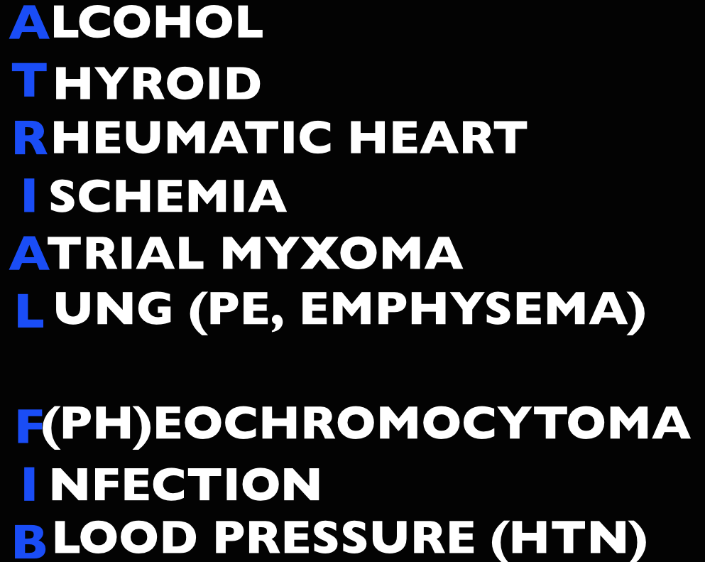
  • Treat the underlying cause (ex: sepsis, pulmonary embolism, hyperthyroidism, etc)
  • May also consider Amiodarone, Digoxin (mean >11 hours to rate control) [3]

In atrial fibrillation with pre-excitation (WPW), an often wide and irregular rhythm with different/changing morphologies to the QRS do NOT treat with an AV Nodal blocking agent as this may result in death (Adenosine, Beta-blocker, Calcium-channel blocker, etc). Treat with procainamide or shock

Disposition – Admit patients that present unstable, with underlying co-morbidities, or those that are not rate controlled.  Depending on the patient’s follow up and local practice patterns, the

Atrial Flutter

How to Avoid Misdiagnosing Atrial Flutter – Dr. Amal Mattu

Management – same as atrial fibrillation

  • More sensitive to electrical cardioversion, less sensitive to chemical cardioversion

Multifocal Atrial Tachycardia

Irregular narrow complex tachycardia with p waves of at least 3 morphologies (this can be difficult to see, so look in multiple leads, particularly V2)

 Etiology -often seen in advanced pulmonary disease

Management  – Treat the underlying cause, do NOT cardiovert MAT

 Learn from the master ECG educator, Dr. Amal Mattu

Generously Donated Rosh Review Questions (Scroll for Answers)

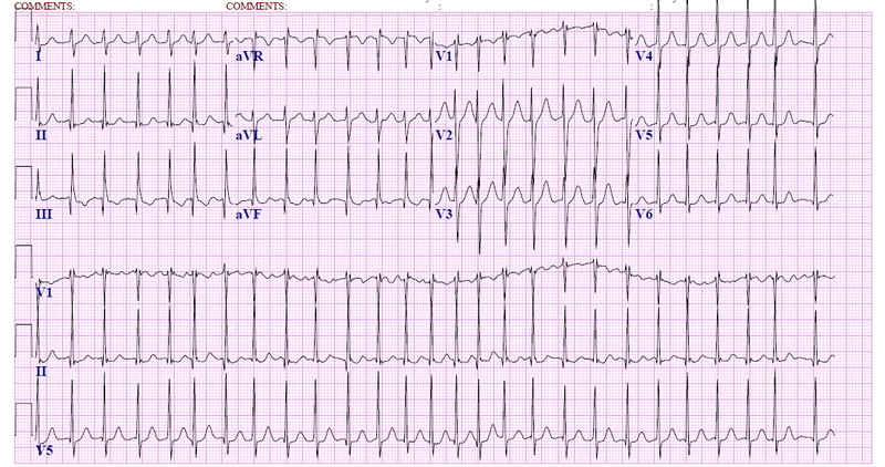
Question 1.  A 72-year-old man with a history of hypertension, diabetes, and congestive heart failure presents to the ED with heart palpitations for the past 4 days. He denies any chest pain, shortness of breath, abdominal pain, or history of similar palpitations. In the ED, his vital signs are BP 135/75, HR 138, RR 14, and oxygen saturation 98% on room air. His ECG is seen below. Which of the following is the most appropriate next step in management?

Rosh Review
Rosh Review

A. Chemical cardioversion

B. Rate Control

C. Synchronized cardioversion

D. Warfarin

Question 2.  When do you worry about giving calcium channel blockers, beta-blockers, or digoxin in a patient with atrial fibrillation?

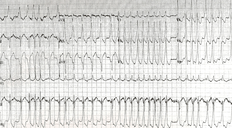
Question 3. An 18-year-old woman presents with palpitations and near syncope. Her vitals are T 98.7F, HR 199, BP 113/66, RR 32, and oxygen saturation 94%. Her ECG is shown below. What treatment is indicated?

Rosh Review
Rosh Review

A. Administer adenosine 6 mg IV

B. Administer diltiazem 10 mg IV

C. Administer lopressor 10 mg IV

D. Administer procainamide 100mg IV

References

1.  Kirkland S, Stiell I, AlShawabkeh T, Campbell S, Dickinson G, Rowe BH. The Efficacy of Pad Placement for Electrical Cardioversion of Atrial Fibrillation/Flutter: A Systematic Review. Acad Emerg Med. 2014;21(7):717–726.

2. Chapter.  Rosen’s Emergency Medicine, 8e.

3.Chapter.  Tintinalli’s Emergency Medicine: A Comprehensive Study Guide, 7e. New York, NY: McGraw-Hill; 2011

4. Cohn BG, Keim SM, Yealy DM. Is Emergency Department Cardioversion of Recent-onset Atrial Fibrillation Safe and Effective? J Emerg Med. 2013;45(1):117–27.

Answers

1.  B. Atrial fibrillation is caused by chaotic, disorderly firing from a second focus within the atria, resulting in uncoordinated atrial contractions. Patients with atrial fibrillation may present with palpitations, chest pain, shortness of breath, or they may be asymptomatic. Atrial fibrillation can be classified as chronic or paroxysmal, with paroxysms lasting minutes to days. On ECG, there are irregularly irregular narrow QRS complexes. In addition, no discernible p-waves are noted, rather fibrillatory waves are seen. Unless the patient is hemodynamically unstable, the mainstay of therapy is rate control. This is achieved through medications that act on the AV node such as calcium channel blockers (eg diltiazem or verapamil), beta-blockers, or digoxin. Due to digoxin’s slow onset of action and side effects, it is considered a second line medication.

If atrial fibrillation has been present for >48 hours, there is an increased risk of atrial thrombus formation. An echocardiogram should be obtained in these patients to exclude thrombus formation prior to rhythm control. Patients with chronic atrial fibrillation usually are placed on warfarin (D) or a similar anticoagulant to prevent thromboembolism.Chemical cardioversion (A) (amiorodone, procainamide or flecainide) can be attempted in patients with paroxysmal atrial fibrillation for less than 48 hours. Synchronized cardioversion (C) is used in patients who are hemodynamically unstable. This can be achieved by administering 50 – 100 J of electricity in synchronization mode.

2.  If a patient has an accessory pathway, such as Wolff-Parkinson-White Syndrome.

3. D. This patient presents with near syncope in the setting of atrial fibrillation with abberant conduction most likely secondary to Wolff-Parkinson-White (WPW) syndrome and should be chemically or electrically cardioverted. WPW syndrome refers to the presence of an accessory pathway between the right atrium and right ventricle. This accessory pathway has a shortened refractory period and can bypass normal conduction down the AV node. Because of the shortened refractory time, the accessory pathway in WPW can conduct atrial impulses much faster than the AV node can allowing for a ventricular rate between 150 and 300 beats per minute. Any tachycardia greater than 200 beats per minute in an adult should raise suspicion for an accessory pathway.

Patients with WPW can be asymptomatic or may present with severe tachydysrhythmias. The most common presenting dysrhythmia is reentrant tachycardia (70-80%) and second is atrial fibrillation (10-30%). In these tachydysrhythmias, the patient can conduct orthodromically (down the AV node and back up the accessory pathway), antidromically (down the accessory pathway and up the AV node) or in both directions. Patients who have any antidromic conduction will present with wide complex tachycardias. In patients with irregularly irregular wide-complex tachycardias, atrial fibrillation with WPW is the most common diagnosis. If the patient is unstable, electrical cardioversion should be pursued immediately as these patients run the risk of degrading into ventricular tachycardia and ventricular fibrillation. If the patient is stable, procainamide can be administered for chemical cardioversion. Procainamide is a class Ia anitdysrhythmic agent. The dose of procainamide (D) is 18-20 mg/kg administered at a rate of 20-30 mg/min.

In patients with WPW, antidysrhythmic agents that block the AV node are contraindicated. Blocking the AV node causes unopposed electrical conduction down the accessory pathway. This can lead to ventricular dysrhythmias. Additionally, the accessory pathway in WPW responds paradoxically to AV nodal blocking agents by further decreasing its refractory time. Adenosine (A), beta-blockers (C), calcium-channel blockers (B) and digoxin all block the AV node.

Episode 12 – Back to Biphasics

(iTunes or listen here)

The Free Open Access Medical education (FOAM)

We review Episode 1 of Dr. Salim Rezaie’s REBEL Cast covering exposing the dogma behind biphasic anaphylaxis reactions.

Traditionally, we’re taught to observe patients in the Emergency Department (ED) for 4-6 hours to watch out for biphasic reactions, as the rate of biphasic reactions can approach 20%.

Grunau BE et al. Incidence of Clinically Important Biphasic Reactions in Emergency Department Patients with Allergic Reactions or Anaphylaxis. Ann of EM 2014; 63(6): 736 – 44.

  • Retrospective chart review of 430,000 visits
  • Anaphylaxis n=496 (2 had biphasic reactions, Allergic reactions n=2323 (3 had biphasic reactions)
  • No deaths, biphasic reactions occurred anywhere from 16 minutes into the ED stay to 6 days later

Rohacek M et al. Biphasic Anaphylactic Reactions: Occurrence and Mortality. Eur J All Clin Imm 2014; 69(6): 791 – 7.

  • Retrospective study in Europe of presentations to one hospital from 2001-2013 of n=1334 allergic reactions, n=524 anaphylaxis reactions
  • 2.3% (n=12) met criteria for clinically important biphasic reactions

Additional FOAM on the topic: The SGEM

The Bread and Butter

We summarize some key topics from the following readings, Tintinalli (7e) Chapter 27 ; Rosen’s 8(e) Chapter 119 but, the point isn’t to just take our word for it. Go enrich your fundamental understanding yourself!

Anaphylaxis

Diagnosis

Two or more systems involved after likely allergen exposure (within hours):

  • Skin – generalized hives, itch-flush, swollen lips-tongue-uvula
  • Pulmonary – dyspnea, wheezing, bronchospasm, stridor
  • Cardiovascular -hypotension or associated symptoms (syncope, incontinence)
  • Gastrointestinal symptoms- crampy abdominal pain, vomiting

Hypotension after exposure to known allergen for that patient (minutes to several hours)also qualifies

Differential Diagnosis -vasovagal (most common mimicker), myocardial ischemia, status asthmaticus, epiglottitis, angioedema, foreign body, carcinoid, vocal cord dysfunction, drug reactions, psychogenic

Treatment

Epinephrine. Commit the dose to memory and look up if, needed as this is a huge source of medication errors [Gaeta et al, Benklefat ].

  • Adults: 0.3mg, Pediatrics 0.01mg/kg of 1:1000 epinephrine intramuscular (IM) to lateral thigh (pediatric patients >30kg get the adult dose).
  • If repeated doses of IM epinephrine required and patient continues to remain hypotensive, start intravenous epinephrine. ALiEM’s post on making the dirty epi drip.

Adjuncts:

  • IV fluids for blood pressure/shock
  • Corticosteroids may help prevent recurrence although they take 4-6 hours to work, so are unhelpful in acute attacks [Choo et al]. Rosen recommends either prednisone 0.5-1mg/kg orally or methylprednisolone 80-125 mg IV. (You don’t have to give IV in all cases)
  • Histamine blockers (H1 and H2 such as diphenhydramine and famotidine, respectively) may help with the dermatologic symptoms and pruritis.
  • Glucagon in patients that aren’t responding or are on beta-blockers. ALiEM post.
  • Give patients that are going home a prescription for an EpiPen (for pediatric patients, have one parent go fill the script during the observation period) and show them how to use the autoinjector. These things are expensive and do expire, and there are some coupons out there to help out.

Disposition – Clearly patients with ongoing symptoms and/or shock should stay in the hospital. However, most patients can be discharged home once they are improved and the effects of the epinephrine have worn off. Tintinalli recommends about 4 hours, referencing a study by Brady et al from 2007. Interestingly, in this study there were 2 biphasic reactions that occurred at 20 hours and 46 hours after the initial ED visit. So, not sure how they came up with 4 hours.

FOAM Pearls Supported by the Literature and Rosenalli –

Iodine Allergy is not a thing.

Shellfish allergy does NOT put a patient at increased risk of contrast allergy more than any other allergen [Kaufman et al].

Cross-reactivity between penicillin and cephalosporins is often quoted at 10-20% but, in reality, is far less and a review demonstrates cross reactivity of 1% in patients reporting a penicillin allergy [Campagna et al]. Rosen’s agrees with this assessment and states that the overall cross reactivity is minimal. ALiEM post on this myth

ACE-Inhibitor Induced Angioedema

Cause: The vasodilation and non-pitting edema of the mucosal, dermal, and subcutaneous tissues thought to be mediated by the build up of bradykinin and substance P. Non-allergic, often asymmetric.

Presentation: Swelling of the lips, tongue, airway most often although it can also involve the genitals and viscera.

Treatment:

  • Stop the ACE-Inhibitor.
  • Active, anticipatory airway management. Perhaps, even an awake airway (The EMCrit Way, The Strayer Way).
  • Epinephrine, corticosteroids, and histamine blockers do not work. While fresh frozen plasma may work for hereditary angioedema, but it doesn’t really work in ACE-inhibitor angioedema and there’s no proven therapy [Winters et al].
  • Investigations underway for icatibant (bradykinin 2 receptor antagonist) Bas et al, Schmidt et al and Ecallantide (reversible kallikrein inhibitor)

Question 1. A 55-year old man who is taking several antihypertensive medications presents to the ED with nausea, vomiting, shortness of breath, and a rash after eating a home-cooked Thai meal at a friend’s house about 1 hour ago. The symptoms began within seconds of the first bite of his meal. Despite the patient being administered 2 doses of intramuscular epinephrine, diphenhydramine, dexamethasone, and crystalloid fluids, his blood pressure remains at 75/38 mm Hg.Which other medication should be considered in this patient?

  • A. Cimetidine
  • B. Glucagon
  • C. Norepinephrine
  • D. Octreotide

Question 2. A 55-year-old woman presents to the ED for swelling of her tongue and lips.

Photo: Rosh Review
Photo: Rosh Review

She recently started a new antihypertensive medication. Which of the following is the direct mediator for her condition?

  • A. Angiotensin
  • B. Bradykinin
  • C. C1-esterase inhibitor
  • D. Histamine

Answers.

1. B. The patient is experiencing an acute anaphylactic reaction, most likely to peanuts that are commonly found in Thai cooking. Although uncommon, patients taking beta-blocking agents for hypertension may exhibit refractory hypotension despite being administered fluids and epinephrine. This is because epinephrine acts by binding to adrenergic receptors, which includes beta-receptors. To circumvent the beta-receptor, glucagon can be administered, which will bypass the beta-adrenergic second messenger system, potentiate the circulating epinephrine, and help restore vasomotor tone.

Cimetidine (A) is an antihistamine. Although it may help in mild allergic reactions, it will not treat hypotension in severe anaphylaxis. In addition, cimetidine prolongs the metabolism of beta-blockers. Octreotide (D) may be used in management of esophageal variceal bleeding control, treatment of carcinoid syndrome, and refractory hypoglycemia after sulfonylurea-induced hypoglycemia. There is no role in anaphylaxis. Norepinephrine (C) also binds adrenergic receptors that may be inhibited in patients who take beta-blocking medications.
2. B. Angioedema is the clinical manifestation of transient, localized, nonpitting swelling of the subcutaneous layer of the skin or submucosal layer of the respiratory or gastrointestinal tracts. There are many cases of angioedema, but the condition is usually divided into hereditary, acquired, and drug-induced causes. Hereditary angioedema (HAE) is caused by deficiency or dysfunction of C1-esterase inhibitor and is usually precipitated by stress or trauma. Acquired angioedema is also due to deficiency or dysfunction of C1-esterase inhibitor, but is not due to a genetic cause; rather, it appears later in life. The exact etiology is unknown, but the condition is exceedingly rare. The most common cause of drug-induced angioedema is due to an adverse reaction from ACE inhibitors. When ACE is inhibited by medications, angiotensin I is not converted to angiotensin II, and bradykinin is not metabolized. It is thought that the increased level of bradykinin is responsible for angioedema induced by ACE inhibitors. Angioedema can result in severe airway compromise or, less commonly, compromise in the GI tract that is associated with abdominal pain. Evaluation should focus on ruling out laryngeal edema and airway compromise. Although direct visualization is best, asking the patient to phonate a high-pitched “E” is one quick way of assessing for laryngeal edema. If the patient is able to phonate a high-pitched “E,” then the presence of laryngeal edema is unlikely. Treatment is mainly supportive with special attention to airway protection. Angioedema caused by deficiency or dysfunction of C1-esterase inhibitor can be treated by replacing C1-esterase inhibitor with fresh frozen plasma or other recombinant agents.

Angiotensin (A) is a peptide hormone that causes vasoconstriction and a subsequent increase in blood pressure. It is part of the renin-angiotensin system, which is a major target for drugs (ACE inhibitors) that lower blood pressure. An elevated level of angiotensin is not responsible for angioedema. C1-esterase inhibitor (C) serves as the main regulator of the kallikrein-kinin system. As a result of decreased amounts of functional C1-INH, when the kallikrein-kinin system is activated, it is not kept in check. This leads to increased formation of bradykinin and the resultant increased vascular permeability and edema formation and is the cause of hereditary angioedema, not ACE-inhibitor induced angioedema. Histamine (D) has many roles in the body, but its primary role is within the immune system. Mast cells release histamine through a process known as degranulation when they have been sensitized with IgE antibodies and then come in contact with an appropriate antigen leading to the development of urticaria and pruritus.


Episode 11 – Ebola and Transmission Precaution Pearls

Episode 11 (iTunes or listen here)

The Free Open Access Medical education (FOAM)

We review Mount Sinai Emergency Medicine Residency’s blog post on Ebola. The Pearls:

  • Signs and symptoms of ebola: Fever (>101.5F, 41C), severe HA, myalgias, vomiting, abdominal pain, unexplained hemorrhage, hypotension plus an epidemiologic risk factor in the past 3 weeks.
  • Risk factors:  contact with blood or other body fluids of a patient known or suspected to have ebola, residence or travel to endemic areas, and direct handling of bats, rodents, or primates from disease endemic areas.
  • CDC recommends screening in those with :
    • percutaneous/mucous membrane exposure or direct skin contact with body fluids of a person with a confirmed or suspected case of ebola without appropriate personal protective equipment
    • laboratory processing of body fluids of suspected or confirmed ebola cases without appropriate PPE or standard biosafety precautions
    • participation in funeral rites or other direct exposure to human remains in the geographic area where the outbreak is occurring without appropriate personal protective equipment.
  • Personal protective equipment is key in prevention of ebola spread.  Ebola is not airborne but due to the case mortality rate, fear, and questionable history of aerosol transmission in the past, we treat it like it is.  Recommended protection in the United States:  fluid impermeable gown, N95 respirator, eye shield and in situations with large amounts of fluids -double gloving, disposable shoe covers, and leg coverings (CDC recommendations).

Check out the CDC website on Ebola

EMDocs post on Ebola

EMCRIT Ebola algorithm


The Bread and Butter

We summarize some key topics from the following readings, Tintinalli (7e) Chapters 157, 148  but, the point isn’t to just take our word for it.  Go enrich your fundamental understanding yourself! Airborne Precautions – used for patients known to be or suspected of being infected with organisms transmitted by airborne droplets and small particle residue (<5 micrometers) of evaporated droplets containing microorganisms that can be spread by air currents.

  • Require special, negative pressure rooms with special ventilation and filtration and the N95 respirators.
  • Limited movement of patient within the health care setting and in the ED they need to be in a room with the door closed.
  • Recommended for: Measles, Varicella, Tuberculosis

Droplet Precautions – used for patients known to have or suspected of having serious illnesses transmitted by large particle droplets (>5 micrometers) produced by the patient during talking, sneezing, or coughing or during procedures.

  • Use a mask and wash your hands.  N95 respirator recommended for procedures like bronchoscopy, suctioning, etc.
  • Non Sterile gown if one anticipates substantial contact with the patient or if the patient is incontinent or has wound drainage not contained by dressings.
  • Limit transportation and movement of the patient – they should wear a mask when transported throughout the hospital.
  • Recommended for: Serious infections, Pertussis, Parvovirus B19, Mumps, Rubella

Varicella – Herpes virus that causes chickenpox (primary infection) and a secondary reactivation (herpes zoster/shingles) as the virus may lie latent in dorsal root ganglia.

  • Use Airborne precautions as it’s spread via respiratory secretions but may also spread (although less infectious) from the fluid of the non-crusted vesicles.
  • Symptoms of chickenpox: fever, malaise, headache and a vesicular rash that appears in crops with lesions at varying stages, including papules, vesicles, and crusted lesions, predominantly on the torso and face.
  • Most infections are minor and self-limited but increased sequelae exist in the immunocompromised and elderly.  These subgroups may benefit from antivirals
  • Immunizations now prevalent although individuals can still get mild chickenpox after immunization.
  • Varicella-zoster immune globulin exists but use as postexposure prophylaxis is essentially limited to non-immune pregnant women and the severely immunosuppressed. Healthy non-immunue individuals can be vaccinated after exposure and, if they are high risk and develop symptoms, they can get antivirals.

Generously Donated Rosh Review Questions

Question 1. [polldaddy poll=8241932]

Question 2.  A 3-year-old boy presents to the ED with 3 days of fever, cough, and runny nose. On exam, you note conjunctival injection and an erythematous, nonblanching, nonvesicular, maculopapular rash behind his ears and on his hairline, with a few spots on his chest. [polldaddy poll=8241939]

References:

Safe Management of Patients with Ebola Virus Disease (EVD) in U.S. Hospitals.”  CDC.  August 6, 2014

Cline DM.  “Chapter 157. Occupational Exposures, Infection Control, and Standard Precautions”  Tintinalli’s Emergency Medicine:  A Comprehensive Study Guide (2011).

Answers

1. Chickenpox is a highly contagious but generally benign and self-limited viral disease caused by the varicella-zoster virus (also known as human herpesvirus 3). The disease is characterized by the sudden onset of fever, malaise, and a pustular maculopapular rash that can occur anywhere on the skin or mucus membranes. The lesions then become vesiculated followed by scabbing over the course of 3-4 days before resolving. Skin lesions appear in crops with multiple lesions of various stages appearing on the skin at the same time. Uncomplicated infection is generally treated with supportive measures, including antipyretic, antipruritic, and pain control medications. Antivirals such as acyclovir, valacyclovir, and foscarnet may also be initiated in severe disease or immunosuppressed individuals. Parents should be cautioned to avoid giving their children aspirin or aspirin containing medications due to the risk of developing Reye’s syndrome.

The lesions of chickenpox appear suddenly rather than gradually (A). Smallpox lesions may appear similar to chickenpox lesions, however they are found in the same stage (B) of development. Rubella (German measles) is associated with the sudden onset of a maculopapular rash that first appears on the face then rapidly spreads inferiorly to the neck, trunk, and extremities and fades by the 3rd day (C).

2. Rubeola, or measles, is associated with fever and rash with cough, conjunctivitis, coryza, and Koplik spots. The characteristic rash is erythematous, nonblanching, and maculopapular. It begins on the head, usually behind the ears and around the hairline, with subsequent spread down the face, to the trunk, and extremities (centrifugal spread). The rash may coalesce into salmon-colored patches and typically disappears within 1 week. Koplik spots or pinpoint-sized white lesions on a red background that appear on the buccal mucosa opposite the molars are pathognomonic.

Roseola (A) is a viral infection with the onset of a rash that occurs upon resolution of a high fever. It is common in ages 6–18 months. Rubella (B) is often referred to as “three-day measles.” It is a mild illness, except for congenital infection, which can cause major birth defects. It is associated with fever, rash, and prominent lymphadenopathy, with tender posterior auricular, cervical, and occipital nodes. Varicella (D) (chicken pox) is associated with a flu-like illness and the formation of macules that progress to fluid-filled vesicles in an erythematous base (“dew drops on a rose petal”). Crops of lesions typically appear at the same time with vesicles in various stages of healing.