Episode 63 – Bradycardia

iTunes or Listen Here

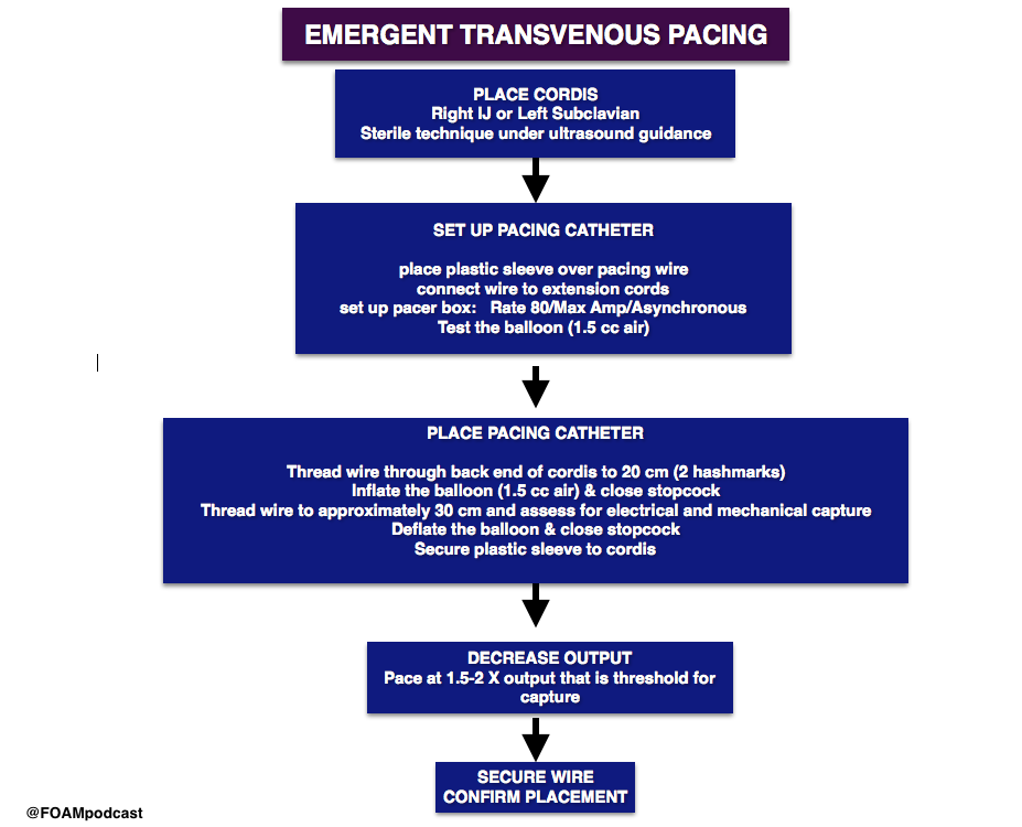
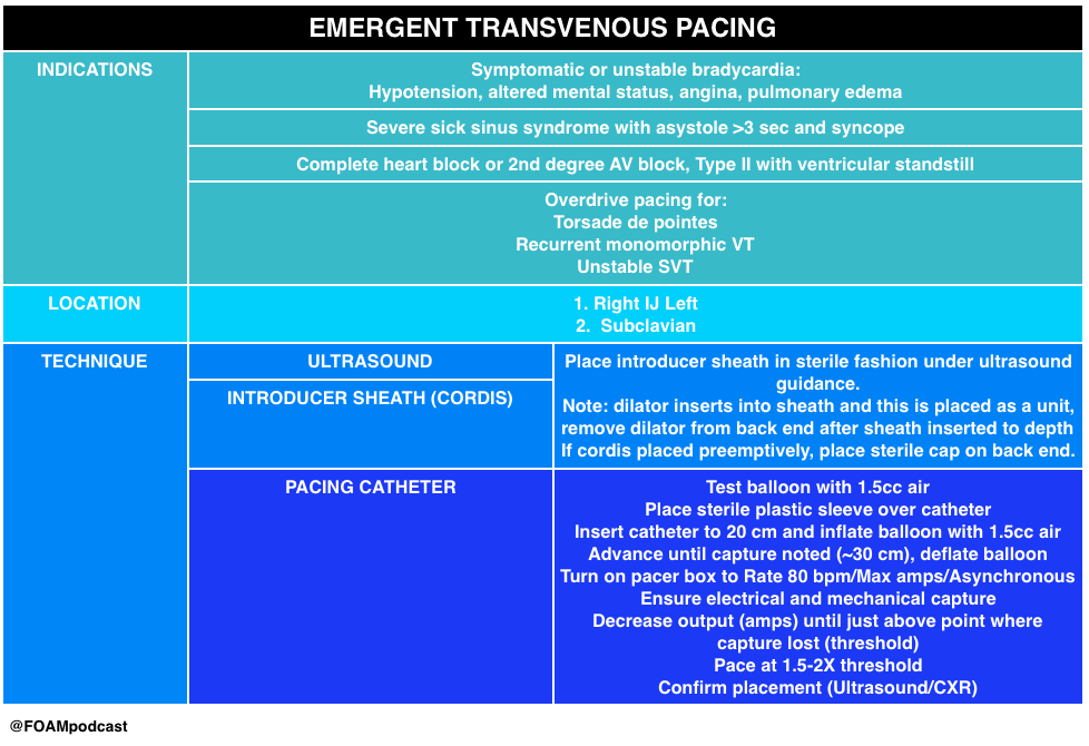
Emergent transvenous pacing is a crucial skill for emergency physicians.  It may be daunting due to the pacing box and various catheters. We have found that  routinely rehearsing the procedure, reviewing where pacing equipment is in our departments, and where

In this episode we review the following videos:

  • Practical Pointers for Pacemaker Placement by Dr. Jason Nomura
  • Transvenous pacing video by Dr. Al Sachetti
  • Pacing 101 (Transcutaneous is Just Stupid) by Dr. Joe Bellezo on the Ultrasound Podcast
    • This podcast lays down the argument that transcutaneous pacing is stupid.  Transcutaneous pacing is difficult – patients are diaphoretic, capture rates may be 40%, and it takes a significant amount of energy. Further, it hurts.
      • However, in our opinion, there may be a role.  In this talk, Dr. Bellezo quotes a 1981 study, in which emergent pacers were placed in “6 minutes.”  Review of this study finds that this was actually 6 minutes, 45 seconds (closer to 7 minutes) [1].  This likely does not reflect the majority of emergency providers experience, certainly not ours, where the range is more often 15-30 minutes for the procedure.  Thus, transcutaneous pacing may be a temporizing measure as one locates the ultrasound, gathers the supplies, and prepares for transvenous pacing in the unstable patient.

transvenous pacing

pacing

Core Content

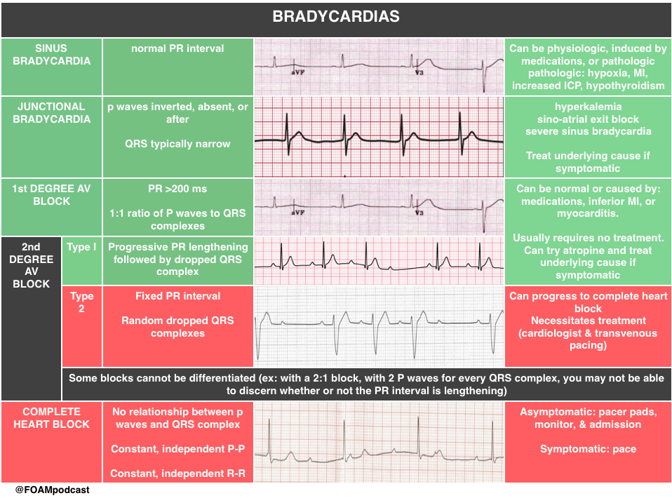
We delve into core content on bradycardias and heart blocks using Rosen’s Emergency Medicine (8th edition) Chapter 79 “Dysrthymias” and Tintinalli’s Emergency Medicine (8th edition) Chapter 18  “Cardiac Rhythm Disturbances” as a guide.

bradycardias

bradycardia

 Rosh Review Emergency Board Review Questions

Question 1a.

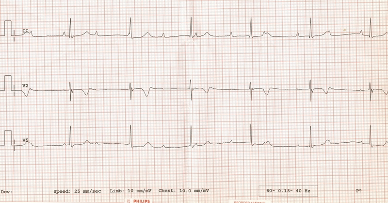
A 71-year-old woman presents after a fall at home. Her electrocardiogram is shown below.

content_ecg_-3rd_degree_heart_block

[accordion]
[toggle title=”Answer” state=”closed”]

Third Degree Heart block.

[/toggle]
[/accordion]

Question 2.

A. Administration of epinephrine

B. Defibrillation

C. Observation

  • D. Placement of transcutaneous pacer pads

[accordion]
[toggle title=”Answer” state=”closed”]

D. The patient has third degree heart block. In third degree atrioventricular (AV) block, also known as complete heart block, there is no conduction through the AV node, and an escape pacemaker is responsible for the ventricular rate. On electrocardiogram, P waves occur at regular intervals, and QRS complexes occur at regular intervals, but there is no association between the P waves and QRS complexes. When the block occurs in the AV node, a junctional escape pacemaker takes over with a rate of 40-60 beats/minute, and the QRS complex is narrow. If the block occurs at the infranodal level, a ventricular escape pacemaker paces at a rate of 40 beats/minute or less. Infranodal blocks results in a wide QRS complex. Patients with third degree heart block require cardiac pacing, as the slow escape rhythm is rarely adequate to maintain cardiac output and tissue perfusion. Transcutaneous pacing should be initiated while arrangements for transvenous pacing are made. Third degree blocks are commonly associated with cardiac ischemia or infarction. A nodal third degree block (narrow QRS complex) is a complication of acute inferior wall myocardial infarction, and may last for several days. Extensive acute anterior wall infarction is associated with infranodal third degree blocks (wide complex QRS), indicating damage to the infranodal conduction system. When a third degree heart block is seen with acute myocardial infarction, mortality is increased.

Administration of epinephrine (A) is incorrect. Defibrillation (B) the treatment for cardiac arrest from ventricular fibrillation or pulseless ventricular tachycardia.  Observation (C) is incorrect since the patient’s slow heart rate is likely not adequate to maintain cardiac output.

[/toggle]
[/accordion]

Question 3.

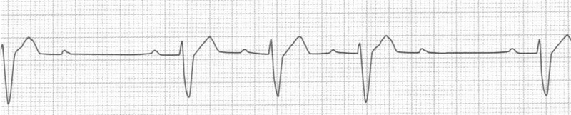
A 56-year-man presents to the ED with right arm pain and some chest discomfort. The day prior to arrival, he tried using heavier weights at the gym. He has a history of hypertension, hyperlipidemia, and COPD. In the ED, his vital signs are BP 136/90, HR 60, RR 16, and oxygen saturation 97% on room air. His rhythm strip is seen below. Which is the most appropriate management for this rhythm?

content_ecg_-_second_degree_heart_block_type_ii

A. Aspirin

B. Cardioversion

C. Observation

D. Temporary pacing

[accordion]
[toggle title=”Answer” state=”closed”]

D. This ECG demonstrates type II second-degree heart block. Second-degree heart block is defined by one or more impulses not reaching the ventricles and is classified as type I and type II. Type II second-degree heart block, also known as Mobitz II, is caused by an infranodal conduction abnormality, most commonly in the bundle of His or the purkinje fibers. ECG findings demonstrate random dropped QRS complexes without any changes in the PR interval. Type II second-degree heart block carries a worse prognosis than type I second-degree heart block and necessitates treatment. Unlike type I, atropine has no effect on the His-Purkinje system and may worsen conduction. Temporary pacing is critical in this case because this rhythm can devolve to complete heart block. In the ED, transcutaneous or transvenous pacing should be instituted if the patient is symptomatic and there should be immediate consultation with a cardiologist. Patients with Mobitz II in the setting of an acute myocardial infarction should be treated with temporary pacing and revascularization; following revascularization most conduction abnormalities will improve or resolve and will not require permanent pacing.

[/toggle]

[/accordion]

Question 4.

A man who presents with syncope is placed on the cardiac monitor. On the monitor you note a repeating trend of 6 P waves, 5 of which are followed by a narrow QRS complex and 1 of which is not followed by a QRS complex. The PR interval during this trend progressively increases. Which of the following is the most likely diagnosis?

A. First-degree AV block

B. Third-degree AV block

C. Type I second-degree AV block

D. Type II second-degree AV block

[accordion]
[toggle title=”Answer” state=”closed”]

C. A key distinction between first-degree and second-degree heart block is that in first-degree block the P wave is always followed by a QRS complex. In other words, the ratio of P waves to QRS complexes is 1:1, or, the electrical signal from the atria always passes to the ventricles. In second-degree AV block, the electrical impulse sometimes gets to the ventricles. There are two main types of second-degree AV block. In Mobitz type I, or Wenckebach, second-degree block, there is a progressive beat-to-beat lengthening of the PR interval until a P wave does not conduct through the AV node. The absent conduction and resultant “missing” QRS complex is called a “dropped” QRS, which represents an absent beat of ventricular contraction. First-degree AV block (A) has a 1:1 ratio of P waves to QRS complexes. Mobitz type II second-degree heart block (D) is characterized by a nonconducted P wave which is not preceded by progressive PR interval prolongation. AV dissociation, or third-degree AV block (B), occurs when none of the P waves conduct through the AV node. This complete AV block occurs with separate atrial and ventricular rates. There is no discrete correlation or trend between P waves and QRS complexes.

[/toggle]

[/accordion]

References:

  1. Lang R, David D, Klein HO, et al. The use of the balloon-tipped floating catheter in temporary transvenous cardiac pacing. Pacing Clin Electrophysiol. 1981;4(5):491-6.
  2. Roberts J and Hedges J. “Emergency Cardiac Pacing.” Roberts and Hedges’ Clinical Procedures in Emergency Medicine

Suviving Sepsis Campaign Guidelines 2017

iTunes or Listen Here

The new Surviving Sepsis Campaign Guidelines are out in Critical Care Medicine.  The biggest change is the change in the definition of sepsis, which now uses the language of Sepsis 3.0, introduced in 2016. Other significant changes include dropping protocolized care (ex: early goal directed therapy), and recommending against combination antibiotic therapy (double coverage) for a single pathogen [1,2].

Bonus discussion on new validation study of qSOFA

Screen Shot 2017-01-19 at 4.02.07 PM

References:

  1. Opal SM, Rubenfeld GD, Poll T Van Der, Vincent J, Angus DC. The Third International Consensus Definitions for Sepsis and Septic Shock (Sepsis-3). 2016;315(8):801–10.
  2. Rhodes A, Evans LE, Alhazzani W, et al. Surviving Sepsis Campaign: International Guidelines for Management of Sepsis and Septic Shock: 2016. Crit Care Med [Internet] 2017;1.
  3. Keh D, Trips E, Marx G, et al. Effect of Hydrocortisone on Development of Shock Among Patients With Severe Sepsis: The HYPRESS Randomized Clinical Trial. JAMA. 2016;316(17):1775-1785.
  4. Seymour CW, Liu VX, Iwashyna TJ, et al. Assessment of Clinical Criteria for Sepsis. Jama [Internet] 2016;315(8):762.
  5. Freund Y, LeMachatti N, Krastinova E.Prognostic Accuracy of Sepsis-3 Criteria for In-Hospital Mortality Among Patients W.ith Suspected Infection Presenting to the Emergency Department. JAMA. 2017;317(3):267-268.

Roundup: Favorite Literature of 2016

(iTUNES OR LISTEN HERE)

Article highlights of 2016

Sepsis 3.0

Singer M, Deutschman CS, Seymour CW, et al: The Sepsis Definitions Task Force The Third International Consensus Definitions for Sepsis and Septic Shock (Sepsis-3).

  • This iteration of sepsis discarded systemic inflammatory response syndrome (SIRS), noting it’s poor discriminatory ability.  Further, these authors sought to define sepsis as a dysregulated response to infection, not just a normal response to infection (i.e. fever and tachycardia are normal responses to infection and therefore are, alone, not evidence of sepsis).

Sepsis 3.0

The quick SOFA score (qSOFA) also came out in hopes it “provides simple bedside criteria to identify adult patients with suspected infection who are likely to have poor outcomes.”  It is not part of the sepsis definition  but may help in identifying those that will die or have a 3 day ICU stay.  Studies looking at the performance of qSOFA applied retrospectively to data sets have not been promising [1,2].

Back Up Head Elevated Intubation

Khandelwal et al. Head-elevated Patient Positioning Decreases Complications of Emergent Tracheal Intubation in the Ward and Intensive Care Unit. Anesthesia and Analgesia. Apr 2016.

back up head elevated intubation

Ketorolac Dose

Motov, S, Yasavolian, M, Likourezos, A, et al. Comparison of Intravenous Ketorolac at Three Single-Dose Regimens for Treating Acute Pain in the Emergency Department. .Ann Emerg Med. 2016 Dec 16.

ketorolac

Pulmonary Embolism in Syncope 

Prandoni et al. Prevalence of Pulmonary Embolism among Patients Hospitalized for Syncope.   N Engl J Med 2016; 375:1524-1531. This trial is probably one of the most over-reacted to trials of 2016.  News outlets, both by lay media and for health professionals overstated the claims of this trial.

image

What the trial actually did…

PESIT

Out of Hospital Cardiac Arrest Prognostication

Jabre et al. Early Identification of Patients With Out-of-Hospital Cardiac Arrest With No Chance of Survival and Consideration for Organ Donation.   Intern Med. 2016 Dec 6;165(11):770-778

OHCA

ALTE (Apparent Life Threatening Event) is out, replaced with a new definition and classification, BRUE (Brief Resolved Unexplained Event).

Tieder JS, Bonkowsky JL, Etzel RA, Franklin WH, Gremse DA, Herman B et al. Subcommittee on Apparent Life Threatening Events. Brief Resolved Unexplained Events (Formerly Apparent Life-Threatening Events) and Evaluation of Lower-Risk Infants: Executive Summary.  Pediatrics. 2016

Screen Shot 2016-05-04 at 9.33.37 AM

References:

  1. Churpek MM, Snyder A, Han X et al. qSOFA, SIRS, and Early Warning Scores for Detecting Clinical Deterioration in Infected Patients Outside the ICU. Am J Respir Crit Care Med. 2016. [article]
  2. Brabrand M, Havshøj U, Graham CA. Validation of the qSOFA score for identification of septic patients: A retrospective study. European Journal of Internal Medicine. 36:e35-e36. 2016. [article]

Episode 62 – Dental Emergencies

iTunes or Listen Here

We review a trick of the trade from Academic Life in Emergency medicine for temporomandibular joint (TMJ) dislocation, the extra-oral reduction.

TMJ dislocation reduction

Traditional Approach to TMJ dislocation and Syringe Technique from Core EM

Core Content

We delve into core content on dental injuries using Rosen’s Emergency Medicine (8th edition) Chapter 70 “Oral Emergencies” and Tintinalli’s Emergency Medicine (8th edition) Chapter 245 “Oral and Dental Emergencies” as a guide.

dental infections

dental trauma

mandible fracture

 Rosh Review Emergency Board Review Questions

A 19-year-old man presents to the Emergency Department with an avulsed tooth. He struck his mouth on the back of another player’s head while playing basketball. He arrives thirty minutes after the injury with his right maxillary central incisor in a bag of cold milk. Which of the following is the most appropriate management?

A. Discharge home with next day dental follow up

B. Fill the alveolar socket with eugenol oil

C. Reimplant the avulsed tooth

D. Scrub the tooth with normal saline

[accordion]
[toggle title=”Answer” state=”closed”]

C. Reimplant the avulsed tooth (B) is the most appropriate next step. Avulsed permanent teeth should be reimplanted as soon as possible, ideally within 30 minutes of the injury. For every minute that the tooth is out of its socket, there is a 1% chance of reimplantation failure. If the tooth is not able to be reimplanted immediately, it should be stored in an appropriate medium. Cold milk is preferable to sterile water or saliva as it has magnesium and calcium. Hank’s solution, a neutral cell culture medium, is ideal. Once the tooth is reimplanted, the tooth should be stabilized until dental follow up is arranged. Avulsed primary teeth should not be reimplanted.

[/toggle]
[/accordion]

A 25 year-old man presents after falling face forward off his bike.  He sustained an abrasion inside his upper lip and complains of a broken front tooth. He brought the fractured fragment with him. On examination, the bony structures of the jaw are non-tender. There is no malocclusion. Tooth #8 has a fracture and in the center of the exposed area is a small pink dot. What is the most appropriate plan for this patient?

A. Dental follow-up within the next 24 hours

B. Irrigation of the tooth

C. Placement of the tooth fragment in Hank’s solution

D. Viscous lidocaine for pain control

[accordion]
[toggle title=”Answer” state=”closed”]

A. This patient has a dental fracture with exposed pulp. This is a dental emergency requiring dental follow-up within the next 24 hours. The most superficial dental fractures involve only the enamel on the surface and treatment is mostly cosmetic and aimed at dulling any sharp edges. Fractures that expose dentin will have an ivory-yellow appearance. In younger patients, there is less dentin relative to the pulp and treatment is aimed at protecting any pulp contamination with placement of a calcium-hydroxide dressing. Younger patients need more urgent follow-up with a dentist. The most significant dental fractures involve the pulp as in this clinical scenario. The tooth should be gently wiped clean with gauze and inspected for a drop of blood or pink blush which represents pulp exposures. The area is usually exquisitely painful. Timely follow-up (within 24 hours) is required for evaluation and possible root canal and extraction of the pulp. If dental follow-up will be delayed, the tooth should be covered with moist cotton and sealed with dry foil or a temporary commercial sealant. In order to clean the tooth, a clean gauze should be used to wipe off the surface, not irrigation of the tooth (B) as the patient will be extremely sensitive to that and all efforts should be maintained to avoid pulp contamination. Hank’s solution (C) is a physiologic solution in which an avulsed tooth may be placed while waiting for reimplantation into the socket. There is no role in a partial tooth fracture. Viscous lidocaine (D) is not an appropriate analgesic for a dental fracture. Oral analgesics or dental block should be provided for pain control.

[/toggle]
[/accordion]

References

  1. Gorchynski J, Karabidian E, Sanchez M. The “syringe” technique: a hands-free approach for the reduction of acute nontraumatic temporomandibular dislocations in the emergency department. The Journal of emergency medicine. 47(6):676-81. 2014. [pubmed]
  2. Caputo ND, Raja A, Shields C, Menke N. Re-evaluating the diagnostic accuracy of the tongue blade test: still useful as a screening tool for mandibular fractures? The Journal of emergency medicine. 45(1):8-12. 2013. [pubmed]
  3. Neiner J, Free R, Caldito G, Moore-Medlin T, Nathan CA. Tongue Blade Bite Test Predicts Mandible Fractures. Craniomaxillofacial trauma & reconstruction. 9(2):121-4. 2016. [pubmed]

Episode 61 – The Elbow

iTunes or Listen Here

We review a podcast from Dr. Tim Horeczko’s Pediatric Emergency Playbook on elbow injuries.

critoe

Core Content

We delve into core content on other elbow adjacent injuries using Rosen’s Emergency Medicine (8th edition) Chapter  and Tintinalli’s Emergency Medicine (8th edition) Chapter  as a guide.

Elbow Trauma

olecranon bursitis

 Rosh Review Emergency Board Review Questions

A 63-year-old man presents with left arm pain after a fall. His X-ray is shown below. What structure is commonly injured with this fracture?

A. Axillary nerve

B. Median nerve

C. Radial nerve

D. Ulnar nerve

[accordion]
[toggle title=”Answer” state=”closed”]

C. Radial nerve injury is the most common nerve injury seen after humeral shaft fractures. These fractures usually occur from a direct blow to the arm and can be seen in falls and motor vehicle collisions. Patients present with severe pain, arm swelling and decreased range of motion. The arm can be shortened or rotated in a complete fracture depending on the location of the fracture. A complete neurovascular exam should be performed as with all fractures and dislocations. The radial nerve may be injured during humeral fracture in up to 20% of patients. The injury is usually a neuropraxia and resolves spontaneously in most patients. However, this recovery can take months. Humeral fractures rarely need specific reduction maneuvers for treatment. They should be placed in a sugar tong splint and placed in a sling. Gravity alone is typically successful in fracture reduction. The axillary nerve (A) may be injured during glenohumeral dislocations. The median nerve (B) may be injured during posterior elbow dislocations. Anterior elbow dislocations can be associated with ulnar nerve injury (D).

[/toggle]
[/accordion]


A 3-year-old girl was walking on the sidewalk with her mom when she fell onto the street. In a panicked state, her mom picked up the little girl by her arm. Immediately after, the little girl refused to move her right arm complaining that it hurt. In the emergency room, the girl is holding her right arm in a flexed, pronated, and adducted position. There is no crepitus, swelling or point tenderness along the entire right arm or clavicle. Which of the following is the next step in management of this patient?

A. Actively supinate and flex the elbow while applying pressure over the radial head

B. Consult orthopedics for casting

C. Obtain an ultrasound

D. Perform a skeletal survey

[accordion]
[toggle title=”Answer” state=”closed”]

A. This child has “nursemaid’s elbow” that is due to subluxation of the annular ligament rather than dislocation of the radial head. The etiology is slippage of the head of the radius under the annular ligament. The distal attachment of the annular ligament covering the radial head is weaker in children than in adults, allowing it to be more easily torn. It occurs in toddlers due to traction via pulling on a pronated and extended arm. The child immediately refuses to the move the arm and often cradles the affected arm. Parents often give a history of a young child with no history of trauma who suddenly refuses to use an arm. The diagnosis is clinical and imaging studies are generally not needed. If reduction is unsuccessful after 2–3 attempts then imaging studies may be warranted. Treatment is manual reduction via supination and flexion or hyperpronation. A palpable click may be felt and the child usually regains immediate movement of the arm and relief of discomfort. A skeletal survey (D) should be obtained in all cases of suspected child abuse to assess for fractures in multiple stages of healing. Child abuse should be on the differential in all pediatric orthopedic cases. Consulting orthopedics for casting (B) is not necessary as this is a dislocation injury. Ultrasonography (C) has been used as a noninvasive modality to assess for annular ligamentous injury and displacement of the radial head from the capitellum. It has also been used to assess progress of treatment for patients with recurrent subluxations, however it is not the first-line diagnostic nor treatment option.

[/toggle]
[/accordion]

Episode 60 – Resuscitative Hysterotomy + First Trimester Emergencies

iTunes or Listen Here

We review a talk by Dr. Sara Gray from SMACC (Social Media and Critical Care) conference in June 2016, Resuscitative hysterotomy, which is the new name for perimortem c-section [1]. In this talk she challenges the “4 minute” rule: if resuscitative efforts following maternal circulatory arrest are unsuccessful, cesarean delivery should be commenced at 4 minutes and completed by 5 minutes to optimize fetal outcome. 

resuscitative hysterotomy

Core Content

We delve into core content on pregnancy emergencies using Rosen’s Emergency Medicine (8th edition) Chapter 98and Tintinalli’s Emergency Medicine (8th edition) Chapter 178 as a guide.

first trimester bleeding

septic abortion

Nausea and vomiting are very common in pregnancy, but few patients have hyperemesis gravidarum (~2%).  Hyperemesis gravidarum is characterized by severe nausea and vomiting with starvation ketosis and significant weight loss and dehydration [2,3].

first trimester vomiting

uti in pregnancy

 Rosh Review Emergency Board Review Questions

A 22-year-old woman presents complaining of vaginal bleeding and cramping for the last 4 hours. She is known to be 14-weeks pregnant. Her cervical os is dilated to 4 cm and she is actively bleeding. Pelvic ultrasound shows the gestational sac in the lower uterine segment near the cervix. Which of the following is the most likely diagnosis?

A. Complete abortion

B. Inevitable abortion

C. Missed abortion

D. Septic abortion

[accordion]
[toggle title=”Answer” state=”closed”]

The patient is experiencing an inevitable abortion, which is characterized by an open cervical os and a gestational sac at the opening of the uterus on ultrasound. The case should be discussed with the patient’s obstetrician as the patient may ultimately require dilatation and curettage if all the products of conception (POC) do not pass spontaneously or the bleeding is not controlled.  A complete abortion (A) occurs when the patient has passed all POC have passed. On examination, the cervix is closed and the uterus is firm and nontender. A missed abortion (C) occurs when a pregnant patient fails to pass the products of conception greater than two months after fetal demise. The pregnancy test will be negative, however ultrasound will show retained POC. A septic abortion (D) occurs when the patient develops foul-smelling discharge, vaginal bleeding, uterine tenderness and peritoneal signs following a spontaneous or induced abortion.

[/toggle]
[/accordion]

[accordion]
[toggle title=”What is the most common cause of bleeding in the primigravid woman?” state=”closed”] 

Threatened abortion

[/toggle]
[/accordion]

References:

  1. Rose CH, Faksh A, Traynor KD, Cabrera D, Arendt KW, Brost BC. Challenging the 4- to 5-minute rule: from perimortem cesarean to resuscitative hysterotomy. American journal of obstetrics and gynecology. 213(5):653-6, 653.e1. 2015. [pubmed]
  2. Heaton, H. “Chapter 98: Ectopic Pregnancy and Emergencies in the First 20 Weeks of Pregnancy.” Tintinalli’s Emergency Medicine: A Comprehensive Review. 8th edition
  3. “Chapter 178: Acute Complications of Pregnancy.”  Rosen’s Emergency Medicine

Episode 59 – Syncope (and the PESIT study)

ITUNES OR LISTEN HERE

The PESIT study in the New England Journal of Medicine stirred up controversy in the FOAM world earlier in October 2016.  In this episode we cover the following posts on this article on pulmonary embolism in syncope:

screen-shot-2016-10-25-at-7-52-02-pm

Core Content

We delve into core content on syncope usingRosen’s Emergency Medicine (8th edition) and Tintinalli’s Emergency Medicine (8th edition) Chapter 52

screen-shot-2016-10-31-at-4-41-19-pm

 Rosh Review Emergency Board Review Questions

An 83-year-old is being evaluated in the emergency department after an episode of syncope. The woman was preparing dinner when she felt her heart start to race. The next thing she remembers is waking up on the floor. She experienced a similar episode about three weeks ago. She has never had anything like this before. Her past medical history is remarkable for hypertension, hyperlipidemia and hypothyroidism. Her medications include lisinopril, atorvastatin and levothyroxine. On physical exam her blood pressure is 142/83, heart rate 76/min, and respiration rate 13/min. Cardiac auscultation reveals no murmur. The remainder of her physical exam is normal. Electrocardiogram reveals normal sinus rhythm with left axis deviation. No cardiac rhythm abnormalities are detected. What is the most likely etiology of this patient’s syncope?

A. Aortic stenosis

B. Cardiac dysrhythmia

C. Orthostatic hypotension

D. Vasovagal

[accordion]
[toggle title=”Answer” state=”closed”]

B. Cardiac dysrhythmia is the most likely cause of this woman’s syncope. Cardiac dysrhythmias are a common cause of syncope in the elderly population. It is characterized by a brief or absent prodrome and palpitations immediately preceding the event. Several episodes over a short period of time in someone with no history of syncope suggest a dysrhythmia. Given this patient’s short prodrome, palpitations and history of a previous similar event makes a cardiac dysrhythmia the most likely etiology.

Aortic stenosis (A) is unlikely the cause of her syncope. Aortic stenosis is associated with a crescendo-decrescendo systolic ejection murmur. Syncope related to aortic stenosis typically occurs during exertion and is associated with very severe disease. This patient’s syncopal episode occurred while stationary. Additionally, she has no systemic symptoms of aortic stenosis.Vasovagal (D) is the most common cause of syncope in the general population. It is usually triggered by provoking factors such a blood draw or an intense emotion. Prodromal symptoms include feeling warm, sweating, nausea, and pallor. This woman does not report any of these symptoms. Orthostatic hypotension (C) causes syncope upon assuming an upright position from supine or sitting. It is often caused by hypovolemia, medications or autonomic nervous system disorders. This woman was standing while preparing dinner making orthostatic hypotension unlikely.


[/toggle]
[/accordion]

An 18-year-old woman presents after having a syncopal episode. She is complaining of a 2-day history of lower abdominal pain and vaginal spotting. Her BP is 86/42, HR is 128, RR is 18 breaths, and oxygen saturation is 99% on room air. She is drowsy, but answers questions appropriately. What is the most appropriate next step in management?


A. Establish large-bore IV access and administer an IV fluid bolus

B. Initiate rapid sequence induction and orotracheal intubation

C. Perform a bedside urine pregnancy testing

D. Perform an ultrasound of the abdomen to assess for free fluid

[accordion]
[toggle title=”Answer” state=”closed”]

A. The patient is hypotensive and tachycardic. She is suffering from hypovolemic shock secondary to a ruptured ectopic pregnancy. Therefore she requires immediate intravenous access and volume resuscitation with Lactated Ringer’s or normal saline.  Emergency Department management of unstable patients includes rapid assessment of the ABC’s (Airway, Breathing, Circulation). This patient is phonating, has a respiratory rate of 18 breaths per minute and an oxygen saturation of 99% on room air.  There is no concern that her airway or breathing is in immediate jeopardy, therefore she would not require immediate rapid sequence induction and orotracheal intubation (B). Although a bedside pregnancy test (C) and abdominal ultrasound (D) would help make a diagnosis of ruptured ectopic pregnancy, the next step would be to resuscitate the patient.

[/toggle]
[/accordion]

References

  1. De Lorenzo RA. “Syncope.” Chapter 15. Rosen’s Emergency Medicine (8 ed). pp 131-145
  2. Chapter 52. Tintinalli’s Emergency Medicine: A Comprehensive Review (8 ed).
  3. Serrano LA, Hess EP, Bellolio MF et al. Accuracy and Quality of Clinical Decision Rules for Syncope in the Emergency Department: A Systematic Review and Meta-analysis. Annals of Emergency Medicine. 56(4):362-373.e1. 2010.

Bell’s Palsy and Burns

(ITUNES OR LISTEN HERE)

We are in Las Vegas at ACEP 2016 thanks to Annals of Emergency Medicine and ACEPnow and discuss high yield and cutting edge lectures each day.

Dr. Megan Osborn – Bell’s Palsy or Stroke?

Traditional teaching: we can differentiate Bell’s palsy (lower motor neuron) from a stroke (upper motor neuron by assessing forehead involvement.  If the patient can wrinkle their forehead? Think stroke.  Dr. Megan Osborn tackled the question: does this actually work all the time in her talk in the New Speakers Forum.

screen-shot-2016-10-19-at-8-19-31-am

Dr. Toree McGowan – Burns

Check out this podcast for more on burns

screen-shot-2016-10-19-at-8-33-56-am

  1. Sherman SC, Thompson TM, Thompson TT. Pontine hemorrhage presenting as an isolated facial nerve palsy. Annals of emergency medicine. 46(1):64-6. 2005. [pubmed]
  2. AAN Bell’s Palsy Guideline Update
  3. Fahimi J, Navi BB, Kamel H. Potential Misdiagnoses of Bell’s Palsy in the Emergency Department. Annals of Emergency Medicine. 63(4):428-434. 2014. [article]
  4. Madhok VB, Gagyor I, Daly F. Corticosteroids for Bell’s palsy (idiopathic facial paralysis). The Cochrane database of systematic reviews. 7:CD001942. 2016. [pubmed]
  5. Gagyor I, Madhok VB, Daly F. Antiviral treatment for Bell’s palsy (idiopathic facial paralysis). The Cochrane database of systematic reviews. 2015. [pubmed]
  6. Wasiak J, Cleland H, Campbell F. Dressings for superficial and partial thickness burns. The Cochrane database of systematic reviews. 2008. [pubmed]
  7. Ringh M, Rosenqvist M, Hollenberg J et al. Mobile-Phone Dispatch of Laypersons for CPR in Out-of-Hospital Cardiac Arrest. N Engl J Med. 372(24):2316-2325. 2015. [article]

Vaccine Side Effects and Placebos

(ITUNES OR LISTEN HERE)

We are in Las Vegas at ACEP 2016 thanks to Annals of Emergency Medicine and ACEPNOW and discuss high yield or cutting edge lectures each day.

Dr. Matthew DeLaney – 21st Century Snake Oil (Placebos)

screen-shot-2016-10-17-at-5-30-30-pm

SGEM http://thesgem.com/2013/03/sgem26-honey-honey/

Dr. Al Sachetti – Immunization Reactions in the Emergency Department

check out the WHO fact sheets

screen-shot-2016-10-18-at-5-34-43-am

Dr. Corey Slovis – Atrial Fibrillation

screen-shot-2016-10-18-at-6-01-08-am

References:

  1. Llor C, Moragas A, Bayona C. Efficacy of anti-inflammatory or antibiotic treatment in patients with non-complicated acute bronchitis and discoloured sputum: randomised placebo controlled trial. BMJ (Clinical research ed.). 347:f5762. 2013. [pubmed]
  2.  Smith et al. Over-the-counter (OTC) medications for acute cough in children and adults in ambulatory settings. Cochrane Database of Systematic Reviews 2012, Issue 8. Art. No.: CD001831. DOI: 10.1002/14651858.CD001831.pub4.22895922
  3. Dobson R. Cough medicines’ effect is mainly placebo. BMJ. 2006 Jan 7; 332(7532): 8. PMCID: PMC1325161
  4. Cohen et al. Effect of Honey on Nocturnal Cough and Sleep Quality: A Double-blind, Randomized, Placebo-Controlled Study Pediatrics; originally published online August 6, 2012; PMID:22869830
  5. Egerton-Warburton D, Meek R, Mee MJ, Braitberg G. Antiemetic use for nausea and vomiting in adult emergency department patients: randomized controlled trial comparing ondansetron, metoclopramide, and placebo. Annals of emergency medicine. 64(5):526-532.e1. 2014. [pubmed]
  6. Furyk JS, Meek RA, Egerton-Warburton D. Drugs for the treatment of nausea and vomiting in adults in the emergency department setting. The Cochrane database of systematic reviews. 2015. [pubmed]
  7. Beadle KL, Helbling AR, Love SL, April MD, Hunter CJ. Isopropyl Alcohol Nasal Inhalation for Nausea in the Emergency Department: A Randomized Controlled Trial. Annals of emergency medicine. 68(1):1-9.e1. 2016. [pubmed]
  8.  Friedman BW, Dym AA, Davitt M. Naproxen With Cyclobenzaprine, Oxycodone/Acetaminophen, or Placebo for Treating Acute Low Back Pain: A Randomized Clinical Trial. JAMA. 314(15):1572-80. 2015. [pubmed]
  9. Derry S, Conaghan P, Da Silva JA, Wiffen PJ, Moore RA. Topical NSAIDs for chronic musculoskeletal pain in adults. The Cochrane database of systematic reviews. 4:CD007400. 2016. [pubmed]
  10. Silberban. Placebos Are Getting More Effective. Drugmakers Are Desperate to Know Why.

Care of transgender patients + more from ACEP

Listen Here or iTunes

We are in Las Vegas at ACEP 2016 and discuss high yield or cutting edge lectures each day.

Dr. Anne Daul – Emergency Care of the Transgender Patient

Most emergency medicine physicians and trainees lack training in caring for LGBTQ patients, let alone transgender patients [1].  Many members of the LGBTQ community may have delays in medical care including 21% of transgender patients in a Canadian survey[2].  Also, according to a 2010 task force, 19% of transgender patients report being denied care in some way [3].

@FOAMpodcast
@FOAMpodcast

Here is a video from SMACC Dublin from Thom O’Neill on caring for LGBT youth

https://www.youtube.com/watch?v=7ZsYgftEv2U&feature=youtu.be

Check out these FOAM resources from EPMonthly and Mayo.

Dr. David Callaway – Active Shooter

2% of active shooter events take place in the health care setting.

Plan of Action in Active Shooter Scenario: AVOID DENY DEFEND TREAT

  • Avoid – stay away from the shooter.
  • Deny – deny them access to you or the area. Lock doors, block pathways, turn off the lights, make it more difficult for them.
  • Defend -.if necessary, defend yourself.
  • Treat – once you are safe, and the scene is safe, treat and care for your patients.

Dr. Kevin Klaur – Lawsuits

Documentation and discharge instructions repeatedly come up in lawsuits.

  • Documentation: If you document after the fact, particularly if there was a bad outcome – be straightforward that you are documenting after the fact. Do not document as though you do now know the outcome
  • Discharge – lawsuits often come up because discharge instructions or documentation were not sufficient. Klauer argues that it is not sufficient to state “patient improved, discharged home.” He urges us to document a repeat exam or show HOW they are improved.

Dr. Klauer also gave some general pearls on lawsuits – high numbers for orthopedics/missed fractures and administration of RhoGham.  An additional pearl he gave was for cauda equina.

  • These patients often have small post void residuals because it’s a neurogenic problem, not a mechanical obstruction.  Thus, if a patient has other features and has a post void residual of 100 cc, it’s not necessarily not cauda equina.


One ACEP16 lecturer talked about magnesium use in alcohol withdrawal – probably not ready for prime time, Cochrane agrees  [4].

References:

  1. Moll J, Krieger P, Moreno-Walton L. The prevalence of lesbian, gay, bisexual, and transgender health education and training in emergency medicine residency programs: what do we know? Academic emergency medicine : official journal of the Society for Academic Emergency Medicine. 21(5):608-11. 2014. [pubmed]
  2. Bauer GR, Scheim AI, Deutsch MB, Massarella C. Reported emergency department avoidance, use, and experiences of transgender persons in Ontario, Canada: results from a respondent-driven sampling survey. Annals of emergency medicine. 63(6):713-20.e1. 2014. [pubmed]
  3. Grant JM, Mottet LA, Tanis JD et al. National Transgender Discrimination Survey Report on health and health care Findings of a Study by the National Center for Transgender Equality and the National Gay and Lesbian Task Force.  October 2010
  4. Tejani SM. Magnesium for the prevention or treatment of alcohol withdrawal syndrome in adults. June 2013